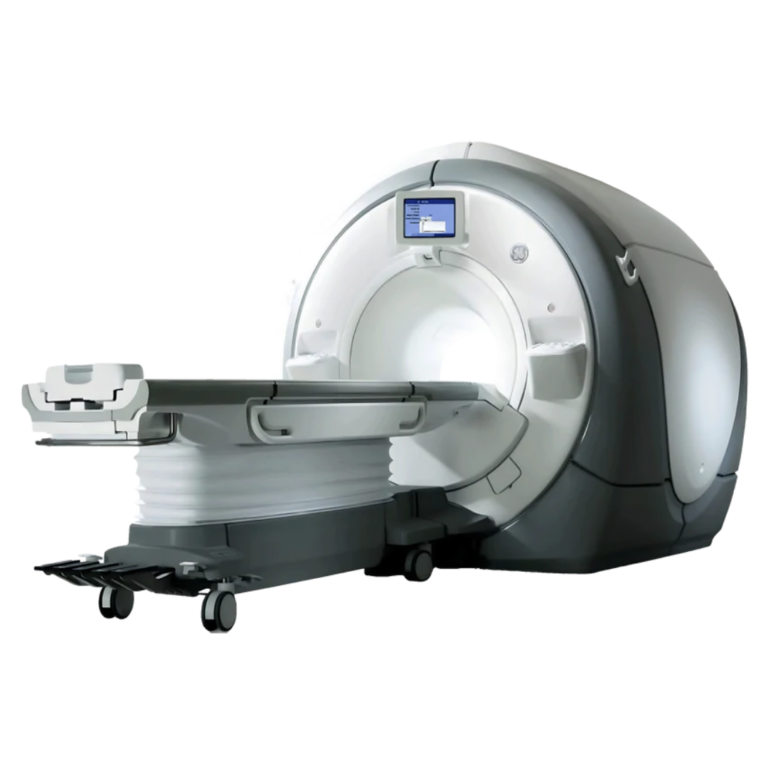
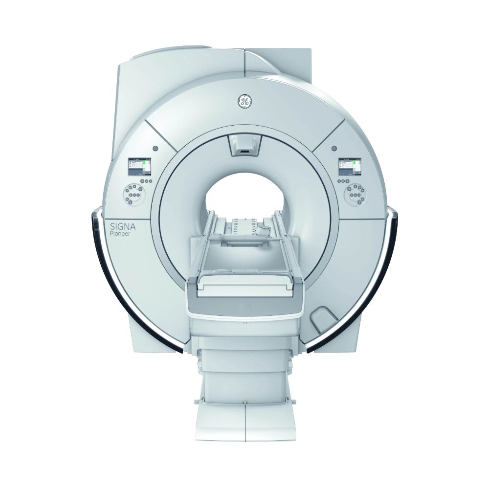
GE SIGNA Pioneer 3T MRI
- Category:
- MRI Systems
- Brand:
- GE
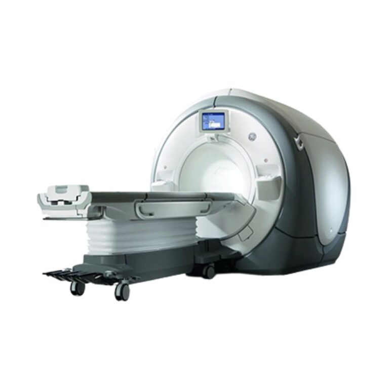
The GE SIGNA Pioneer 3T MRI is a wide-bore scanner well suited for hospitals, imaging centers, and specialty practices seeking detailed imaging with efficient operation. Its 70 cm bore, energy-efficient gradient technology, and patient-friendly design combine advanced diagnostic capabilities with comfortable positioning and reduced operating demands.
SIGNA Pioneer MRI Highlights & Benefits
The GE SIGNA Pioneer combines 3T imaging performance with an emphasis on patient comfort, efficient workflows, and lower operating demands. Its Ultra High Efficiency gradients, digital imaging architecture, and wide-bore design support comprehensive diagnostic examinations in a space-conscious system.
Key Features & Benefits
-
- 3T High-Resolution Imaging: A superconducting magnet supports detailed neurological, musculoskeletal, body, and advanced clinical examinations.
- 70 cm Wide Bore: Provides more patient space than conventional 60 cm MRI scanners, with feet-first positioning for compatible examinations.
- Ultra High Efficiency Gradients: Reduce electrical power requirements while supporting detailed imaging and advanced scan techniques.
- Digital RF Technology: Total Digital Imaging (TDI) supports multichannel signal acquisition and detailed anatomical imaging.
- Patient-Friendly Table: Compatible Comfort Plus configurations support easier patient access and positioning, with capacity up to 550 lb (250 kg).
- Advanced Imaging & Comfort: Available AIR Coils, AIR Recon DL, and quieter scanning technologies support flexible positioning, image quality, and examination efficiency.
SIGNA Pioneer 3T Imaging Performance & Image Detail
The SIGNA Pioneer combines a 3T superconducting magnet, energy-efficient gradients, and digital signal acquisition to deliver detailed anatomical imaging. Its hardware supports routine diagnostic examinations and advanced clinical applications while helping control energy demands.
3T Magnet, Imaging Coverage, & Anatomical Detail
The scanner uses a 3.0 Tesla superconducting magnet designed for whole-body MRI.
Its magnetic field supports detailed visualization of the brain, spine, joints, and soft tissues. The high-field design also supports advanced diffusion, vascular, and neurological examinations.
A maximum field of view of 50 × 50 × 48 cm (19.7 × 19.7 × 18.9 in) provides coverage for larger anatomical regions and off-center structures such as shoulders and hips when supported by the selected acquisition.
Compatible image correction technologies help maintain consistent image quality and anatomical accuracy across the examination area.
Ultra High Efficiency Gradient Performance
The SIGNA Pioneer features GE’s Ultra High Efficiency (UHE) gradient architecture, designed to deliver detailed MRI examinations with lower electrical power demands.
A representative Pioneer configuration provides 36 mT/m maximum gradient amplitude and 150 T/m/s maximum slew rate.
Gradient amplitude determines how precisely the scanner locates signals within the body, while slew rate measures how quickly gradients change.
Together, these capabilities support detailed anatomical imaging, diffusion studies, and advanced acquisition techniques.
The UHE design emphasizes electrical and thermal efficiency rather than relying solely on greater gradient power to achieve imaging performance.
Actual gradient specifications depend on the installed hardware configuration.
Digital Signal Acquisition with Total Digital Imaging
The SIGNA Pioneer uses GE’s Total Digital Imaging (TDI) architecture, with a 97-channel receive system available in a representative configuration.
The radiofrequency (RF) system collects signals from imaging coils and converts them into the data used to create MRI images.
Compatible Direct Digital Interface (DDI) technology digitizes incoming signals close to the receive hardware, helping preserve signal quality during acquisition.
The multichannel architecture supports anatomical coil combinations, detailed imaging coverage, and advanced acquisition methods.
RF channel configuration and compatible coil connections depend on the installed system hardware.
SIGNA Pioneer Wide-Bore Patient Access & Comfort
The SIGNA Pioneer combines wide-bore access, flexible coil technology, and patient-friendly positioning options. These features help accommodate different body types, examination requirements, and mobility needs.
70 cm Bore & Feet-First Examinations
The scanner features a 70 cm (27.6 in) bore, providing approximately 10 cm (3.9 in) more diameter than conventional 60 cm MRI scanners.
The wider opening provides additional space for patients with broader body dimensions and examinations requiring specialized coil arrangements.
Feet-first imaging is available for compatible examinations, reducing the need for head-first entry when clinically appropriate.
Comfort Plus Table: Patient Positioning & Weight Capacity
The SIGNA Pioneer supports GE’s Comfort Plus Patient Table, designed to improve patient access and positioning.
Its wider patient surface accommodates different body types, while the lower table height helps patients get on and off the scanner more comfortably.
Applicable table configurations support a maximum patient weight of 550 lb (250 kg).
These features help facilities accommodate a broad range of patients while simplifying examination preparation.
Quieter Scans, Flexible Coils, & Patient Comfort
Compatible GE AIR Coils use lightweight, flexible construction that conforms to patient anatomy.
This helps reduce the bulk of traditional rigid coil housings while providing adaptable coverage for supported examinations.
Additional patient-focused technologies include:
-
- Silent Scan Technology: Reduces acoustic noise during compatible examinations, helping improve the patient experience.
- Free-Breathing Imaging: Supports selected body examinations without repeated breath-holding instructions.
- AIR Recon DL: Uses deep-learning reconstruction to improve image sharpness and support shorter acquisitions on compatible systems.
Quiet-scanning and advanced reconstruction capabilities depend on the installed software and imaging options.
Clinical Environments & Applications
The GE SIGNA Pioneer supports routine diagnostic MRI and advanced imaging applications across multiple specialties. Its 3T imaging performance, 70 cm wide bore, and efficient gradient technology make it well suited to facilities providing comprehensive imaging services while balancing patient comfort and operational efficiency.
Clinical Environments
-
- Hospitals and diagnostic imaging departments
- Outpatient imaging centers
- Multispecialty medical practices
- Specialty diagnostic and surgical facilities
- Oncology and cancer care centers
- Academic medical centers and research facilities
Clinical Specialties
-
- Radiology: Routine and advanced brain, spine, musculoskeletal, abdominal, and pelvic MRI examinations.
- Neurology & Neurosurgery: Brain and spine imaging, diffusion studies, and advanced neurological examinations with compatible software.
- Orthopedics: Joint, cartilage, ligament, tendon, and soft-tissue imaging for injury assessment and surgical planning.
- Oncology: Tumor characterization, disease monitoring, and follow-up imaging.
- Cardiology: Cardiac anatomy, function, and selected advanced cardiac examinations with compatible coils and software.
- Vascular Medicine: Magnetic resonance angiography (MRA), including supported non-contrast vascular imaging techniques.
- Gastroenterology & Hepatology: Abdominal imaging, liver assessment, and quantitative liver fat analysis with compatible applications.
- Urology: Kidney, prostate, pelvic, and urinary tract imaging with appropriate protocols.
- Breast Imaging: Contrast-enhanced breast MRI using dedicated coils and clinical software.
- Pediatric Radiology: Age-appropriate neurological, musculoskeletal, and body MRI examinations using suitable coils and protocols.
Common MRI Applications
-
- Brain, head, and spine MRI
- Musculoskeletal, joint, cartilage, and extremity imaging
- Abdominal and pelvic MRI
- Liver and pancreatic imaging
- Prostate and renal imaging
- Breast MRI with compatible coils and software
- Cardiac MRI with appropriate clinical applications
- MR angiography and selected non-contrast vascular studies
- Diffusion-weighted and advanced neurological imaging
- Oncology evaluation and follow-up
- Quantitative liver fat assessment with compatible software
- Functional MRI and spectroscopy when equipped
- Pediatric MRI with appropriate protocols and coils
SIGNA Pioneer Exam Setup & Scan Reconstruction
The SIGNA Pioneer combines streamlined examination planning, accelerated acquisition, and advanced image reconstruction to help technologists manage scanning efficiently. Compatible workflow tools reduce repetitive steps, while reconstruction technologies support image quality and shorter examinations.
SIGNA Works Exam Planning & Coil Selection
GE’s SIGNA Works platform provides anatomy-specific protocols and workflow tools for routine and advanced MRI examinations.
Compatible technologies include:
-
- AIR Touch: Automatically selects appropriate coil elements based on patient positioning and anatomy, reducing manual coil setup.
- AIR x: Uses anatomical recognition to assist with scan planning and standardize positioning for supported brain and knee examinations.
- Free-Breathing Imaging: Uses motion-management techniques to support selected abdominal examinations without repeated breath-holding.
These capabilities simplify examination preparation and support more consistent workflows when installed.
Scan Acceleration & Advanced Image Reconstruction
The Pioneer supports reconstruction and acquisition technologies designed to improve image detail, reduce motion artifacts, and shorten compatible examinations.
-
- AIR Recon DL: Uses deep learning to reduce image noise and improve sharpness. Compatible systems can support shorter scans or higher-resolution imaging.
- HyperSense: Accelerates selected examinations by acquiring less scan data and reconstructing the remaining information.
- HyperBand: Collects information from multiple slices simultaneously to accelerate supported neurological examinations.
- PROPELLER: Reduces the effects of patient motion during suitable sequences.
- Cube: Acquires three-dimensional image volumes that can be reviewed from different directions.
- MAGiC: Generates multiple image contrasts from one acquisition, reducing the need for separate scans during compatible neurological examinations.
Available technologies depend on installed software, licenses, and supported upgrades.
Advanced Clinical MRI & Image Management
Beyond routine anatomical MRI, the SIGNA Pioneer supports specialized imaging applications for neurological, abdominal, and vascular assessment. Compatible software and processing tools extend its clinical capabilities and support image analysis.
Brain Perfusion, Liver Fat, & Vascular Imaging
Compatible clinical applications support advanced measurements and examinations beyond conventional anatomical scans.
-
- 3D Arterial Spin Labeling (3D ASL): Measures cerebral blood flow without injected contrast material.
- IDEAL IQ: Provides quantitative liver fat assessment using specialized MRI acquisition and processing.
- Inhance Suite: Supports selected vascular examinations without injected contrast material.
These capabilities help facilities expand neurological, abdominal, and vascular imaging services when equipped with the appropriate options.
Image Review, Post-Processing, & PACS Connectivity
The scanner supports DICOM-based image transfer to compatible picture archiving and communication systems (PACS) for storage, review, and distribution.
Available workstations and software support multiplanar reconstruction, advanced visualization, and quantitative analysis.
Specialized processing functions depend on the installed software and clinical applications.
SIGNA Pioneer Configurations, Coils, & Available Options
The SIGNA Pioneer is a configurable 3T MRI platform. Hardware generations, coil packages, and clinical software can be matched to different imaging requirements, with installed capabilities varying by configuration.
Hardware Configurations & Upgrade Compatibility
The Pioneer platform combines a 3T magnet, 70 cm wide bore, and Ultra High Efficiency gradients.
Configuration differences can include:
-
- Gradient and RF hardware
- Software generation and reconstruction capabilities
- Coil connections and compatible coil families
- AIR imaging and workflow technology
- Patient table and positioning accessories
- Advanced clinical application packages
Compatible upgrades can extend imaging functionality and support additional clinical services.
Upgrade eligibility depends on the installed hardware, software generation, and available upgrade path.
RF Coil Options & Anatomical Coverage
The SIGNA Pioneer supports coils designed for different anatomical examinations.
Compatible options include:
-
- Head Coils: For neurological and brain imaging.
- Head & Neck Coils: For combined brain and cervical examinations.
- Spine Coils: For cervical, thoracic, and lumbar imaging.
- Body & Anterior Array Coils: For abdominal, pelvic, and general body examinations.
- AIR Coils: Lightweight, flexible options for adaptable anatomical coverage.
- Extremity Coils: For joints and specialized musculoskeletal imaging.
- Breast Coils: For dedicated breast MRI.
- Cardiac Coil Arrangements: For supported cardiovascular examinations.
Compatibility depends on RF hardware, coil connectors, and installed software.
The coils included with an available system may differ from the platform’s complete range of compatible options. MXR Imaging can help match a coil package to the facility’s planned examinations.
MXR Imaging also provides MRI coil repair services for supported equipment.
Clinical Software & Optional Imaging Packages
The Pioneer supports clinical software for routine and specialized MRI examinations.
Available options include:
-
- Neurology: Advanced diffusion, functional imaging, spectroscopy, and quantitative brain imaging.
- Musculoskeletal Imaging: Three-dimensional acquisition and specialized cartilage and joint examinations.
- Body Imaging: Free-breathing techniques, motion correction, and quantitative liver applications.
- Cardiac Imaging: Cardiac anatomy, function, and supported advanced techniques.
- Breast Imaging: Dedicated acquisition and analysis tools.
- Vascular Imaging: Contrast-enhanced and supported non-contrast angiography.
- Reconstruction & Acceleration: AIR Recon DL, HyperSense, HyperBand, and related technologies.
- Patient Experience: Silent Scan and compatible motion-reduction options.
Advanced software requires compatible hardware and the appropriate licenses.
SIGNA Pioneer Magnet Cooling & Helium Requirements
The SIGNA Pioneer uses a liquid-helium-cooled superconducting magnet. Its refrigeration system maintains the low temperatures needed for magnet operation while supporting helium conservation during normal use.
Magnet status, helium condition, refrigeration performance, and service history are important considerations during equipment selection and relocation.
MXR Imaging provides MRI refrigeration and helium services for compatible systems.
SIGNA Pioneer MRI Installation & Operating Requirements
The Pioneer’s space-efficient hardware design can benefit facilities planning a 3T MRI installation within an existing imaging department or a new suite. Installation requirements depend on the scanner configuration, available facility space, and supporting infrastructure.
MRI Room Layout, Shielding, & Site Planning
Installation planning includes:
-
- Magnet room dimensions and service clearances
- Equipment room and operator area
- Magnet delivery routes and building access
- Structural support and floor loading
- RF shielding and magnetic field safety boundaries
- Cryogenic venting and emergency provisions
- Equipment placement and maintenance access
Exact room dimensions depend on the installed equipment and facility layout.
MXR Imaging supports MRI projects through custom MRI shielding solutions and turnkey imaging project management.
Electrical Power, Equipment Cooling, & Energy Efficiency
The Pioneer’s Ultra High Efficiency gradient architecture helps reduce electrical power consumption while supporting 3T imaging.
The system can consume approximately 50% less power than a contemporary conventional MRI system under the conditions used for the manufacturer’s comparison. Actual savings depend on scan volume, examination types, installed equipment, and facility infrastructure.
The scanner requires dedicated electrical service, equipment cooling, HVAC, and magnet refrigeration appropriate for its configuration.
This combination supports detailed imaging while helping facilities manage long-term operating expenses.
GE SIGNA Pioneer vs. Other SIGNA MRI Models
The GE SIGNA family includes 3T and 1.5T systems with different imaging capabilities, hardware generations, and installation requirements. Comparing the Pioneer with other SIGNA models can help identify the right balance of imaging performance, patient access, clinical flexibility, and operating efficiency.
Pioneer, Architect, & Voyager: 3T and 1.5T Differences
-
- SIGNA Pioneer 3T: Well suited for facilities prioritizing energy-efficient 3T imaging, a 70 cm wide bore, patient comfort, and streamlined clinical operation.
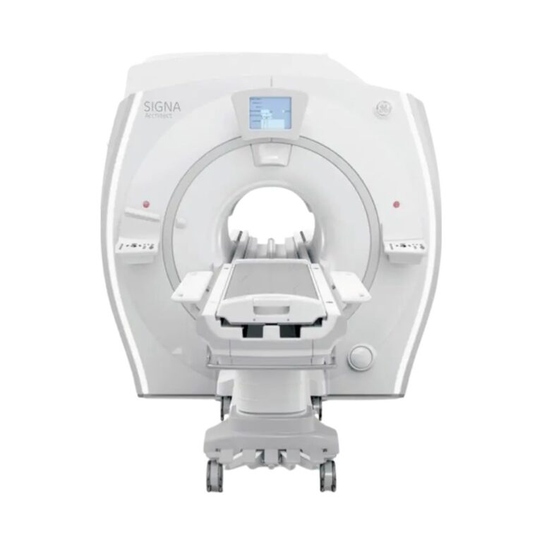
- SIGNA Architect 3T: Well suited for facilities seeking advanced 3T imaging, flexible coil configurations, expanded digital RF capabilities, and specialized imaging workflows.
- SIGNA Voyager 1.5T: Well suited for facilities prioritizing versatile 1.5T imaging, wide-bore patient access, space-efficient installation, and efficient operation.
SIGNA HD, HDx, & HDxt: Earlier 1.5T Systems
MXR Imaging also offers established 1.5T MRI platforms from GE’s earlier SIGNA HD family. These systems provide additional options for facilities seeking proven diagnostic imaging capabilities with different hardware and software configurations.
-
- GE SIGNA HD 1.5T: Features high-definition imaging, an 8-channel RF architecture, and a detachable patient table for routine whole-body examinations.
- GE SIGNA HDx 1.5T: Builds on the earlier HD platform with updated image reconstruction, RF technology, and operator workflow.
- GE SIGNA HDxt 1.5T: Expands the HD platform with advanced image reconstruction, high-density coils, and compatible Cube and IDEAL imaging applications.
Choosing the Right SIGNA MRI System
The Pioneer and Architect provide 3T imaging with different hardware architectures and clinical priorities, while the Voyager offers a newer-generation 1.5T alternative.
The HD, HDx, and HDxt models provide additional 1.5T options, with differences in software generation, coil connectivity, reconstruction, and imaging capabilities.
MXR Imaging can help match the appropriate MRI platform and configuration to your planned examinations, patient volume, facility requirements, and budget.
Available SIGNA Pioneer System Configuration
GE SIGNA Pioneer systems can be supplied in different hardware, software, and accessory configurations, allowing MXR Imaging to align the equipment with clinical and operational requirements.
Included Hardware, Coils, & Software
Configurations may differ in:
-
- Model year and hardware generation
- Gradient and RF architecture
- Installed reconstruction and workflow software
- Included head, spine, body, and specialty coils
- Advanced neurological, cardiac, vascular, and body imaging applications
- Patient table and positioning accessories
- Operator workstation and post-processing software
- Magnet, helium, and refrigeration service history
- Additional hardware upgrades and accessories
MXR Imaging works with healthcare providers to identify a Pioneer configuration suited to planned examinations, patient needs, clinical workflow, and facility infrastructure.
SIGNA Pioneer MRI Price & Availability
MXR Imaging offers GE SIGNA Pioneer 3T MRI systems with guidance on equipment selection, clinical configurations, installation planning, and ongoing service.
Pricing depends on the scanner’s hardware configuration, installed coils and software, included accessories, and installation requirements.
Our team can help match equipment capabilities to your clinical needs, operating goals, budget, and facility infrastructure.
MXR Imaging also provides MRI maintenance and repair services to support long-term equipment operation.
Contact MXR Imaging to discuss SIGNA Pioneer pricing, available configurations, and installation planning.
GE SIGNA Pioneer Specifications
The specifications below summarize the platform’s core hardware and representative equipment configurations. Gradient, RF, patient table, coil, and software details may vary by generation.
Magnet System
-
- Field Strength: 3.0T
- Magnet Type: Superconducting
- System Design: Whole-body, wide-bore MRI
- Bore Diameter: 70 cm (27.6 in)
- Bore Length: Approximately 163 cm (64.2 in) for the applicable configuration
- Maximum Field of View: Up to 50 × 50 × 48 cm (19.7 × 19.7 × 18.9 in), depending on the acquisition
- Cryogenic Cooling: Liquid helium with mechanical refrigeration
- Magnet Homogeneity: High-uniformity magnet design
Gradient System
-
- Gradient Technology: Ultra High Efficiency (UHE)
- Representative Maximum Gradient Amplitude: 36 mT/m
- Representative Maximum Slew Rate: 150 T/m/s
- Gradient Configuration: Dependent on installed hardware
- Efficiency: Lower electrical power demands than conventional gradient designs under comparable operating conditions
RF System
-
- RF Technology: Total Digital Imaging (TDI)
- Representative Receive Architecture: 97-channel configuration
- Signal Digitization: Compatible Direct Digital Interface (DDI) technology
- Coil Architecture: Multichannel anatomical coils
- Flexible Coil Technology: Compatible GE AIR Coils
Bore & Patient Table
-
- Bore Diameter: 70 cm (27.6 in)
- Patient Table: Comfort Plus option
- Maximum Patient Weight: Up to 550 lb (250 kg) in the applicable table configuration
- Patient Positioning: Feet-first imaging for compatible examinations
- Table Height & Travel: Per installed table specification
Coils & Accessories
-
- Compatible Coils: Head, head/neck, spine, body, extremity, breast, and cardiac arrangements
- Flexible Coil Technology: Compatible GE AIR Coils
- Included Coils: Based on selected equipment package
- Optional Equipment: Specialized clinical accessories and positioning supports
Siting & Utilities
-
- Electrical Service: Per installed system requirements
- Gradient Architecture: Ultra High Efficiency
- Cooling: Dedicated equipment cooling and magnet refrigeration
- RF Shielding: Required for MRI installation
- Cryogen Safety: Appropriate quench venting and emergency provisions
- Room Dimensions: Based on the applicable installation configuration
- Service Clearances: Dependent on magnet and equipment cabinet layout
System Dimensions & Weight
-
- Bore Diameter: 70 cm (27.6 in)
- Magnet Dimensions: Configuration-specific
- Magnet Weight: Based on installed magnet configuration
- Installed System Weight: Dependent on magnet, patient table, equipment cabinets, and accessories
- Installation Footprint: Determined by equipment dimensions and required service clearances
Final dimensions, weights, and facility requirements are matched to the selected system.