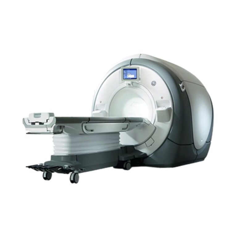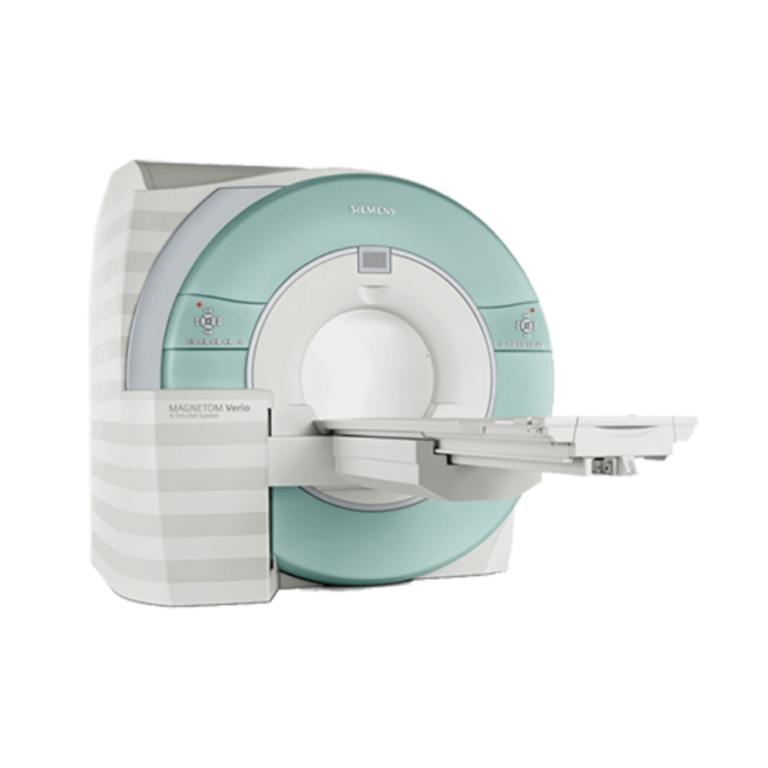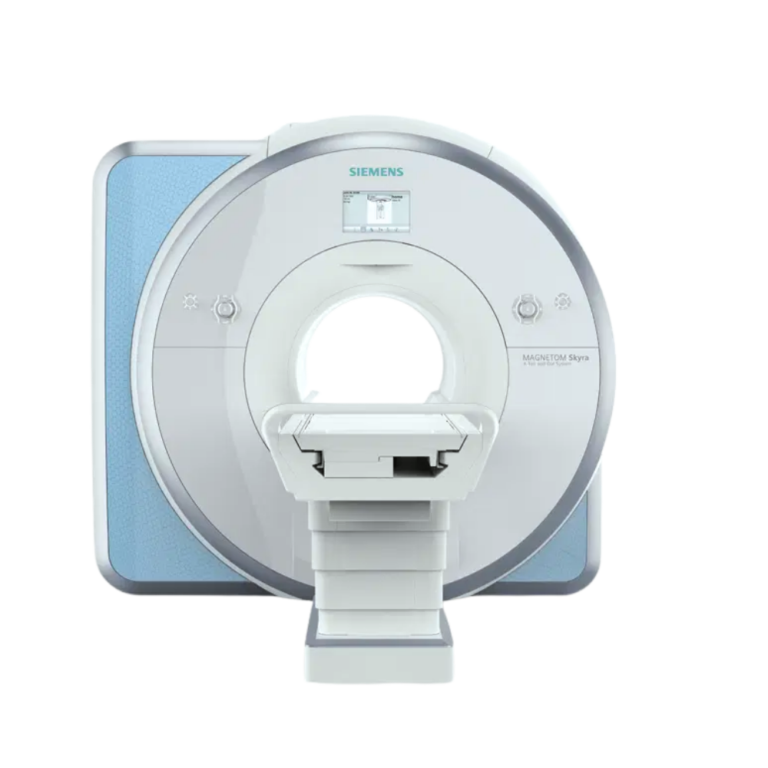
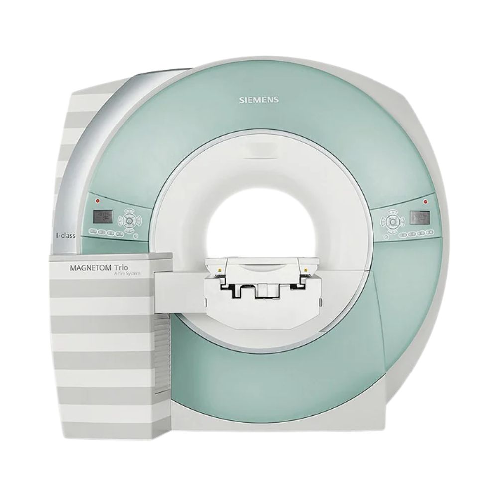
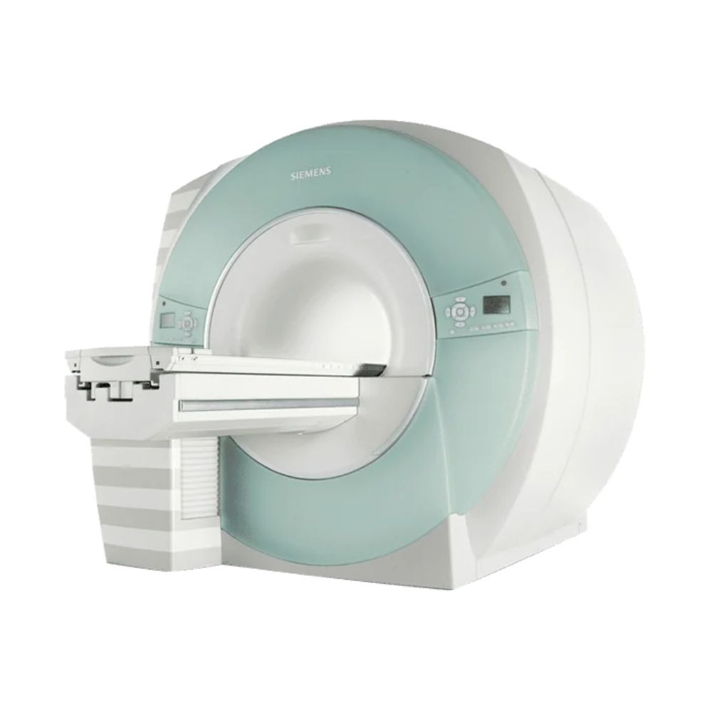
Siemens MAGNETOM Trio 3T MRI
- Category:
- MRI Systems
- Brand:
- Siemens
The Siemens MAGNETOM Trio 3T is a whole-body MRI scanner suited for hospitals, outpatient imaging centers, and advanced neuroimaging programs. Its 60 cm bore, high-performance gradients, and available Tim coil technology support detailed anatomical imaging, accelerated examinations, and specialized applications including functional MRI and diffusion imaging.
MAGNETOM Trio MRI Highlights & Benefits
The Siemens MAGNETOM Trio combines 3T imaging performance with a powerful gradient system and advanced clinical capabilities. Available Tim configurations extend its versatility through multichannel signal acquisition, faster compatible scans, and broader anatomical coverage, supporting routine diagnostic imaging and specialized neurological examinations.
Key Benefits
-
- 3T Imaging Performance: Provides strong imaging signals for detailed brain, spine, musculoskeletal, and body examinations.
- TQ Gradient System: Up to 45 mT/m gradient strength and 200 T/m/s slew rate support high-resolution and advanced diffusion imaging.
- Tim Coil Technology: Available configurations support up to 102 coil elements and 32 independent receiver channels for flexible imaging coverage and accelerated scanning.
- 60 cm Short-Bore Design: Supports whole-body MRI with feet-first positioning available for appropriate examinations.
- AudioComfort Noise Reduction: Reduces acoustic noise during supported scans to improve patient comfort.
- Advanced Neuroimaging: Supports functional MRI, diffusion tensor imaging, perfusion imaging, and MR spectroscopy with compatible software.
MAGNETOM Trio Imaging Performance
3T Magnet for High-Resolution Imaging
The MAGNETOM Trio uses a 3 Tesla superconducting magnet designed for detailed whole-body MRI. Its strong magnetic field provides the signal needed for high-resolution anatomical examinations and advanced imaging techniques.
The magnet’s field uniformity supports consistent image quality across the imaging area. Depending on configuration, the Trio supports a field of view of approximately 40 cm (15.7 in), with up to 50 cm (19.7 in) available on certain I-class configurations.
The larger imaging field supports broader anatomical coverage during body, vascular, and musculoskeletal examinations.
TQ Gradients for Advanced MRI
The Trio’s TQ gradient system controls the rapid magnetic-field changes used to capture MRI images.
Applicable configurations provide up to 45 mT/m gradient strength and 200 T/m/s slew rate. Gradient strength helps support fine anatomical detail, while slew rate measures how quickly the magnetic fields can change.
Together, these capabilities support high-resolution imaging, diffusion-weighted examinations, and demanding neurological studies that require precise signal acquisition.
Tim Technology for Flexible Imaging Coverage
Available Tim (Total Imaging Matrix) technology combines multiple receiving coils into a coordinated imaging system.
The MAGNETOM Trio a Tim System supports up to 102 connected coil elements and as many as 32 independent receiver channels, depending on configuration.
Coil elements collect signals from different anatomical locations, while receiver channels determine how many independent signals the scanner can process simultaneously. Higher channel counts support more flexible coil combinations and faster compatible acquisitions.
Tim technology also allows selected multiregion examinations to be completed with fewer coil exchanges or patient-positioning adjustments. Certain I-class configurations support extended imaging coverage of up to 181 cm (71.3 in) without repositioning the patient.
MAGNETOM Trio Patient Access & Comfort
60 cm Bore with Short-Bore Construction
The Siemens Trio 3T features a 60 cm (23.6 in) bore with a short-bore magnet design.
Its construction reduces the enclosed length of the scanner compared with traditional long-bore systems. Feet-first positioning is also supported for appropriate examinations, allowing the patient’s head to remain outside the magnet during selected studies.
The 60 cm opening should be considered when evaluating patient access, especially for larger individuals or patients who require additional positioning space.
Patient Table & Positioning
Documented Trio a Tim configurations support patients weighing up to 200 kg (441 lb), with motorized table movement for accurate anatomical positioning.
Compatible systems may also include an exchangeable patient tabletop and transport trolley. These options allow patient preparation and transfer outside the scanner when appropriate, helping technologists manage patients who require additional assistance.
Table height, movement range, and included transport accessories depend on the installed configuration.
AudioComfort for Quieter MRI Exams
Siemens AudioComfort technology reduces acoustic noise generated during MRI scanning through noise-reduction measures built into the scanner and its imaging operation.
On supported configurations, AudioComfort can reduce acoustic noise by up to 90% under specified comparison conditions. Actual reductions depend on the examination and scanning sequence.
Tim-equipped systems also support lightweight coil designs that can improve patient comfort. For example, the compatible Body Matrix coil weighs approximately 950 g (2.1 lb) without its cable, reducing the weight placed over the patient during selected body examinations.
MAGNETOM Trio Clinical Environments & Applications
The MAGNETOM Trio supports routine diagnostic imaging and specialized applications that benefit from 3T field strength, high-performance gradients, and multichannel receiving coils.
Its strengths include advanced neurological imaging, detailed musculoskeletal examinations, and academic research. Other clinical applications depend on the installed software, coils, and accessories.
Clinical Environments
-
- Hospitals and health systems
- Outpatient diagnostic imaging centers
- Neurology and neuroscience centers
- Orthopedic and musculoskeletal imaging facilities
- Oncology and specialty imaging programs
- Cardiac imaging centers with appropriate configurations
- Academic medical centers and MRI research facilities
- Veterinary specialty hospitals and academic veterinary imaging programs with suitable equipment
Clinical Specialties & Applications
-
- Neurology & Neurosurgery: Brain and spine imaging, stroke-related diffusion examinations, functional brain mapping, and white-matter assessment.
- Orthopedics & Sports Medicine: Knee, shoulder, hip, wrist, cartilage, ligament, tendon, and soft-tissue examinations using appropriate musculoskeletal coils.
- Podiatry & Foot/Ankle Imaging: Foot and ankle examinations, including joint, ligament, tendon, and soft-tissue assessment with compatible extremity coils.
- Abdominal & Pelvic Imaging: Liver, kidney, abdominal organ, and pelvic examinations using supported body coils and sequences.
- Urology: Prostate and pelvic MRI, including multiparametric prostate examinations when appropriate acquisition and processing capabilities are installed.
- Breast Imaging: Diagnostic breast MRI using compatible dedicated breast coils and clinical software.
- Cardiology & Vascular Medicine: Cardiac anatomy, ventricular function, blood-flow evaluation, and MR angiography with appropriate software and monitoring equipment.
- Oncology: Imaging of tumors and cancer-related abnormalities involving the brain, spine, abdomen, pelvis, breast, and other supported regions.
- Pediatric Radiology: Selected neurological, musculoskeletal, and body examinations using suitable protocols, coils, and positioning equipment.
- Veterinary Imaging: Documented applications include canine brain MRI and feline musculoskeletal examinations in specialty and research settings.
Advanced applications require compatible coils, imaging software, accessories, and clinical protocols.
Trio Technologies for Faster Scans
The MAGNETOM Trio supports Siemens technologies that shorten image acquisition and help maintain image quality during demanding examinations. Their availability depends on installed hardware and software.
Parallel Imaging & Scan Acceleration
- iPAT (Integrated Parallel Acquisition Techniques): Uses signals from multiple receiving coils to reduce the amount of scan data needed, shortening compatible examinations.
- GRAPPA: Reconstructs images from accelerated acquisitions using information collected by multiple coils, helping balance scan speed and image quality.
- Tim Matrix Imaging: Combines compatible coil elements across larger anatomical regions, supporting extended coverage with fewer positioning changes.
These capabilities help technologists manage examination time while maintaining the image detail needed for the clinical application.
Motion Reduction During MRI
Patient movement and breathing can reduce image sharpness, particularly during neurological and abdominal examinations.
The Trio supports motion-management techniques on appropriately configured systems:
- BLADE: Collects imaging data in overlapping patterns to reduce certain motion-related artifacts, particularly during neurological and musculoskeletal examinations.
- PACE: Uses respiratory movement information to coordinate compatible scans with breathing, helping reduce motion artifacts during body examinations.
- HASTE: Captures images rapidly using a single-shot technique, reducing sensitivity to movement during selected examinations.
These technologies provide additional options when patients have difficulty remaining still or holding their breath.
Advanced Trio Neuroimaging Technologies
The Trio’s combination of 3T field strength and high-performance gradients makes it particularly valuable for specialized neurological examinations.
Diffusion Imaging & White-Matter Assessment
-
- Diffusion-Weighted Imaging (DWI): Measures water movement within tissue, helping identify abnormalities including changes associated with acute ischemic stroke.
- Diffusion Tensor Imaging (DTI): Measures the direction of water movement to provide information about brain white-matter organization.
- Fiber Tractography: Uses diffusion imaging data to create visual representations of white-matter pathways for specialized neurological evaluation and research.
Functional & Metabolic Brain Imaging
-
- BOLD Functional MRI: Detects changes in blood oxygenation associated with brain activity, supporting functional brain mapping and neuroscience research.
- Perfusion Imaging: Assesses blood delivery to tissue, providing additional information during selected neurological examinations.
- MR Spectroscopy: Measures chemical characteristics of tissue, complementing anatomical MRI during specialized neurological and oncology examinations.
These applications require compatible acquisition sequences, processing software, and, in certain cases, additional equipment.
Trio MRI Workflow & Image Management
syngo Exam Planning & Processing
The MAGNETOM Trio uses Siemens’ syngo MR operating environment to manage examination protocols, scan settings, image acquisition, and review.
Technologists can select stored imaging protocols, adjust acquisition parameters, and review reconstructed images from the operator console.
Available workflow capabilities may include inline image processing, protocol management, and specialized post-processing applications, depending on software generation.
These functions support more consistent examinations and help reduce manual processing steps during routine and advanced imaging.
Image Reconstruction & PACS Connectivity
The Trio supports Digital Imaging and Communications in Medicine (DICOM) for transferring studies to compatible Picture Archiving and Communication Systems (PACS).
Depending on the installed software and network configuration, available functions include image transfer, multiplanar reconstruction, advanced image review, and specialized neurological processing.
Workstation hardware and installed licenses determine which post-processing features are available.
MAGNETOM Trio Configurations, Coils, & Options
Original Trio and Trio a Tim Configurations
The MAGNETOM Trio platform includes the original Trio and later MAGNETOM Trio a Tim System configurations, sometimes referred to as the Tim Trio.
Both use 3T superconducting magnet technology, but their receiving architecture and compatible coil configurations differ.
Tim-equipped systems expanded the platform through integrated multichannel coil technology. Documented configurations include 8-, 18-, and 32-channel receiving systems, with the available receiver architecture depending on hardware generation.
Certain Trio systems were also offered with I-class capabilities, which extended imaging coverage, coil integration, and workflow performance.
The I-class designation identifies a configuration of the Trio platform rather than a separate field-strength model.
These distinctions matter when comparing pre-owned systems because the model name alone does not establish the installed gradient package, receiver channels, coils, or software capabilities.
Tim Coils for Anatomical Imaging
The Trio platform supports a variety of anatomical and specialty coil options:
-
- Head & Head/Neck Coils: For brain, neurological, and cervical examinations.
- Spine Matrix Coils: For cervical, thoracic, and lumbar spine imaging.
- Body Matrix Coils: For abdominal, pelvic, and other body examinations.
- Knee, Shoulder & Wrist Coils: For dedicated joint and musculoskeletal imaging.
- Foot/Ankle & Extremity Coils: For detailed lower-extremity and small-joint examinations.
- Breast & Cardiac Coils: For compatible specialty imaging applications.
- Flexible Surface Coils: For adaptable positioning around selected anatomical regions.
Tim-equipped systems can combine compatible coils to support broader coverage and accelerated imaging.
Coil compatibility depends on the receiver architecture, and the coils included with an available system may differ from the platform’s complete range of supported options.
Software & Clinical Options
The Trio supports Siemens clinical applications that extend its capabilities beyond routine anatomical MRI.
Depending on installed software, available options include advanced diffusion imaging, functional MRI, perfusion imaging, MR spectroscopy, cardiac imaging, vascular imaging, and specialized post-processing.
Systems may also include motion-management and workflow technologies such as BLADE, PACE, and additional syngo applications.
MXR Imaging can help match an available system’s software and coil package to a facility’s clinical requirements.
Trio Magnet, Helium, & Site Requirements
Superconducting Magnet & Helium Cooling
The MAGNETOM Trio uses a superconducting magnet cooled by liquid helium and supported by mechanical refrigeration.
The cooling system maintains the extremely low temperatures required for magnet operation. On the Trio a Tim System, gradual helium boil-off occurs during normal operation, requiring periodic replenishment.
Magnet condition, helium level, refrigeration performance, and service history are important considerations when evaluating a pre-owned MRI scanner.
MXR Imaging provides MRI refrigeration and helium services to support long-term magnet operation.
MRI Room Layout & Shielding
The Trio requires a dedicated MRI environment with appropriate magnetic-field containment, RF shielding, structural support, and access for patients and service personnel.
The magnet assembly weighs approximately 12,000 kg (26,455 lb), making structural floor loading and equipment delivery routes important installation considerations.
Historical Trio installation layouts include compact configurations with approximately 33 m² (355 sq ft) of required space under specific planning conditions. Actual room dimensions depend on the selected equipment, magnetic-field limits, shielding design, and service clearances.
MXR Imaging offers custom MRI shielding solutions to support new installations and facility modifications.
Electrical Power & Cooling Requirements
The Trio requires dedicated three-phase electrical service, equipment cooling, and magnet refrigeration appropriate for the installed configuration.
Historical systems support electrical supply configurations including 380, 400, 420, 440, and 480 V. Final power, cooling, and ventilation requirements depend on the selected equipment and supporting infrastructure.
Facility planning must also account for cryogen venting, magnetic-field safety, equipment access, and ongoing maintenance.
MXR Imaging’s turnkey imaging project management services can help coordinate site preparation, equipment relocation, delivery, and installation.
Available MAGNETOM Trio System Configuration
MXR Imaging can help identify a Siemens MAGNETOM Trio configuration suited to a facility’s imaging workload, patient volume, and installation requirements.
Available equipment may include original Trio or Tim-equipped systems with different gradient packages, receiver-channel counts, coils, software generations, and clinical application licenses.
Individual systems may also differ in patient tables, workstations, accessories, installed upgrades, and magnet condition.
MXR Imaging works with healthcare facilities of all sizes to match available hardware, software, and accessories to their intended examinations. Installed features and accompanying equipment are confirmed during system selection.
Siemens MAGNETOM Trio Price & Availability
MXR Imaging offers pre-owned Siemens MAGNETOM Trio 3T MRI scanners with equipment selection, installation planning, and ongoing technical support.
Pricing depends on the system’s hardware configuration, condition, software, included coils, accessories, and installation requirements.
Platinum Certified Reconditioning
Eligible systems may be available through MXR Imaging’s Platinum Certified program.
Platinum Certified MRI systems undergo a comprehensive 100-point refurbishment process following ISO 13485-certified quality management procedures. The program includes a full one-year warranty, with project planning, installation support, and clinical applications training.
Additional service options are available for facilities requiring ongoing maintenance and technical support.
Nationwide MRI Service & Support
Family-owned since 1964, MXR Imaging supports healthcare facilities nationwide through imaging equipment sales, installation, preventive maintenance, repair, replacement parts, and medical imaging supplies.
The company’s ISO 13485:2016-certified quality management system supports controlled processes for equipment refurbishment, parts, installation, service, and repair.
MXR Imaging also provides MRI maintenance and repair and MRI coil repair services to support long-term equipment performance.
System availability is subject to inventory.
Siemens MAGNETOM Trio 3T MRI Specifications
Specifications vary between original Trio and Trio a Tim configurations. The values below describe documented platform capabilities and available options.
Magnet System
-
- Manufacturer: Siemens
- Model: MAGNETOM Trio
- Field Strength: 3 Tesla (3T)
- System Type: Whole-body MRI
- Magnet Type: Superconducting
- Bore Diameter: 60 cm (23.6 in)
- Maximum Field of View: Approximately 40 cm (15.7 in) on earlier configurations; up to 50 cm (19.7 in) on applicable I-class configurations
- Magnet Cooling: Liquid helium with mechanical refrigeration
- Magnetic Shielding: Actively shielded
Gradient System
-
- Gradient Architecture: Whole-body gradient system
- Gradient Package: TQ on applicable configurations
- Maximum Gradient Amplitude: Up to 45 mT/m
- Maximum Slew Rate: Up to 200 T/m/s
- Gradient Cooling: Water-cooled on applicable configurations
- Advanced Imaging: Diffusion imaging, DTI, and high-resolution neurological acquisitions with appropriate software
RF System
-
- RF Architecture: Multichannel receiving system
- Tim Receiver Options: Documented 8-, 18-, and 32-channel configurations
- Maximum Tim Coil Elements: Up to 102 on compatible configurations
- Parallel Imaging: iPAT and GRAPPA on supported systems
- Coil Compatibility: Siemens anatomical, matrix, and specialty coils appropriate to the installed configuration
Bore & Patient Table
-
- Bore Diameter: 60 cm (23.6 in)
- Patient Weight Capacity: Up to 200 kg (441 lb) on documented Trio a Tim table configurations
- Patient Positioning: Motorized table
- Feet-First Imaging: Supported for appropriate examinations
- Exchangeable Tabletop: Available on compatible systems
- Table Travel: Configuration-dependent
- Table Height: Dependent on the installed patient table
Coils & Accessories
Compatible coil options include:
-
- Head and head/neck coils
- Spine Matrix coils
- Body Matrix coils
- Flexible surface coils
- Knee, shoulder, and wrist coils
- Foot/ankle and extremity coils
- Breast and cardiac coils
- Physiological monitoring accessories
- Specialized functional MRI equipment
Included coils and accessories depend on the available system.
Siting & Utilities
-
- Electrical Service: Three-phase
- Historical Supply Voltages: 380 / 400 / 420 / 440 / 480 V, depending on configuration
- Equipment Cooling: Dedicated cooling infrastructure
- Magnet Refrigeration: Required
- RF Shielding: Required
- Cryogen Venting: Required
- Historical Installation Area: Approximately 33 m² (355 sq ft) in certain documented compact configurations; actual requirements are site-specific
System Dimensions & Weight
-
- Approximate Magnet Assembly Dimensions: 221 cm H × 244 cm W × 213 cm D (87.0 × 96.1 × 83.9 in) for a documented Trio a Tim configuration
- Approximate Magnet Assembly Weight: 12,000 kg (26,455 lb)
- Approximate Magnet and Helium Weight: 10,000 kg (22,046 lb)
- Installation Clearances: Dependent on equipment configuration and approved site design
Final dimensions, weights, utilities, and installation requirements are verified for the selected system.