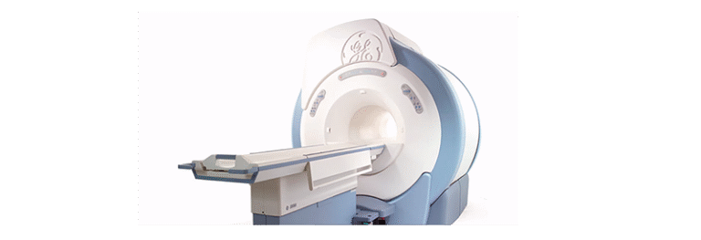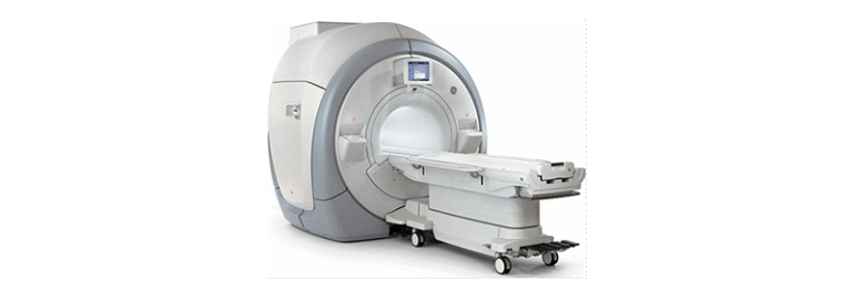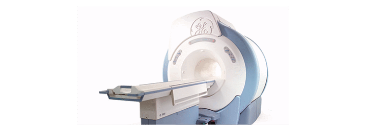
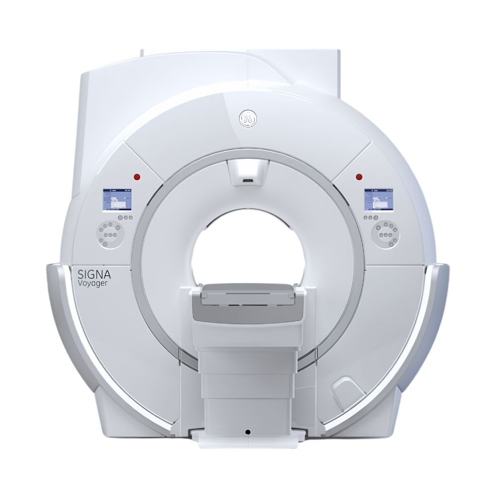
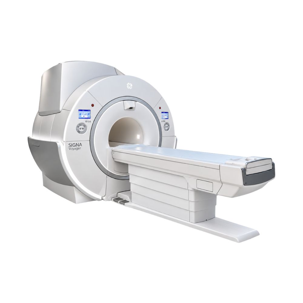
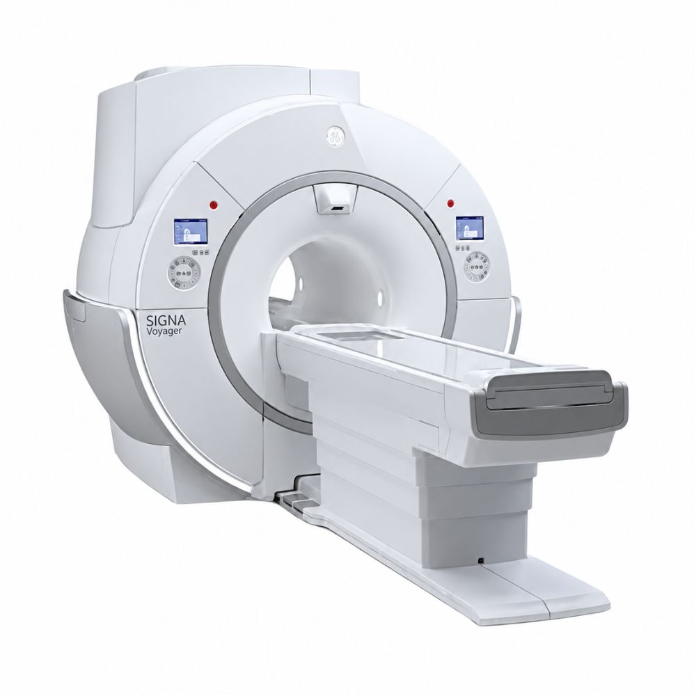
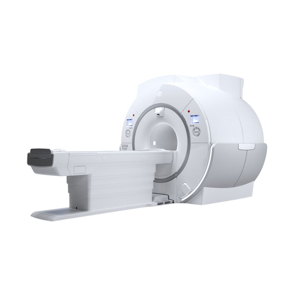
GE SIGNA Voyager 1.5T MRI
- Category:
- MRI Systems
- Brand:
- GE
The GE SIGNA Voyager 1.5T MRI is a wide-bore scanner well suited for hospitals, imaging centers, and specialty practices seeking versatile diagnostic imaging. Its spacious 70 cm bore, compact design, energy-efficient gradients, and available advanced imaging technology support patient comfort, detailed scans, and efficient clinical workflows.
SIGNA Voyager MRI Highlights & Benefits
The GE SIGNA Voyager combines the clinical versatility of a 1.5T MRI scanner with wide-bore patient access and a compact, energy-efficient design. Its digital imaging architecture supports routine and advanced diagnostic examinations, while available GE AIR technologies expand image quality, patient comfort, and workflow capabilities.
Key Features & Benefits
-
- 1.5T Whole-Body Imaging: Supports neurological, musculoskeletal, abdominal, pelvic, vascular, and other diagnostic MRI examinations.
- 70 cm Wide Bore: Provides additional patient space compared with conventional 60 cm MRI scanners, helping accommodate different body types and positioning needs.
- Ultra High Efficiency Gradients: Support detailed imaging and responsive scanning performance with lower electrical demands than earlier conventional gradient designs.
- Compact MRI Design: Combines wide-bore patient access with a space-efficient magnet and equipment architecture suited to new installations and selected replacement projects.
- Patient-Friendly Scanning: Compatible Comfort Plus patient tables, quieter scanning techniques, and motion-management software help improve examination comfort.
- Available AIR Imaging Technology: Compatible AIR Coils, AIR Recon DL, and automated workflow tools support flexible positioning, sharper images, and shorter examinations on appropriately equipped systems.
SIGNA Voyager MRI Video Preview
See the GE SIGNA Voyager 1.5T MRI in action, including its wide-bore design, advanced imaging capabilities, and patient-focused technologies.
SIGNA Voyager 1.5T Imaging Performance & Image Detail
The SIGNA Voyager combines a superconducting 1.5T magnet, GE’s Ultra High Efficiency gradient technology, and digital signal acquisition. These components support consistent anatomical imaging, advanced clinical applications, and efficient MRI operation across a broad range of examinations.
1.5T Magnet, Imaging Coverage, & Anatomical Detail
The Voyager uses a 1.5 Tesla superconducting magnet designed for whole-body MRI.
Its magnetic field supports detailed visualization of the brain, spine, joints, soft tissues, and internal organs. The 1.5T platform accommodates a broad range of established imaging protocols, including examinations requiring diffusion imaging, fat suppression, and specialized anatomical coverage.
A representative maximum field of view of 50 × 50 × 48 cm (19.7 × 19.7 × 18.9 in) provides coverage for larger anatomical regions, including suitable off-center examinations of shoulders and hips.
The magnet’s field uniformity supports consistent anatomical imaging and fat suppression, helping distinguish different tissue types during body, breast, and musculoskeletal examinations.
This combination makes the Voyager a versatile wide-bore MRI scanner for facilities performing both routine diagnostic studies and selected advanced examinations.
Field-of-view specifications can differ by system generation and acquisition.
Ultra High Efficiency Gradient Performance
The SIGNA Voyager uses GE’s Ultra High Efficiency (UHE) gradient architecture, designed to deliver detailed MRI acquisition with lower electrical power requirements.
A representative original Voyager configuration provides:
-
- Maximum gradient amplitude: 36 mT/m
- Maximum slew rate: 150 T/m/s
Gradient amplitude affects the scanner’s ability to locate signals precisely, while slew rate describes how quickly the gradient fields change.
Together, these capabilities support diffusion imaging, motion-sensitive examinations, and detailed anatomical scans.
The UHE architecture is designed to provide responsive gradient performance while consuming less electrical power than earlier conventional designs. This can help reduce demands on supporting electrical and cooling infrastructure.
Later Voyager configurations may provide different gradient capabilities. The applicable hardware specifications are matched to the selected system rather than treated as interchangeable.
Total Digital Imaging & RF Signal Acquisition
The Voyager uses GE’s Total Digital Imaging (TDI) technology to acquire signals from compatible MRI coils.
Its digital radiofrequency (RF) architecture helps preserve image information as signals travel from the patient to the reconstruction system.
Original Voyager configurations include 33-channel and 65-channel receive architectures, depending on installed hardware.
The receive-channel configuration determines how signals from multiple coil elements can be collected and processed during an examination. This influences supported coil combinations, imaging coverage, and acquisition capabilities.
GE’s Direct Digital Interface technology converts incoming signals into digital information close to the receiving hardware, helping preserve signal quality before reconstruction.
Later hardware configurations can offer different receive capabilities and compatibility with newer flexible MRI coils.
SIGNA Voyager Wide-Bore Patient Access & Positioning
The SIGNA Voyager combines a spacious 70 cm bore, adaptable positioning, and available comfort-focused scanning technologies. These capabilities help facilities accommodate different patient sizes, mobility needs, and examination requirements while supporting efficient daily MRI workflows.
70 cm Bore & Feet-First Examinations
The scanner features a 70 cm (27.6 in) bore, approximately 10 cm (3.9 in) wider than conventional 60 cm MRI systems.
The additional space supports patients with broader body dimensions and examinations requiring specialized coil arrangements.
Head-first and feet-first entry are available for compatible examinations, reducing the need for head-first positioning when clinically appropriate.
The wide-bore design can also help patients who experience discomfort or anxiety in more confined scanning environments.
Patient communication, ventilation, and adjustable lighting features further support the examination experience.
Comfort Plus Patient Table & Weight Capacity
The Voyager supports GE’s Comfort Plus Patient Table, designed to simplify patient access and positioning.
Compatible configurations provide:
-
- 550 lb (250 kg) Patient Capacity: Accommodates patients across a broad range of body types.
- Low Table Entry Height: Helps patients get on and off the table more comfortably.
- Powered Table Positioning: Supports controlled movement during preparation and scanning.
- Integrated Posterior Imaging Array: Incorporates coil elements beneath the patient, reducing the need to position a separate posterior coil during supported examinations.
Compatible detachable patient-table configurations can provide additional flexibility for patient preparation and transfer, depending on the system’s installed hardware.
These features can be particularly valuable for outpatient imaging centers, hospitals, and facilities serving patients with limited mobility.
Table height, travel, and positioning accessories depend on the installed configuration. MXR Imaging can help match the appropriate table arrangement to patient-access and examination requirements.
SIGNA Voyager Patient Comfort & Motion Reduction
The Voyager supports patient-focused technologies that help reduce scanning noise, movement-related image distortion, and unnecessary interruptions. These capabilities can make examinations more manageable for patients who are sensitive to noise, have difficulty remaining still, or struggle with repeated breath-holding.
Quieter MRI Examinations with Silent Scan
Conventional MRI scanners produce loud knocking and tapping sounds as their gradient coils rapidly switch magnetic fields during image acquisition. The Voyager supports GE’s Silent Scan technology, which reduces this noise through specialized scanning techniques.
-
- Silent Scan: Uses specialized MRI pulse sequences that minimize the rapid gradient switching responsible for much of the noise heard during conventional scanning. Instead of simply reducing the volume of the scanner’s existing sequences, Silent Scan changes how compatible images are acquired, allowing selected neurological examinations to be performed at sound levels much closer to the surrounding room environment.
Silent Scan can make examinations more comfortable for patients who are sensitive to noise, particularly during brain imaging. Available silent imaging sequences may also support spine and musculoskeletal examinations, depending on the installed software package.
2D & 3D Motion Correction for Clearer MRI Images
Patient movement can cause blurred images and repeat scanning. The Voyager supports different motion-correction technologies designed to address this challenge according to the type of examination.
-
- PROPELLER: Collects MRI information in overlapping, rotating strips rather than using only conventional sequential sampling. Because these strips repeatedly sample shared information, the scanner can identify and reduce certain motion-related effects during reconstruction. This is particularly useful for selected brain, spine, and musculoskeletal examinations.
- 3D PROMO: Uses prospective motion correction to monitor changes in head position during compatible three-dimensional brain scans. When movement is detected, the scanner adjusts its imaging coordinates to follow the patient’s position rather than continuing to acquire data using the original alignment. This helps preserve anatomical detail during examinations where slight head movements could otherwise compromise image quality.
PROPELLER primarily reduces motion effects through its acquisition and reconstruction method, while 3D PROMO adjusts imaging in response to detected movement. Both help reduce the likelihood of repeat scans, although neither eliminates the need for patients to remain as still as possible.
Free-Breathing Body Imaging & Flexible AIR Coils
Breath-holding and uncomfortable coil positioning can make MRI examinations more difficult, particularly during abdominal imaging or longer studies. The Voyager offers compatible technologies that address these challenges in different ways.
-
- Free-Breathing Imaging with Auto Navigator: Tracks the movement of the diaphragm as a patient breathes and coordinates MRI acquisition with the respiratory cycle. This allows selected abdominal examinations to collect image information during appropriate breathing phases rather than requiring repeated breath-holds. It can be particularly useful for patients who have difficulty following breath-holding instructions or maintaining consistent breathing patterns.
- AIR Coils: Use lightweight, flexible coil elements that can conform more closely to the patient’s body rather than requiring the anatomy to fit within a rigid coil structure. The coil elements receive MRI signals from the area being examined, while their flexible construction allows technologists to position them around different body shapes and anatomical regions. This supports comfortable positioning and suitable signal coverage during compatible examinations.
Together, these technologies help accommodate different patient needs while supporting consistent image acquisition and efficient examinations.
Silent Scan, PROPELLER, 3D PROMO, Auto Navigator, and AIR Coils depend on compatible hardware, installed software, and available accessories.
Clinical Environments & Applications
The GE SIGNA Voyager supports routine diagnostic MRI and advanced examinations across multiple specialties. Its 1.5T imaging capabilities, 70 cm wide bore, and space-efficient design make it well suited to facilities providing comprehensive imaging services while prioritizing patient access, workflow efficiency, and operational flexibility.
Clinical Environments
-
- Hospitals and diagnostic imaging departments
- Outpatient imaging centers
- Multispecialty medical practices
- Specialty diagnostic and surgical facilities
- Oncology and cancer care centers
- Academic medical centers and research facilities
- Veterinary hospitals and specialty referral centers
Clinical Specialties
-
- Radiology: Routine and advanced brain, spine, abdominal, pelvic, and musculoskeletal MRI examinations.
- Neurology & Neurosurgery: Brain and spine imaging, diffusion studies, and neurological assessment with compatible applications.
- Orthopedics: Joint, cartilage, ligament, tendon, and soft-tissue examinations for injury assessment and surgical planning.
- Oncology: Tumor characterization, disease monitoring, and follow-up MRI.
- Cardiology: Cardiac anatomy, function, and specialized cardiovascular examinations with suitable coils and software.
- Vascular Medicine: Magnetic resonance angiography (MRA) and selected non-contrast vascular examinations.
- Gastroenterology & Hepatology: Liver, pancreas, abdominal, and quantitative fat-assessment imaging with compatible applications.
- Urology: Kidney, prostate, urinary tract, and pelvic MRI examinations.
- Breast Imaging: Dedicated breast MRI with appropriate coils and clinical software.
- Pediatric Radiology: Age-appropriate neurological, musculoskeletal, and body examinations using suitable positioning, coils, and protocols.
- Veterinary Medicine: Neurological, spinal, and musculoskeletal MRI examinations in companion animals using appropriate veterinary coils, positioning, anesthesia, and monitoring protocols.
Common MRI Applications
-
- Brain, head, and spine MRI
- Joint, cartilage, and musculoskeletal imaging
- Abdominal and pelvic examinations
- Liver, pancreas, and renal imaging
- Prostate MRI
- Breast MRI with compatible coils and software
- Cardiac MRI when appropriately configured
- Magnetic resonance angiography
- Non-contrast vascular imaging with compatible applications
- Diffusion-weighted neurological examinations
- Oncology evaluation and follow-up
- Quantitative liver fat assessment when equipped
- Functional MRI and spectroscopy with suitable options
- Pediatric MRI using appropriate protocols and accessories
- Veterinary brain, spine, and musculoskeletal MRI in appropriately equipped facilities
SIGNA Voyager Exam Planning & Motion Management
The GE SIGNA Voyager combines automated examination planning and motion-management technology to simplify MRI workflows. Available SIGNA Works applications reduce manual setup, improve positioning consistency, and help manage patient movement during scanning.
Automated Exam Planning & Coil Selection
SIGNA Works brings together GE’s clinical imaging protocols and automated workflow tools. Rather than requiring technologists to configure every aspect of an MRI examination manually, compatible applications assist with coil selection and anatomical positioning.
-
- AIR Touch: Automatically identifies which connected coil elements provide suitable signal coverage for the selected anatomy and imaging area. Instead of manually choosing coil channels, the system selects an appropriate combination based on patient positioning and the planned scan. This simplifies setup and supports consistent signal coverage, particularly when using flexible AIR Coils.
- AIR x: Uses deep-learning-based anatomical recognition to identify structures in the brain or knee and automatically position image slices. This reduces manual adjustments traditionally needed to align scans with anatomical landmarks, helping technologists obtain consistent imaging planes across patients and repeat examinations.
- Anatomy-Specific Protocols: SIGNA Works provides preset scanning techniques for different body regions and clinical applications. These protocols organize acquisition settings, image contrasts, and scan sequences, reducing the need to build examinations from scratch while allowing patient-specific adjustments.
These workflow applications help technologists prepare examinations more efficiently and maintain consistency across routine and specialized studies.
Breathing & Patient Motion Management
Patient movement can reduce MRI image quality, particularly during longer examinations and abdominal imaging.
The Voyager offers compatible motion-management technologies that address respiratory movement and other patient motion through different acquisition methods.
-
- Auto Navigator: Tracks the patient’s diaphragm during breathing and coordinates image acquisition with respiratory movement. Unlike conventional breath-hold imaging, this allows selected abdominal examinations to proceed while the patient breathes normally. The scanner uses respiratory information to time data collection and reduce movement-related distortion.
- PROPELLER: Collects MRI data in overlapping, rotating strips rather than relying entirely on conventional sequential sampling. These overlapping measurements provide information that helps correct some movement-related effects during reconstruction. This is useful for selected brain, spine, and musculoskeletal examinations where movement could otherwise produce blurred images or repeat scans.
These applications support imaging consistency while helping facilities accommodate patients who have difficulty remaining still or repeatedly holding their breath.
AIR Touch, AIR x, Auto Navigator, PROPELLER, and other workflow capabilities depend on installed software and compatible system hardware.
SIGNA Voyager Image Reconstruction & Advanced Scanning
The Voyager supports technologies that address examination time, image clarity, and specialized acquisition requirements. Different applications improve reconstructed images, acquire more anatomical information, or capture clinical details that conventional techniques may require several separate scans to obtain.
Deep-Learning Reconstruction & Scan Acceleration
AIR Recon DL and HyperSense improve imaging efficiency through different approaches: enhancing the quality of acquired MRI data and reducing the amount of data needed to reconstruct an image.
-
- AIR Recon DL: Uses a deep-learning algorithm during reconstruction to reduce image noise and improve sharpness. By processing acquired MRI data before the final image is formed, it can produce clearer images without relying solely on longer acquisitions to improve signal quality. Technologists can use this capability to shorten compatible examinations or acquire finer anatomical detail.
- HyperSense: Uses compressed sensing, a technique that collects fewer measurements than a conventional MRI scan and mathematically reconstructs the image using the available information. Specialized sampling patterns and iterative reconstruction recover image detail, allowing compatible examinations to be completed faster or with improved spatial resolution within a similar scanning period.
These technologies are complementary rather than interchangeable. AIR Recon DL improves reconstruction quality, while HyperSense primarily accelerates data acquisition.
Both can support more efficient MRI workflows when installed on compatible Voyager configurations.
3D Imaging & Multiple Image Contrasts
Cube and MAGiC provide different ways to obtain more diagnostic information from MRI acquisitions.
-
- Cube: Acquires a high-resolution three-dimensional volume instead of a conventional series of separate two-dimensional slices. The resulting dataset can be reconstructed into different viewing planes without repeating the scan. This is useful for detailed joint, spine, and neurological imaging when anatomy needs to be evaluated from multiple directions.
- MAGiC: Uses a specialized acquisition to collect information about tissue magnetic properties and generate multiple image contrasts from one scan. Compatible brain examinations can produce T1, T2, proton-density, and selected fluid-suppressed images from the same dataset. Image contrast can also be adjusted after acquisition, reducing the need for additional scanning when different tissue views are required.
Cube emphasizes flexible three-dimensional anatomical review, while MAGiC provides multiple tissue contrasts from a single acquisition.
Dynamic Contrast Imaging for Body Examinations
Dynamic contrast-enhanced MRI captures how contrast material moves through tissue over time.
-
- DISCO: Uses a specialized sampling technique that updates portions of a three-dimensional MRI dataset during contrast enhancement. Instead of repeating a complete conventional acquisition for every phase, DISCO collects information across successive phases as contrast material moves through tissue.
This supports dynamic liver, breast, and prostate imaging, where the timing and pattern of enhancement can help characterize tissue and abnormalities.
AIR Recon DL, HyperSense, Cube, MAGiC, and DISCO expand the range of available scanning techniques, but their availability depends on the installed software, licenses, and hardware generation.
Certain applications may require compatible AIR Edition upgrades.
Advanced Clinical MRI & Image Management
Beyond routine anatomical imaging, the SIGNA Voyager supports specialized neurological, abdominal, vascular, and cardiovascular applications when equipped with appropriate clinical software.
These capabilities help facilities expand diagnostic services without moving to a different MRI platform.
Brain Perfusion, Liver Fat, & Vascular Imaging
Compatible clinical applications include:
-
- 3D Arterial Spin Labeling (3D ASL): Measures cerebral blood flow using magnetically labeled blood rather than injected contrast material. This provides additional information about brain perfusion.
- IDEAL IQ: Separates water and fat signals to support quantitative liver fat assessment, providing measurements that can assist in evaluating fatty liver disease.
- Inhance Suite: Supports selected magnetic resonance angiography examinations without injected contrast material, expanding options for appropriate vascular studies.
- Diffusion Imaging: Measures the movement of water within tissues, providing additional information during neurological, oncology, and other supported examinations.
- Cardiac Imaging Applications: Support evaluation of heart anatomy, function, and selected cardiovascular measurements with appropriate coils and acquisition software.
These advanced MRI applications depend on compatible software and equipment packages.
Image Review, Post-Processing, & PACS Connectivity
The Voyager supports DICOM image transfer to compatible picture archiving and communication systems (PACS), allowing examinations to be stored, reviewed, and distributed within an imaging department.
Available post-processing tools support multiplanar reconstruction, image review, and specialized clinical analysis.
Compatible READYView technology provides additional visualization and processing capabilities when installed.
The workstation, software generation, and purchased clinical options determine which processing functions are available.
SIGNA Voyager Configurations, Coils, & Available Options
The SIGNA Voyager is available in different hardware generations, software packages, and compatible upgrade configurations.
Understanding these differences helps buyers distinguish its core 1.5T imaging platform from advanced imaging technologies that may require additional hardware or software.
Original Voyager & AIR Edition Configurations
The GE SIGNA Voyager AIR is an updated version of the original SIGNA Voyager 1.5T MRI platform, retaining its 70 cm wide bore and energy-efficient design while introducing expanded AIR imaging capabilities. Depending on the configuration, these advancements include flexible AIR Coils, AIR Recon DL for improved image quality and faster scanning, and automated exam-planning technologies. GE also offers modernization pathways for compatible original Voyager systems.
The original SIGNA Voyager and later Voyager AIR Edition configurations belong to the same broader model family, but do not necessarily contain identical hardware or software.
The original Voyager established the platform’s 1.5T magnet, 70 cm wide bore, efficient gradient architecture, and digital RF system.
Later AIR Edition configurations introduced or expanded compatibility with GE’s AIR imaging technologies, including flexible AIR Coils, advanced image reconstruction, and automated workflow applications.
An original Voyager may support selected upgrades depending on its hardware generation and installed software. However, purchasing an AIR-related application does not automatically provide every AIR Edition capability.
Important configuration differences include:
-
- AIR Recon DL: Requires compatible image reconstruction software and system hardware.
- AIR Coils: Require supported RF connections, coil compatibility, and appropriate software.
- AIR Touch & AIR x: Are separate workflow applications with individual compatibility requirements.
- Gradient Performance: Depends on installed gradient hardware rather than the AIR Edition designation alone.
- Advanced Clinical Applications: Depend on software licenses and supported acquisition technology.
- Patient Table & Accessories: Can vary independently of the imaging software package.
Later modernization packages and region-specific Voyager editions may introduce further differences.
For healthcare providers comparing original Voyager and AIR Edition MRI scanners, the practical distinction is the installed technology package and the examinations it can support, not simply the edition name.
MXR Imaging can help align the appropriate Voyager configuration with the facility’s clinical requirements and operating goals.
RF Coil Options & Anatomical Coverage
The Voyager supports a range of MRI coils for whole-body and specialty imaging.
Compatible options include:
-
- Head Coils: Neurological and brain MRI.
- Head & Neck Coils: Combined cranial and cervical examinations.
- Spine Coils: Cervical, thoracic, and lumbar imaging.
- Body Array Coils: Abdominal, pelvic, and general body examinations.
- AIR Coils: Lightweight, flexible imaging options on supported configurations.
- Extremity Coils: Joint and musculoskeletal studies.
- Breast Coils: Dedicated breast MRI.
- Cardiac Coil Arrangements: Supported cardiovascular examinations.
GE AIR Coils use flexible, lightweight construction designed to conform more closely to patient anatomy. This can simplify positioning and improve comfort compared with conventional rigid coils.
Supported combinations depend on the installed RF system, coil connections, and software.
The included coil package can be matched to planned examinations rather than assuming every system contains the same specialty equipment.
MXR Imaging also provides MRI coil repair services for supported equipment.
Clinical Software & Optional Imaging Packages
Available software packages can expand the Voyager’s clinical capabilities.
Options include:
-
- Neurology: Diffusion imaging, brain perfusion, and advanced neurological techniques.
- Musculoskeletal Imaging: Three-dimensional acquisitions and specialized joint studies.
- Body Imaging: Free-breathing techniques, dynamic contrast imaging, and quantitative liver assessment.
- Cardiac Imaging: Cardiac function and selected advanced cardiovascular examinations.
- Breast Imaging: Dedicated acquisition and analysis applications.
- Vascular Imaging: Contrast-enhanced and supported non-contrast angiography.
- Reconstruction & Acceleration: Compatible AIR Recon DL, HyperSense, and related technologies.
- Patient Comfort: Silent Scan and available motion-reduction applications.
Software features depend on installed licenses, compatible hardware, and the system’s upgrade pathway.
SIGNA Voyager Magnet Cooling & Helium Requirements
The SIGNA Voyager uses a superconducting magnet maintained at very low temperatures through a liquid-helium cooling system.
Mechanical refrigeration helps maintain the conditions required for magnet operation while limiting helium losses during normal use.
The Voyager’s magnet and refrigeration design emphasizes efficient long-term operation, making helium management an important part of ownership and service planning.
For pre-owned systems, magnet condition, refrigeration performance, helium history, and service records can affect equipment value and ongoing maintenance requirements.
MXR Imaging provides MRI refrigeration and helium services for compatible systems.
SIGNA Voyager MRI Installation & Operating Requirements
The Voyager combines a 70 cm wide bore with a compact magnet and space-conscious equipment design.
This can make it attractive to hospitals and outpatient imaging centers planning a new MRI suite, replacing an existing scanner, or evaluating equipment for a constrained installation area.
Compact Magnet Design, Room Layout, & Shielding
The Voyager’s compact architecture helps accommodate wide-bore 1.5T MRI installations within appropriately planned imaging suites.
For a published Voyager AIR Edition configuration, GE provides the following representative minimum room dimensions:
-
- Magnet Room: 3.7 × 5.8 m (12.1 × 19.0 ft)
- Equipment Room: 1.8 × 2.7 m (5.9 × 8.9 ft)
- Control Room: 1.5 × 2.1 m (4.9 × 6.9 ft)
- Minimum Ceiling Height: 2.5 m (8.2 ft)
These figures are configuration-specific planning examples rather than universal installation requirements.
Additional considerations include:
-
- Building access and magnet delivery routes
- Floor loading and structural support
- RF shielding and magnetic field safety boundaries
- Cryogenic venting and emergency procedures
- Equipment placement and maintenance access
- Patient preparation and circulation space
The final MRI suite design must accommodate the selected scanner, applicable safety requirements, and necessary service clearances.
MXR Imaging supports MRI installations through custom MRI shielding solutions and turnkey imaging project management.
Electrical Power, Equipment Cooling, & Energy Efficiency
The SIGNA Voyager’s Ultra High Efficiency gradient architecture helps lower electrical demands compared with earlier conventional gradient systems.
Reduced gradient power requirements can help manage energy consumption and equipment cooling needs, particularly in facilities performing substantial daily scan volumes.
The scanner still requires dedicated electrical service, HVAC, cooling infrastructure, and magnet refrigeration.
Actual operating expenses depend on examination volume, equipment configuration, facility utilities, and local energy costs.
For facilities comparing MRI equipment costs, the Voyager’s energy-efficient design, compact installation requirements, and configurable software can contribute to long-term operating value beyond the initial purchase price.
MXR Imaging can help evaluate the selected scanner’s electrical, cooling, and installation requirements during equipment planning.
GE SIGNA Voyager vs. Other SIGNA MRI Models
The GE SIGNA lineup includes multiple 1.5T and 3T MRI systems with different imaging capabilities, hardware generations, and facility requirements.
Comparing these scanners helps identify where the Voyager offers the strongest balance of clinical versatility, patient comfort, and operational efficiency.
Voyager, Pioneer, Architect, & Premier: Imaging Priorities
-
- SIGNA Voyager 1.5T: Emphasizes versatile 1.5T imaging, a 70 cm wide bore, efficient operation, and a compact system design.
- SIGNA Pioneer 3T: Offers high-field MRI with Ultra High Efficiency gradients, wide-bore patient access, and an emphasis on energy-efficient operation.
- SIGNA Architect 3T: Combines advanced 3T imaging with flexible RF technology, specialty clinical capabilities, and configurable imaging workflows.
- SIGNA Premier 3T: Emphasizes advanced 3T imaging performance, high-gradient capabilities, AIR technology, and demanding specialty examinations.
SIGNA HD, HDx, & HDxt: Earlier 1.5T Systems
MXR Imaging also offers established 1.5T platforms from GE’s earlier SIGNA HD family.
-
- GE SIGNA HD 1.5T: An earlier-generation 1.5T scanner with high-definition imaging technology and routine whole-body capabilities.
- GE SIGNA HDx 1.5T: Builds on the HD platform with updated reconstruction, RF architecture, and imaging workflow capabilities.
- GE SIGNA HDxt 1.5T: Expands the HD family with additional reconstruction, coil, and specialized clinical imaging options.
These earlier MRI scanners provide alternative 1.5T configurations with different equipment generations, installed capabilities, and ownership considerations.
Choosing Between Voyager & Other SIGNA Systems
The Voyager is well suited to facilities seeking the clinical versatility of 1.5T MRI, wide-bore patient access, and efficient daily operation.
For many routine brain, spine, joint, abdominal, and pelvic examinations, 1.5T offers established diagnostic performance. It can also provide advantages during selected examinations affected by magnetic susceptibility artifacts, including some imaging near metallic implants when appropriate techniques are used.
A 3T MRI scanner generally provides greater signal potential, which can support higher-resolution imaging and specialized examinations. However, higher field strength can also introduce different artifact, scan-planning, safety, and operating considerations.
MRI examinations involving implants must follow the specific medical device’s MR safety labeling and operating conditions. Neither 1.5T nor 3T is universally appropriate for every implant.
Pioneer, Architect, and Premier provide different 3T capabilities, while Voyager offers a versatile 1.5T alternative for facilities emphasizing routine diagnostic coverage, patient comfort, and manageable operating demands.
The HD, HDx, and HDxt systems offer additional pre-owned 1.5T options, with suitability influenced by equipment generation, available upgrades, and service history.
MXR Imaging can help compare these scanners according to clinical applications, patient volume, facility infrastructure, and budget.
Available SIGNA Voyager System Configuration
GE SIGNA Voyager MRI systems can be supplied in different hardware, software, and accessory packages.
MXR Imaging works with healthcare providers to align the scanner’s capabilities with planned examinations, imaging workflows, and installation requirements.
Included Hardware, Coils, & Software
The selected equipment package can include different combinations of:
-
- Original Voyager or later AIR-equipped hardware
- Model year and software generation
- Gradient and RF architecture
- Installed AIR Recon DL and other reconstruction applications
- Head, spine, body, and specialty MRI coils
- Clinical application packages and software licenses
- Patient table and positioning accessories
- Operator workstation and post-processing software
- Magnet, helium, and refrigeration service history
- Compatible upgrades and additional accessories
The included hardware, coils, and software determine which routine and advanced examinations the scanner can support.
The included hardware, coils, and software determine which routine and advanced examinations the scanner can support.
For buyers evaluating a used or pre-owned GE SIGNA Voyager, these configuration details are important when comparing equipment capabilities, overall value, and long-term service needs.
MXR Imaging can help identify a scanner and accessory package suited to the facility’s intended clinical services.
SIGNA Voyager MRI Price & Availability
MXR Imaging offers GE SIGNA Voyager 1.5T MRI systems with guidance on equipment selection, clinical configurations, installation planning, and long-term service.
Pricing depends on the scanner’s hardware generation, installed software, coil package, equipment condition, and installation requirements.
Original Voyager and later AIR-equipped systems may differ in their reconstruction technologies, workflow capabilities, included accessories, and equipment value.
Beyond the initial MRI scanner price, purchasing considerations include magnet and refrigeration condition, service history, coil availability, software compatibility, and site preparation.
MXR Imaging works with facilities to match equipment capabilities and configurations to planned examinations, patient needs, operating requirements, and budget.
Our team also provides MRI maintenance and repair services to support ongoing equipment operation.
Contact MXR Imaging to discuss GE SIGNA Voyager pricing, available configurations, and installation planning.
GE SIGNA Voyager Specifications
The specifications below describe the original Voyager platform and representative later configurations where identified. Gradient, RF, patient table, and software capabilities vary by hardware generation and installed options.
Magnet System
-
- Field Strength: 1.5T
- Magnet Type: Superconducting
- System Design: Whole-body, wide-bore MRI
- Patient Bore Diameter: 70 cm (27.6 in)
- Representative Magnet Length: 179 cm (70.5 in)
- Usable Bore Length: Approximately 163 cm (64.2 in)
- Original Voyager Maximum Field of View: Up to 50 × 50 × 48 cm (19.7 × 19.7 × 18.9 in)
- Magnet Shielding: Active
- Cooling: Liquid helium with mechanical refrigeration
Gradient System
-
- Gradient Technology: Ultra High Efficiency (UHE)
- Original Voyager Peak Gradient Amplitude: 36 mT/m
- Original Voyager Peak Slew Rate: 150 T/m/s
- Later Configuration Gradient Amplitude: Up to 45 mT/m in applicable published configurations
- Later Configuration Slew Rate: Up to 200 T/m/s in applicable published configurations
- Gradient Duty Cycle: Up to 100% in supported configurations
- Installed Gradient Performance: Dependent on gradient hardware and system generation
RF System
-
- RF Technology: Total Digital Imaging (TDI)
- Original Voyager Receive Configurations: 33 or 65 channels, depending on hardware
- Digital Signal Acquisition: Direct Digital Interface technology
- Compatible Coil Architecture: Multichannel anatomical coils
- AIR Coil Compatibility: Available on appropriately configured or upgraded systems
- RF Hardware: Configuration-specific
Bore & Patient Table
-
- Bore Diameter: 70 cm (27.6 in)
- Patient Entry: Head-first or feet-first for supported examinations
- Patient Table Options: Comfort Plus and compatible detachable configurations
- Maximum Patient Weight: Up to 550 lb (250 kg) on applicable table configurations
- Patient Positioning: Powered table movement on compatible configurations
- Integrated Posterior Coil: Available with compatible Comfort Plus configurations
- Patient Comfort Features: In-bore communication, ventilation, and lighting
Coils & Accessories
-
- Compatible Coil Categories: Head, head/neck, spine, body, extremity, breast, and cardiac
- Flexible Coil Technology: GE AIR Coils on supported configurations
- Included Coils: Based on selected equipment package
- Patient Accessories: Positioning supports and specialized examination accessories
Siting & Utilities
-
- Representative Magnet Room: 3.7 × 5.8 m (12.1 × 19.0 ft)
- Representative Equipment Room: 1.8 × 2.7 m (5.9 × 8.9 ft)
- Representative Control Room: 1.5 × 2.1 m (4.9 × 6.9 ft)
- Representative Minimum Ceiling Height: 2.5 m (8.2 ft)
- Electrical Service: Configuration-specific
- Equipment Cooling: Dedicated cooling infrastructure
- Magnet Cooling: Cryogenic refrigeration
- RF Shielding: Required
- Magnet Safety: Controlled access and appropriate emergency provisions
- Cryogenic Venting: Required according to installation specifications
System Dimensions & Weight
-
- Representative Magnet Length: 179 cm (70.5 in)
- Patient Bore Diameter: 70 cm (27.6 in)
- Representative Magnet Width: Approximately 210 cm (82.7 in) in a published AIR Edition configuration
- Representative Magnet Height: Approximately 240 cm (94.5 in) in the same configuration
- Representative Magnet Weight with Cryogens: Approximately 7,275 lb (3,300 kg) in the applicable AIR Edition configuration
- Installed System Weight: Includes magnet, table, cabinets, and accessories
- Installation Footprint: Determined by selected equipment dimensions and required safety and service clearancesyers evaluating a used or pre-owned GE SIGNA Voyager, these configuration details are important when comparing equipment capabilities, overall value, and long-term service needs.
MXR Imaging can help identify a scanner and accessory package suited to the facility’s intended clinical services.