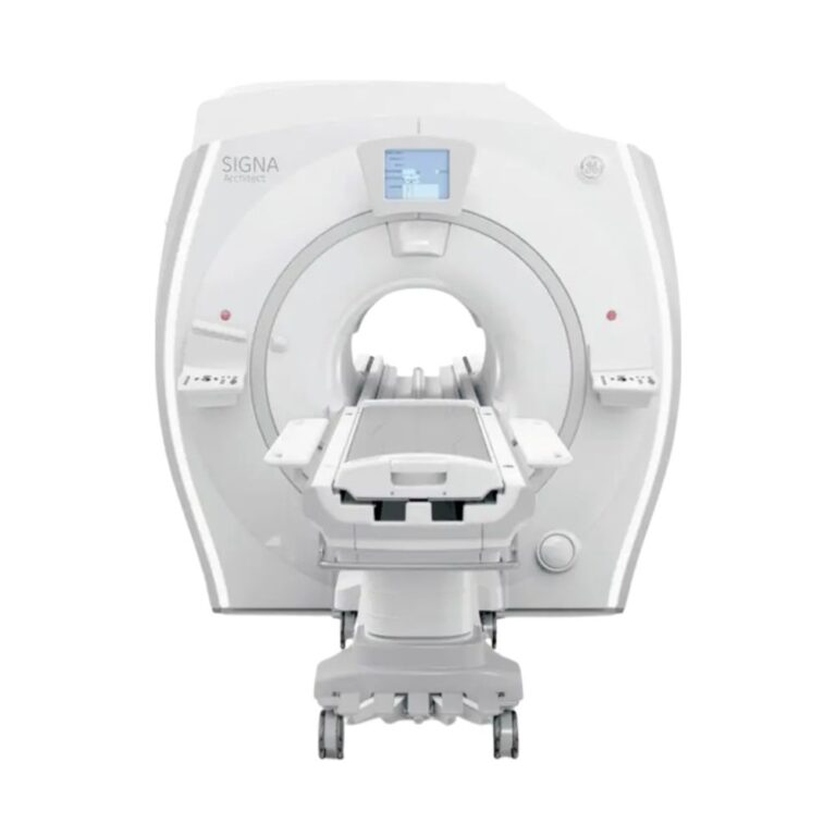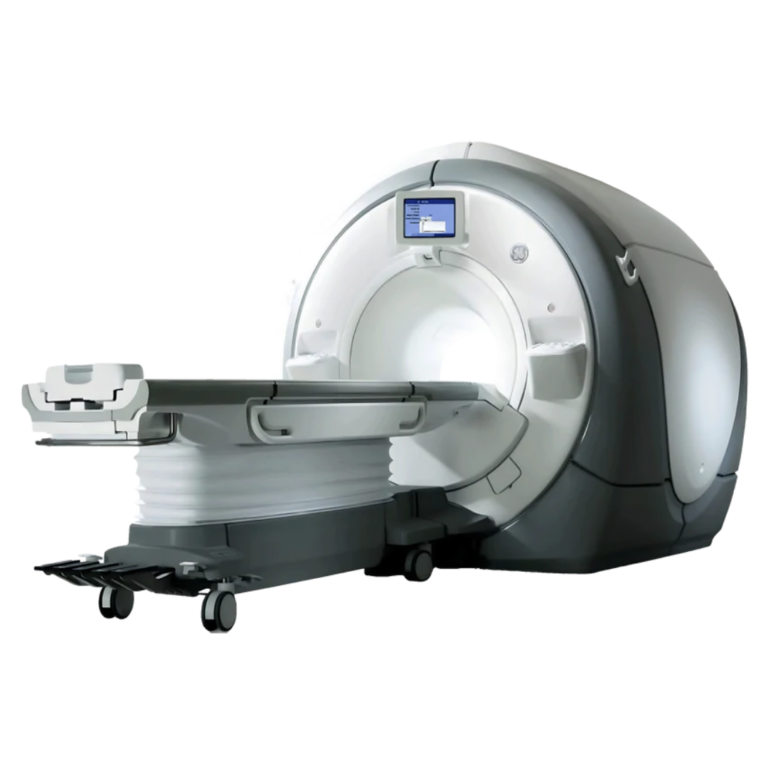
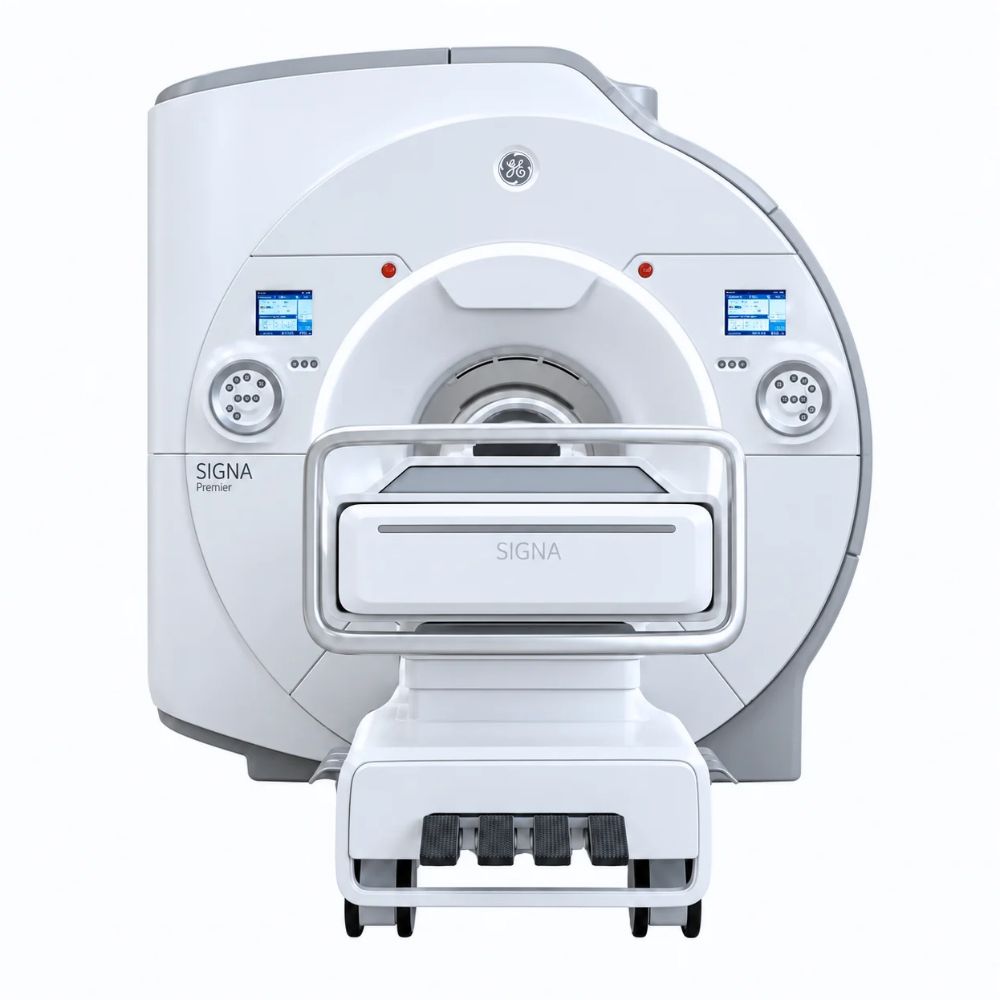
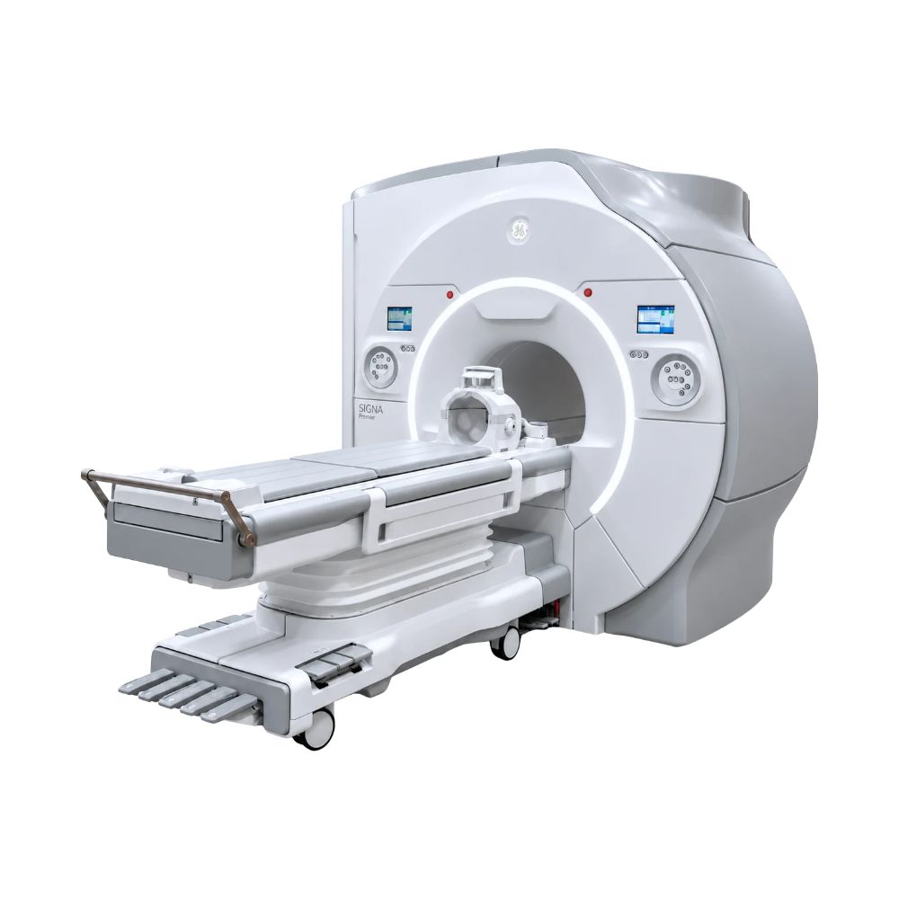
GE SIGNA Premier 3T MRI
- Category:
- MRI Systems
- Brand:
- GE
The GE SIGNA Premier 3T MRI is a 70 cm wide-bore scanner well suited for hospitals, imaging centers, and research institutions. Its powerful SuperG gradients and advanced digital imaging system deliver detailed images, efficient scanning, and the performance needed for specialized clinical examinations and neuroscience research.
SIGNA Premier MRI Highlights & Benefits
The GE SIGNA Premier is a high-performance 3T MRI scanner designed for comprehensive clinical imaging and advanced research. It combines powerful imaging technology with a 70 cm wide bore, giving facilities the flexibility to perform routine diagnostic examinations and demanding specialty studies while accommodating a broad range of patients.
Its key advantage is the ability to deliver detailed imaging and advanced scanning performance without sacrificing the patient space of a wide-bore MRI system.
Key Features & Benefits
-
- Powerful SuperG Gradient Technology: Uses an advanced magnetic field control system to capture fine anatomical detail and support demanding brain imaging and research examinations.
- 146-Channel Digital Imaging System: Collects signals from multiple receiving coils to support detailed images, extensive anatomical coverage, and efficient scanning.
- 70 cm Wide Bore: Provides more room for patients than conventional 60 cm MRI scanners, with flexible positioning and supported feet-first examinations.
- Consistent 3T Image Quality: Combines a strong magnetic field with MultiDrive technology, which helps distribute imaging signals more evenly for consistent image quality across different body regions.
- Flexible Coils & Advanced Image Processing: Available AIR Coils adapt to different body shapes, while AIR Recon DL uses deep learning to help produce sharper images with less background noise.
- Versatile Clinical & Research Imaging: Supports routine diagnostic MRI as well as advanced brain imaging, studies of nerve pathways, and specialized neuroscience research.
SIGNA Premier SuperG Gradient & 3T Imaging Performance
The Premier combines a 3T superconducting magnet, SuperG 80/200 gradients, and a 146-channel digital radiofrequency (RF) system. Together, these technologies support detailed whole-body imaging and demanding specialty examinations.
3T Magnet Performance & Whole-Body Imaging
The Premier’s 3 Tesla magnetic field supports detailed imaging of the brain, spine, joints, abdomen, pelvis, and other anatomical regions.
A 3T magnet can produce stronger MRI tissue signals than a 1.5T system under comparable conditions. That additional signal can support finer anatomical detail, faster compatible acquisitions, and advanced imaging techniques.
Magnetic field correction, known as shimming, helps maintain consistent image quality and tissue contrast across the examination area. The Premier also supports a maximum published field of view of 50 × 50 × 50 cm (19.7 × 19.7 × 19.7 in), providing extensive anatomical coverage.
SuperG 80/200 Gradient Technology
SuperG is a defining feature of the SIGNA Premier. Its maximum gradient amplitude of 80 mT/m and slew rate of 200 T/m/s support precise anatomical imaging, advanced diffusion studies, and demanding research protocols.
Gradients are controlled magnetic fields that tell the scanner where MRI signals originate. Gradient amplitude describes their strength, while slew rate describes how quickly they change.
SuperG uses internally water-cooled hollow conductors to remove heat generated during scanning. Its force-balanced construction also reduces vibration from rapidly changing magnetic fields.
This design helps maintain stable, high-performance acquisitions within the Premier’s spacious 70 cm bore, combining demanding neurological imaging capabilities with wide-bore patient access.
146-Channel RF System & MultiDrive Transmission
The Premier’s Total Digital Imaging (TDI) architecture provides up to 146 receive channels for collecting signals from compatible imaging coils.
Its multichannel design supports extensive anatomical coverage and parallel imaging, which uses information from multiple coil elements to shorten compatible scans.
MultiDrive complements this receiving architecture through two independently controlled RF transmit channels. By adjusting how radiofrequency energy is distributed through the body, MultiDrive helps reduce variations in image brightness and tissue contrast, particularly during 3T body imaging.
The number of active receiving elements depends on the installed coils and selected examination.
SIGNA Premier Wide-Bore Patient Access
The Premier combines high-performance 3T imaging with patient-friendly positioning and a spacious 70 cm bore. These features help facilities accommodate different patient sizes, mobility needs, and clinical examinations.
70 cm Bore & Feet-First Examinations
The Premier’s 70 cm (27.6 in) bore provides approximately 10 cm (3.9 in) more diameter than conventional 60 cm MRI scanners.
Its dual-flared design provides additional space near the bore openings, while supported feet-first examinations reduce the need for head-first entry when clinically appropriate.
The approximately 163 cm (64.2 in) bore length, patient communication system, adjustable lighting, and ventilation further support examination comfort.
Comfort Plus Patient Table & Weight Capacity
The Premier supports GE’s Comfort Plus Patient Table, designed for accessible positioning and efficient examination preparation.
Representative original configurations include:
-
- 550 lb (250 kg) Patient Capacity: Accommodates a broad range of patient sizes.
- Adjustable Table Height: Approximately 53.5–93 cm (21.1–36.6 in) for patient access and positioning.
- Powered Table Movement: Provides controlled vertical and longitudinal positioning.
- Integrated Posterior Coil Array: Collects MRI signals beneath the patient during supported examinations.
The original published scannable range is approximately 168 cm (66.1 in). Compatible later tables may have different specifications, and detachable eXpress configurations provide additional patient-transfer options.
SIGNA Premier Patient Comfort During Scanning
The Premier supports technologies that reduce common challenges associated with MRI examinations, including rigid coil positioning, scanner noise, patient movement, and repeated breath-holding.
Flexible AIR Coils & Patient Positioning
Compatible GE AIR Coils use lightweight, flexible receiving elements that conform more closely to patient anatomy than traditional rigid coils.
These elements collect MRI signals while allowing technologists to position the coil around different body shapes. AIR Anterior Arrays and multipurpose coils support flexible positioning during suitable body and musculoskeletal examinations.
This can improve comfort without sacrificing appropriate signal coverage.
Quieter MRI Scanning & Motion Correction
The Premier’s force-balanced SuperG gradients help reduce vibration associated with rapid gradient switching. Compatible quiet-scanning sequences further reduce noise by changing how selected MRI measurements are acquired.
Available motion-management technologies address patient movement through different methods:
-
- PROPELLER: Collects overlapping, rotating strips of MRI data to identify and reduce certain motion-related effects during reconstruction.
- PROMO: Detects head movement during compatible brain scans and adjusts subsequent acquisition to preserve image alignment.
These techniques help reduce motion-related image distortion and the potential need for repeat scans.
Free-Breathing Abdominal MRI
Compatible respiratory-navigation technology tracks breathing-related movement, typically through diaphragm position, and coordinates image acquisition with the respiratory cycle.
This allows selected abdominal examinations to proceed without repeated breath-holding, supporting patients who have difficulty maintaining consistent breath-holds.
Quiet scanning, motion correction, and respiratory-navigation capabilities depend on installed software and supported protocols.
Clinical Environments & Applications
The GE SIGNA Premier supports comprehensive diagnostic MRI and advanced clinical research. Its 3T field strength, SuperG gradients, and wide-bore design make it well suited to facilities providing routine examinations alongside demanding neurological and specialty imaging.
Clinical Environments
-
- Hospitals and diagnostic imaging departments
- Outpatient diagnostic imaging centers
- Multispecialty medical practices
- Neurology and neuroscience imaging centers
- Oncology and cancer care facilities
- Orthopedic and sports medicine centers
- Academic medical centers and teaching hospitals
- Clinical research institutions and neuroscience research facilities
Clinical Specialties
-
- Radiology: Brain, spine, musculoskeletal, body, and vascular MRI.
- Neurology & Neurosurgery: Advanced brain imaging, diffusion studies, functional MRI, and spinal examinations.
- Orthopedics: Cartilage, ligament, tendon, joint, and peripheral nerve imaging.
- Oncology: Tumor characterization, treatment assessment, and follow-up imaging.
- Cardiology: Cardiac anatomy and function with compatible applications.
- Vascular Medicine: Contrast-enhanced and selected non-contrast vascular imaging.
- Gastroenterology & Hepatology: Abdominal, liver, pancreatic, and quantitative tissue imaging.
- Urology: Prostate, renal, and pelvic examinations.
- Breast Imaging: Dedicated breast MRI using appropriate coils and software.
- Pediatric Radiology: Neurological, musculoskeletal, and body MRI with suitable protocols.
- Neuroscience & Clinical Research: Advanced diffusion imaging, functional brain mapping, and connectivity research.
Common MRI Applications
-
- Brain and spine MRI
- Diffusion-weighted and diffusion tensor imaging
- Functional MRI and brain connectivity studies
- Joint, cartilage, and musculoskeletal imaging
- Peripheral nerve examinations
- Abdominal and pelvic MRI
- Liver, pancreatic, and prostate imaging
- Breast and cardiac MRI with compatible options
- Magnetic resonance angiography
- Oncology assessment and follow-up
- High-resolution three-dimensional imaging
- Selected bone imaging using zero-echo-time techniques
- Specialized neuroscience research
SIGNA Premier Exam Planning & Workflow Automation
The Premier supports automated applications that simplify MRI examination preparation, improve positioning consistency, and help technologists manage daily scanning.
Automated Coil Selection & Anatomical Planning
Compatible workflow technologies include:
-
- AIR Touch: Automatically selects suitable receiving coil elements based on the connected coils and examination area, reducing manual setup while maintaining appropriate signal coverage.
- AIR x: Uses deep-learning anatomical recognition to identify structures in the brain or knee and automatically align imaging slices. This helps produce consistent scan positioning across patients and follow-up examinations.
- SIGNA Flow: Organizes examination protocols, scan preparation, and compatible worklist information, reducing repetitive setup while allowing patient-specific adjustments.
These technologies address different parts of examination preparation, helping facilities maintain consistent workflows without removing technologist control.
Image Review & PACS Connectivity
The Premier supports DICOM-based image transfer to compatible picture archiving and communication systems (PACS) for storage, review, and distribution.
Compatible workstations provide multiplanar reconstruction, three-dimensional review, quantitative analysis, and specialized visualization.
Available workflow and processing functions depend on the installed console and software.
SIGNA Premier Image Reconstruction & Scan Acceleration
The Premier supports advanced technologies that improve reconstructed image quality, shorten compatible acquisitions, and expand imaging options.
Deep-Learning Reconstruction & Faster MRI Scans
Different technologies address image quality and acquisition time in different ways:
-
- AIR Recon DL: Uses deep learning during image reconstruction to reduce noise and improve sharpness, supporting finer detail or shorter compatible scans.
- HyperSense: Uses compressed sensing to acquire fewer measurements and mathematically reconstruct the missing information, shortening suitable examinations.
- Sonic DL: Uses newer deep-learning-based acceleration methods to reconstruct images from accelerated acquisitions on compatible systems.
These technologies are not interchangeable. AIR Recon DL primarily improves reconstructed image quality, while HyperSense and Sonic DL use their respective methods to accelerate supported acquisitions.
Sonic DL and other newer applications depend on the software generation and installed licenses.
Three-Dimensional & Multi-Contrast Imaging
The Premier also supports technologies that reduce the need for separate acquisitions or provide more flexible image review.
-
- Cube: Acquires a three-dimensional image volume that can be reconstructed into different viewing planes.
- HyperCube: Combines compatible volumetric imaging with accelerated sampling to shorten detailed 3D examinations.
- MAGiC: Measures tissue properties during one specialized acquisition and generates multiple image contrasts, including compatible T1, T2, and fluid-suppressed images.
These applications are particularly useful for neurological, spine, and musculoskeletal imaging when equipped with the appropriate software.
SIGNA Premier Advanced Diffusion & Brain Imaging
The Premier’s SuperG gradient performance makes it particularly suited to demanding neurological imaging and neuroscience research. Its high-channel-count RF architecture and available acquisition options support detailed investigations of brain structure and activity.
Advanced Diffusion Imaging & White-Matter Pathways
Diffusion MRI measures water movement within tissue, helping characterize the organization of structures such as white-matter fibers in the brain.
Compatible technologies include:
-
- Diffusion Tensor Imaging (DTI): Measures water movement in multiple directions to characterize the orientation of white-matter pathways.
- Multi-Shell Diffusion Imaging: Collects measurements at several diffusion-weighting strengths to support more detailed tissue modeling.
- HyperBand: Acquires information from multiple slices simultaneously and separates the signals during reconstruction, accelerating compatible diffusion studies.
The Premier’s 80/200 SuperG gradients support these demanding measurements, making advanced diffusion imaging one of its principal differentiators.
Functional MRI & Brain Connectivity Research
Functional MRI (fMRI) measures changes in MRI signals associated with blood oxygenation, providing information about activity in different brain regions.
Resting-state fMRI evaluates relationships between these signal changes while the patient or research participant is not performing a specific task.
Together with diffusion imaging, these methods support Human Connectome research investigating the structural and functional connections within the brain.
Research-specific applications require appropriate software, receiving coils, reconstruction tools, and validated protocols.
SIGNA Premier Specialized Clinical Imaging
The Premier extends beyond conventional anatomical MRI through compatible applications for bone imaging, contrast-enhanced studies, blood flow assessment, and tissue characterization.
Bone Imaging & Dynamic Contrast Studies
-
- oZTEo: Uses near-zero-echo-time acquisition to collect signals from tissues, particularly bone, that conventional MRI sequences have difficulty capturing. This provides additional information about bone morphology alongside conventional soft-tissue MRI, although CT remains preferable for certain clinical questions.
- DISCO: Updates portions of an MRI dataset during contrast enhancement, allowing successive phases to be reconstructed without repeating a complete conventional scan each time. This supports selected liver, breast, prostate, and other dynamic examinations.
Brain Perfusion, Liver Fat, & Vascular Imaging
Compatible clinical applications include:
-
- 3D Arterial Spin Labeling: Magnetically labels blood and compares measurements to estimate cerebral blood flow without injected contrast material.
- IDEAL IQ: Separates water and fat signals using measurements at different echo times, supporting quantitative tissue fat assessment.
- Non-Contrast MR Angiography: Uses blood-flow or magnetic signal differences to visualize selected vessels without injected contrast material.
These technologies expand neurological, abdominal, and vascular imaging services when appropriately equipped.
Tissue Chemistry & Specialized MRI Research
Magnetic resonance spectroscopy examines signal frequencies to provide information about selected tissue metabolites rather than anatomy alone.
Specialized research configurations can support investigations involving other atomic nuclei, including multinuclear spectroscopy and hyperpolarized carbon-13 applications.
These capabilities require dedicated hardware, software, coils, and research infrastructure. They are not standard features of every Premier system.
SIGNA Premier Configurations, Coils, & Available Options
The Premier provides a high-performance 3T foundation, with installed software, receiving coils, and clinical options determining which additional capabilities are available.
System Configurations
A purpose-built Premier combines the 3T magnet, SuperG 80/200 gradients, and 146-channel RF architecture.
Configurations can vary by software generation, coil package, patient table, workstation, and specialty applications.
GE also offers SIGNA Premier Evo as a modernization pathway for certain earlier 3T systems. An upgraded Evo configuration should not automatically be considered identical to a purpose-built Premier.
MXR Imaging helps facilities identify the included capabilities and select appropriate options for their planned examinations.
RF Coils
-
- Compatible coil categories include:
- Head & Neck: Neurological and cervical examinations
- Spine Arrays: Cervical, thoracic, and lumbar imaging
- AIR Anterior Arrays: Flexible abdominal, pelvic, and body coverage
- AIR Multi-Purpose Coils: Adaptable joint and musculoskeletal imaging
- Posterior Arrays: Signal collection beneath the patient
- Extremity Coils: Dedicated joint examinations
- Breast Coils: Specialized breast MRI
- Cardiac Arrangements: Supported cardiovascular examinations
The original Premier configuration includes a published 32-channel integrated posterior spine array. Certain later compatible AIR table configurations incorporate up to 60 posterior channels.
Coil compatibility and included accessories depend on the installed RF hardware.
MXR Imaging provides MRI coil repair services for supported equipment.
Software & Clinical Options
Compatible options support advanced neurological, musculoskeletal, cardiac, vascular, breast, and body imaging, along with reconstruction and scan acceleration.
AIR Recon DL, HyperSense, HyperBand, AIR Touch, AIR x, and other advanced applications require compatible software and hardware. Newer technologies such as Sonic DL may require later system generations or supported upgrades.
Research-specific capabilities can require additional equipment beyond standard clinical software packages.
SIGNA Premier Magnet Cooling & Helium Management
The Premier uses a superconducting 3T magnet maintained at extremely low temperatures by liquid helium and mechanical refrigeration.
Its original published configuration incorporates zero-boil-off technology intended to limit routine helium evaporation under specified normal conditions. This does not eliminate maintenance or all potential helium requirements.
For pre-owned systems, magnet condition, refrigeration performance, helium history, and service records remain important ownership considerations.
MXR Imaging provides MRI refrigeration and helium services for compatible equipment.
SIGNA Premier MRI Installation & Operating Requirements
Premier installations require coordinated planning for the magnet, supporting equipment, electrical utilities, cooling, and magnetic safety environment. Requirements depend on the selected configuration and facility layout.
MRI Room Dimensions, Access, & Shielding
The original Premier magnet’s published dimensions with its enclosure are approximately:
-
- Length: 209 cm (82.3 in)
- Width: 252 cm (99.2 in)
- Height: 250 cm (98.4 in)
- Weight with Cryogens: 14,060 lb (6,378 kg)
These are equipment dimensions, not minimum MRI room dimensions.
Site planning must also address patient-table clearance, equipment cabinets, delivery access, structural support, RF shielding, magnetic safety boundaries, and cryogenic venting.
MXR Imaging supports installations through custom MRI shielding solutions and turnkey imaging project management.
Electrical Power, Cooling, & Long-Term Operation
The Premier requires electrical service, equipment cooling, HVAC, and magnet refrigeration appropriate for its configuration.
Internally cooled SuperG gradients help manage heat during demanding examinations, while compatible accelerated scanning techniques can shorten acquisition times and improve workflow efficiency.
Operating expenses depend on examination volume, installed equipment, facility utilities, and maintenance requirements.
GE SIGNA Premier vs. Other SIGNA MRI Models
The GE SIGNA MRI family includes multiple 3T and 1.5T scanners with different imaging architectures, patient-access features, and performance priorities.
Comparing these systems helps facilities select equipment aligned with their clinical workload and specialized imaging needs.
Premier, Architect, Pioneer, & Voyager: Imaging Priorities
-
- SIGNA Premier 3T: Emphasizes SuperG 80/200 gradients, a 146-channel RF architecture, high-performance clinical imaging, and demanding neuroscience applications.
- SIGNA Architect 3T: Provides a versatile 70 cm wide-bore platform with 128-channel digital RF technology and broad specialty clinical capabilities.
- SIGNA Pioneer 3T: Emphasizes energy-efficient 3T imaging and an alternative gradient architecture suited to comprehensive diagnostic examinations.
- SIGNA Voyager 1.5T: Combines wide-bore patient access, 1.5T imaging, and efficient clinical operation for a broad range of diagnostic services.
Premier’s SuperG Performance vs. Other SIGNA Models
Premier is particularly well suited for facilities requiring strong gradient performance for advanced diffusion imaging, functional MRI, and neuroscience research.
Its SuperG 80/200 architecture provides greater maximum gradient amplitude than the published 44 mT/m configuration of SIGNA Architect, while both platforms support high-field 3T clinical MRI.
Premier’s 146-channel receiving architecture also differs from Architect’s published 128-channel configuration.
These distinctions can matter when planning demanding imaging programs, although actual examination performance also depends on protocols, receiving coils, reconstruction software, and the installed system.
Architect and Pioneer remain relevant choices for facilities prioritizing versatile clinical 3T imaging.
Voyager provides a 1.5T alternative for facilities seeking wide-bore access and broad diagnostic coverage without requiring Premier’s advanced 3T gradient platform.
The appropriate system depends on planned examinations, patient needs, operating costs, infrastructure, and budget.
MXR Imaging can help compare the available platforms and identify a configuration suited to the facility’s clinical priorities.
Available SIGNA Premier System Configuration
A pre-owned GE SIGNA Premier system may differ from another Premier in its software generation, installed applications, receiving coils, patient table, and service history.
Included Hardware, Coils, & Software
Available equipment packages may vary in:
-
- Hardware and software generation
- Included anatomical and specialty receiving coils
- AIR Recon DL and other reconstruction applications
- Advanced neurological and diffusion software
- Functional MRI and research capabilities
- Musculoskeletal, cardiac, vascular, breast, and body packages
- Patient-table configuration
- Operator workstation and post-processing applications
- Magnet and refrigeration condition
- Service history and compatible upgrades
Facilities performing routine diagnostic examinations may require a different combination of clinical options than those conducting advanced neurological research.
MXR Imaging works with buyers to identify configurations that support their intended examinations and operating requirements.
SIGNA Premier MRI Price & Availability
MXR Imaging helps healthcare facilities source GE SIGNA Premier 3T MRI systems and select configurations suited to their clinical imaging requirements. Our team can assist with equipment options, pricing, installation planning, and long-term service support.
SIGNA Premier MRI pricing depends on the scanner’s age, condition, hardware and software generation, installed clinical options, receiving coils, and included accessories.
Systems equipped for advanced neuroscience research may have different capabilities and values than configurations intended primarily for routine diagnostic imaging.
Total project costs may also include equipment transportation, installation, site preparation, shielding, utilities, and ongoing service requirements.
For facilities replacing an existing scanner, the available infrastructure can influence the scope and cost of the project.
MXR Imaging helps hospitals, imaging centers, specialty practices, and research institutions evaluate the available equipment alongside their clinical needs and budgets.
Our team also provides MRI maintenance and repair services for compatible systems.
Contact MXR Imaging to discuss GE SIGNA Premier MRI pricing, available equipment, and installation requirements.
GE SIGNA Premier Specifications
The following specifications describe the original GE SIGNA Premier 3T platform and representative compatible configurations. Software, receiving coils, table options, and research capabilities may vary.
Magnet System
-
- Field Strength: 3T
- Magnet Type: Superconducting
- Scanner Design: Whole-body, wide-bore MRI
- Bore Diameter: 70 cm (27.6 in)
- Bore Length: Approximately 163 cm (64.2 in)
- Patient Aperture at Magnet Flare: Approximately 74 cm (29.1 in)
- Maximum Field of View: 50 × 50 × 50 cm (19.7 × 19.7 × 19.7 in)
- Magnet Shielding: Active
- Magnetic Field Correction: Higher-order shimming
- Cooling: Liquid helium with mechanical refrigeration
- Cryogen Management: Zero-boil-off design under specified normal conditions
SuperG Gradient System
-
- Gradient Technology: SuperG
- Maximum Gradient Amplitude: 80 mT/m
- Maximum Slew Rate: 200 T/m/s
- Published Maximum Duty Cycle: 100%
- Cooling Architecture: Internally water-cooled hollow conductors
- Gradient Heat Removal: Up to approximately 45 kW in GE’s published design
- Mechanical Design: Force- and torque-balanced
- Supported Applications: Advanced diffusion, functional MRI, high-resolution imaging, and specialized research
RF System
-
- RF Architecture: Total Digital Imaging (TDI)
- Receive Channels: Up to 146
- RF Transmit Technology: MultiDrive
- Published Transmit Architecture: Two independently controlled 15 kW channels
- Peak Combined RF Power: 30 kW
- Receiving Coils: Compatible multichannel anatomical arrays and AIR Coils
Bore & Patient Table
-
- Bore Diameter: 70 cm (27.6 in)
- Patient Entry: Head-first and supported feet-first examinations
- Compatible Tables: Comfort Plus and supported eXpress configurations
- Maximum Patient Weight: Up to 550 lb (250 kg) on compatible tables
- Original Comfort Plus Table Height: Approximately 53.5–93 cm (21.1–36.6 in)
- Original Comfort Plus Scannable Range: Approximately 168 cm (66.1 in); later configurations may differ
- Original Comfort Plus Cradle Dimensions: Approximately 245 × 56 cm (96.5 × 22.0 in)
- Table Movement: Powered vertical and longitudinal positioning
- Patient Comfort: Communication, adjustable lighting, and ventilation
Coils & Accessories
-
- Compatible Coil Categories: Head, head/neck, spine, body, extremity, breast, and cardiac
- Original Posterior Array: Published 32-channel TDI integrated spine array
- Compatible Later AIR Posterior Arrays: Up to 60 channels
- AIR Coil Options: Flexible anterior arrays and multipurpose coils
- Included Coil Package: Configuration-dependent
- Positioning Accessories: Compatible supports and specialty equipment
Imaging Software & Research Options
-
- Deep-Learning Reconstruction: AIR Recon DL when installed
- Scan Acceleration: Compatible HyperSense, HyperBand, and Sonic DL applications
- 3D Imaging: Cube and compatible HyperCube techniques
- Multi-Contrast Imaging: MAGiC when installed
- Advanced Neurological Imaging: DTI, multi-shell diffusion, and functional MRI
- Bone Imaging: oZTEo when equipped
- Quantitative Imaging: Compatible IDEAL IQ and perfusion applications
- Specialized Research: Multinuclear spectroscopy and related applications with appropriate hardware
Siting & Utilities
-
- RF Shielding: Required
- Cryogenic Refrigeration: Required
- Equipment Cooling: Dedicated infrastructure
- Electrical Service: Configuration-specific
- Cryogenic Venting: Required according to the applicable installation specifications
- Structural Support: Based on equipment weight and site layout
- Safety Planning: Controlled magnetic field access and appropriate emergency provisions
System Dimensions & Weight
-
- Magnet Length with Enclosure: Approximately 209 cm (82.3 in)
- Magnet Width with Enclosure: Approximately 252 cm (99.2 in)
- Magnet Height with Enclosure: Approximately 250 cm (98.4 in)
- Magnet Weight with Cryogens: Approximately 14,060 lb (6,378 kg)
- Magnet Dimensions Without Enclosure: Approximately 174 × 212 × 240 cm (68.5 × 83.5 × 94.5 in)
- Patient Table Dimensions: Dependent on configuration
- Installation Footprint: Determined by room layout, supporting equipment, safety boundaries, and service clearances
Dimensions and weight are representative of the published original Premier configuration. Final site requirements should be matched to the specific equipment and applicable installation documentation.