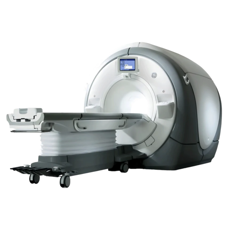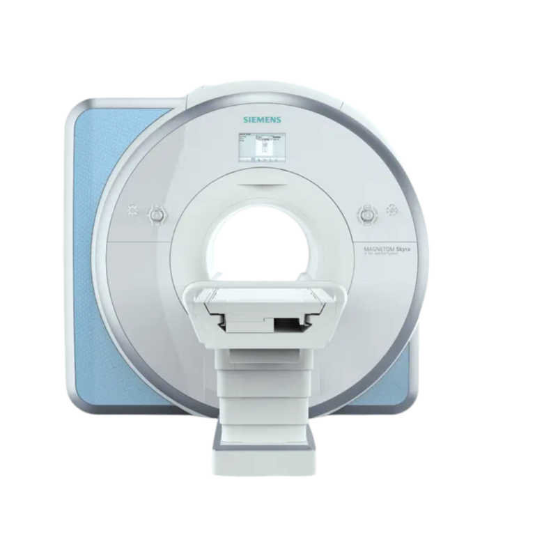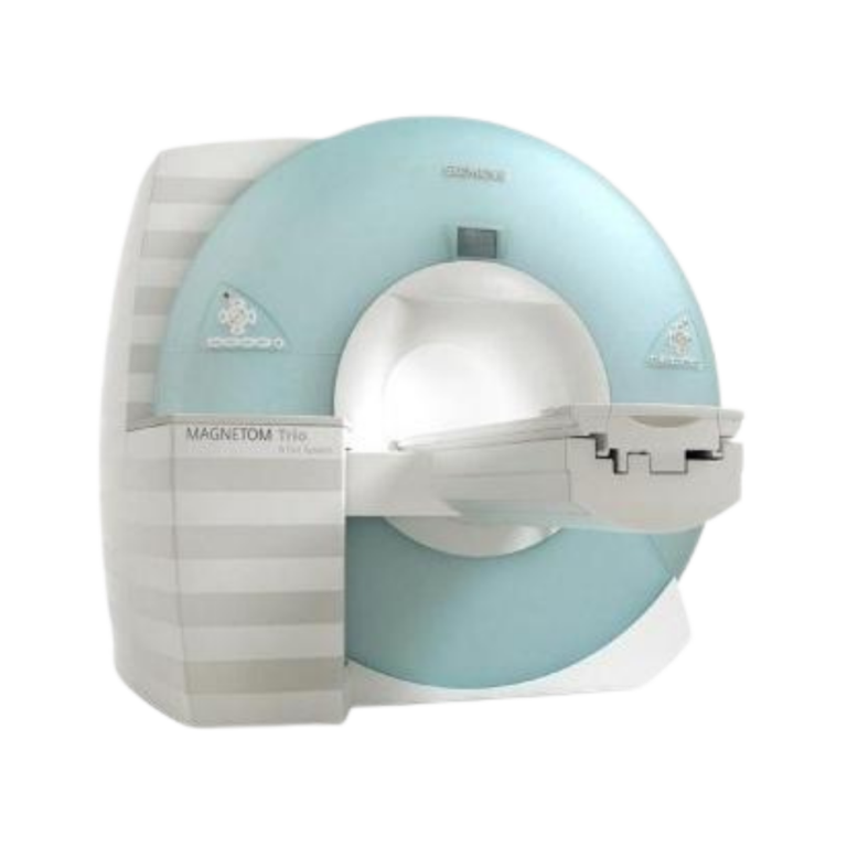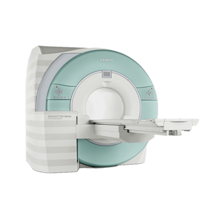
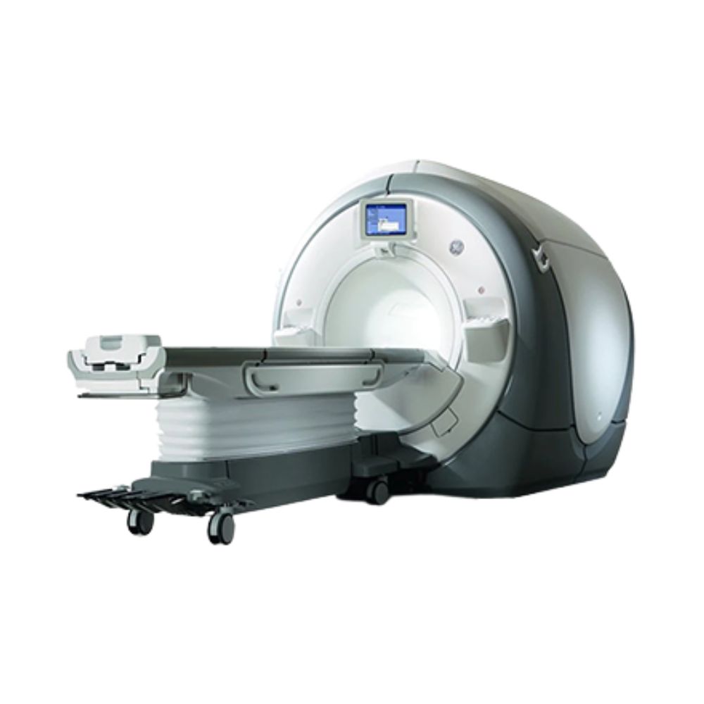
GE Discovery MR750 3T MRI
- Category:
- MRI Systems
- Brand:
- GE
The GE Discovery MR750 is a 3T whole-body MRI scanner best suited for organizations that need high-resolution routine and advanced imaging. Its 60 cm short-bore design, broad 48 cm imaging field, and high-performance image acquisition support demanding neurological, musculoskeletal, body, cardiac, vascular, breast, and oncology exams.
GE Discovery MR750 Highlights & Benefits
The GE Discovery MR750 combines a 60 cm short-bore 3T magnet, a broad 48 cm imaging field, a high-performance gradient system for demanding diffusion and high-resolution exams, and a detachable 500 lb-capacity patient table. Its signal-processing and positioning technologies are designed to support detailed imaging while keeping exam setup and image reconstruction efficient.
MR750 Key Benefits
-
- 60 cm short-bore 3T design: Provides high-field MRI performance in a shorter patient opening with head-first or feet-first positioning.
- 48 cm imaging field: Supports whole-body examinations and broad anatomical coverage.
- 50 mT/m gradients with 200 T/m/s slew rate: Support demanding diffusion, cardiac, vascular, functional, and high-resolution imaging.
- 32-channel OpTix RF system: Converts received MRI signals to digital optical data in the scan room to help preserve signal quality before reconstruction.
- Detachable Express patient table: Supports up to 227 kg (500 lb) and can assist with patient preparation, positioning, and transport.
- IntelliTouch and ARC workflow tools: IntelliTouch automates routine positioning steps, while ARC uses data from multiple coil elements to accelerate compatible MRI acquisitions.
MRI Performance & Image Quality
3T Field Strength & Magnet Design
The Discovery MR750 uses a short-bore 3.0T superconducting magnet designed to maintain a uniform magnetic field across the imaging area. Consistent field uniformity supports reliable image quality across routine and advanced examinations.
The system provides a maximum 48 × 48 × 48 cm (18.9 × 18.9 × 18.9 in.) imaging field of view for whole-body MRI.
The magnet also uses active shielding, which helps limit how far the magnetic field extends beyond the scanner. Magnet hardware can vary between MR750 configurations, so installation requirements should be matched to the specific system.
Gradient Performance
The MR750 gradient system delivers up to 50 mT/m amplitude and 200 T/m/s slew rate per axis.
MRI gradients rapidly change the magnetic field to locate signals within the body. Gradient amplitude describes their maximum strength, while slew rate describes how quickly they can change.
The MR750’s gradient performance supports demanding applications including diffusion imaging, functional MRI, cardiac and vascular studies, and high-resolution 3D imaging.
The gradient system is water-cooled to help manage the heat generated during demanding or repeated acquisitions.
OpTix RF & 32-Channel Signal Architecture
OpTix Optical RF converts the MRI signals received from the patient coils into digital optical data within the scan-room environment. This helps reduce electrical noise and signal degradation before the data reaches the reconstruction system.
The MR750 supports up to 32 simultaneous receive channels on applicable configurations. These channels provide separate pathways for signals collected by multi-channel MRI coils, supporting detailed imaging and faster compatible acquisitions.
PERFORM 2.0 provides patient-specific management of specific absorption rate, or SAR. SAR measures the amount of radiofrequency energy absorbed by the body during an MRI examination. PERFORM 2.0 manages RF energy for the individual patient while maintaining 3T imaging performance.
Patient Access & Exam Experience
60 cm Short-Bore Patient Access
The Discovery MR750 uses a 60 cm (23.6 in.) bore with a short design that widens toward both ends of the scanner.
The patient opening measures approximately 70 cm long × 60 cm wide × 60 cm high (27.6 × 23.6 × 23.6 in.). Head-first and feet-first positioning can be used depending on the examination and coil configuration.
The short-bore design reduces the length of the patient opening compared with longer conventional MRI systems while maintaining 3T imaging performance.
Express Patient Table & Positioning
The detachable Express patient table supports patients up to 227 kg (500 lb) and can be prepared outside the scan room when appropriate.
Powered vertical and longitudinal movement assists with patient positioning, while the detachable design allows the table to be docked, undocked, and used for patient transport.
The table provides approximately 205 cm (80.7 in.) of scannable range. IntelliTouch can automate routine positioning steps, including identifying the anatomical reference position, selecting a compatible coil, centering the patient, and moving the table into scanning position.
Short-Bore Exam Experience
The short-bore design reduces the length of the patient opening and provides access from either end of the scanner.
Applicable configurations include adjustable in-bore lighting and ventilation and two-way communication between the patient and technologist.
Dual-sided scanner controls also allow technologists to work from either side of the patient table during setup.
GE Discovery MR750 Clinical Environments & Applications
The GE Discovery MR750 supports routine and advanced 3T MRI across human and veterinary imaging settings. Clinical capability depends on the coils, software, application licenses, and supporting equipment included with the individual system.
Clinical Environments
-
- Hospitals and health systems
- Outpatient imaging centers
- Independent diagnostic imaging centers
- Academic medical centers
- Research imaging programs
- Specialty imaging centers
- Neurology and orthopedic imaging centers
- Cardiovascular imaging programs
- Oncology and radiation oncology programs when appropriately configured
- Veterinary hospitals, specialty referral centers, and advanced veterinary imaging programs
Clinical Specialties
-
- Diagnostic Radiology
- Neuroradiology
- Neurology & Neurosurgery
- Orthopedics & Musculoskeletal Imaging
- Cardiology
- Vascular Imaging
- Oncology
- Breast Imaging
- Abdominal & Pelvic Imaging
- Pediatric Radiology
- Radiation Oncology when configured for applicable MR treatment-planning workflows
- Veterinary Radiology & Advanced Veterinary Imaging
Common MRI Applications
-
- Brain, head, neck, and temporomandibular joint (TMJ) MRI
- Spine and spinal cord imaging
- Diffusion and perfusion imaging for evaluating tissue and blood-flow characteristics
- Functional MRI for mapping activity within the brain when supported
- Diffusion tensor imaging for evaluating white-matter pathways when supported
- Shoulder, hip, knee, wrist, hand, ankle, foot, and other musculoskeletal MRI
- Abdominal and pelvic MRI
- Liver imaging and MR elastography for evaluating tissue stiffness when appropriately equipped
- Prostate and other pelvic MRI
- Breast MRI with an appropriate dedicated coil
- Cardiac imaging for heart structure, function, viability, and blood flow
- MR angiography for blood-vessel imaging with or without contrast when supported
- Peripheral vascular imaging
- Oncology imaging and treatment follow-up
- Imaging around orthopedic implants with compatible metal artifact reduction software
- Pediatric MRI using appropriate coils and protocols
- Veterinary neurological, spinal, musculoskeletal, body, and cardiac MRI with appropriate coils, positioning, anesthesia, and monitoring
- MR spectroscopy for evaluating tissue chemistry when the required software and hardware are installed
- MRI-based simulation and image acquisition for applicable radiation treatment-planning workflows
MRI Workflow, Reconstruction & Imaging Technology
Exam Setup & Scan Automation
The Discovery MR750 uses IntelliTouch, Express Exam, ProtoCopy, and in-room operator controls to reduce repetitive steps during patient setup and exam preparation.
IntelliTouch can automate routine patient landmarking and table positioning. Express Exam organizes acquisition, image processing, and networking steps within the selected examination protocol.
ProtoCopy allows complete exam protocols to be transferred between compatible systems, helping facilities maintain consistent scan setup across technologists or scanners.
High-Resolution In-Room Operator Control, or iROC, displays supported patient and scan information near the magnet so technologists can complete setup tasks while remaining close to the patient.
Image Reconstruction & Acceleration
ARC is GE’s parallel imaging technology. It uses signals from multiple MRI coil elements to accelerate compatible acquisitions without requiring a separate calibration scan.
The MR750 also uses a high-speed volume reconstruction engine to process the large datasets produced during multi-channel and 3D MRI examinations.
Together, ARC and the reconstruction system help reduce acquisition or processing time for compatible exams while supporting the spatial resolution required for demanding neurological, vascular, cardiac, and body imaging.
Post-Processing & Connectivity
The MR750 supports Digital Imaging and Communications in Medicine (DICOM), the standard used to exchange medical images and related information between imaging systems.
DICOM connectivity allows the scanner to send images to compatible picture archiving and communication systems (PACS) for storage and review and to work with supported electronic worklists.
The installed workstation and software determine which additional analysis tools are available for neurological, cardiac, vascular, body, functional, quantitative, and 3D imaging.
MR750 Configurations, Coils & Available Options
System Configurations
GE’s official model designation is Discovery MR750 3.0T. The system may also appear as Discovery 750 in equipment listings, particularly for earlier installations, but the MR750 designation identifies the underlying 3T platform.
Discovery MR750 systems can differ by model year, magnet configuration, software release, RF hardware, coil package, patient table, workstation, clinical application licenses, and installed upgrades.
Eligible systems may include MultiDrive RF Transmit, a GE technology that adjusts radiofrequency transmission to improve signal uniformity across different patients and anatomy at 3T. MultiDrive should be verified on the individual scanner rather than assumed to be included with every MR750.
Other differences between available systems can include installed clinical software, RF hardware, workstation capabilities, and coil packages, making the individual configuration important when matching an MR750 to a facility’s planned examinations.
RF Coils
MRI receiver coils collect signals from the anatomy being examined. The Discovery MR750 supports a broad range of multi-channel coils designed for specific body regions and clinical applications.
Compatible configurations can include:
-
- 32-channel brain coils
- Head, neck, and spine coils
- Torso and body arrays
- Cardiac coils
- Breast coils
- Knee and other extremity coils
- Shoulder coils
- Hand and wrist coils
- Foot and ankle coils
- Flexible surface coils
- Specialty coils, including compatible prostate imaging equipment
Supported coils are not necessarily included with an available system. The actual coil package should be matched to the examinations the facility intends to perform.
Software & Clinical Options
Software capabilities vary by software generation, installed licenses, and upgrade history.
Depending on configuration, available applications can include:
-
- MAGiC for generating multiple neurological image contrasts from a compatible acquisition
- BrainWave for functional brain imaging
- Advanced diffusion imaging tools
- IDEAL and IDEAL IQ for separating and quantifying fat and water signals
- MAVRIC SL for reducing image distortion caused by metal implants
- DISCO and Turbo LAVA for dynamic and accelerated body imaging
- VIBRANT Flex for breast MRI
- PROPELLER for reducing the effects of patient motion on compatible examinations
- SilentSuite for quieter compatible MRI sequences
- MR elastography for evaluating tissue stiffness
- MR spectroscopy for evaluating tissue chemistry
- Cardiac and vascular imaging packages
Eligible upgraded systems may also support later GE SIGNA Works applications. Installed software and upgrade eligibility should be verified for the individual system.
Magnet, Cryogen & Site Requirements
Magnet & Helium System
The Discovery MR750 uses a liquid-helium-cooled 3T superconducting magnet with active shielding and a dedicated cryocooler.
Applicable configurations support zero-boil-off operation under normal conditions, helping minimize routine helium loss.
For an available system, helium level, cryocooler condition, magnet service history, and prior magnet work should be reviewed because they can affect installation planning and long-term service needs.
MXR provides MRI refrigeration and helium services for supported MRI systems.
Room & Installation Requirements
MR750 installation requires coordinated planning for the magnet room, equipment room, and control room.
Site planning should account for:
-
- RF shielding
- Magnetic fringe field and 5-gauss line
- Cryogen venting
- Structural loading
- Magnet delivery and equipment access
- Vibration and electromagnetic interference
- Service clearances
The 5-gauss line marks the boundary where the surrounding magnetic field reaches 5 gauss and is used during MRI room planning.
MXR’s custom MRI shielding solutions can support RF and magnetic shielding design and installation.
Power, Cooling & Facility Requirements
Depending on the installation, the MR750 can use 380, 400, 415, or 480 VAC three-phase power.
Typical configurations require approximately 114 L/min (30 GPM) of facility cooling-water flow and up to 70 kW of heat removal to support the scanner and associated equipment.
Final electrical, cooling, ventilation, and facility requirements should be confirmed for the specific system before construction or installation begins.
MXR also provides turnkey imaging project management for facilities coordinating site preparation, delivery, installation, and system activation.
Available GE Discovery MR750 Configuration
Available MR750 systems can differ even when they share the same model designation. The installed hardware, software, coils, and magnet history determine the examinations and workflows a particular system can support.
Configuration Variations
-
- Model year and system history
- Installed magnet configuration
- Software release
- RF and receiver hardware
- MultiDrive status when applicable
- Included head, body, musculoskeletal, breast, cardiac, vascular, and specialty coils
- Express patient-table configuration
- Installed clinical application licenses
- Workstation and post-processing package
- Magnet and helium status
- Cryocooler condition
- Hardware and software upgrade history
- Included monitoring equipment and accessories
MXR can help match an available MR750 configuration to the facility’s planned examinations, workflow, site requirements, and clinical priorities.
GE Discovery MR750 vs. MR750w
The GE Discovery MR750 and MR750w are both 3T whole-body MRI systems, but they are designed around different priorities. The MR750 combines a 60 cm short bore with higher gradient performance, while the MR750w provides a wider 70 cm bore for greater patient access and positioning flexibility.
Discovery MR750
The MR750 combines a 60 cm (23.6 in.) short bore with 50 mT/m gradients and a 48 cm (18.9 in.) imaging field of view. It is well suited for facilities prioritizing advanced neurological, diffusion, cardiac, vascular, musculoskeletal, research, and other demanding 3T MRI applications.
Discovery MR750w
The MR750w increases the bore diameter to 70 cm (27.6 in.), providing more room for larger patients, positioning equipment, and anatomy that must be positioned away from the center of the scanner. Compatible coil and patient-table configurations can also simplify positioning and multi-region imaging.
Facilities prioritizing wider patient access and positioning flexibility should also consider the GE Discovery MR750w 3T MRI Scanner.
GE Discovery MR750 Price & Availability
GE Discovery MR750 pricing varies based on system year, hardware and software configuration, included coils, clinical applications, magnet and helium condition, accessories, installation scope, and service requirements.
An MXR representative can review current availability and help identify a configuration suited to the facility’s clinical requirements, workflow, site, and budget.
Ongoing MRI maintenance and repair options are also available for supported systems, including preventative maintenance, repair, and customizable service coverage.
GE Discovery MR750 Specifications
Magnet System
-
- Field strength: 3.0T
- Magnet type: Superconducting
- Magnet design: Short-bore, actively shielded
- Bore diameter: 60 cm (23.6 in.)
- Maximum imaging field of view: 48 × 48 × 48 cm (18.9 × 18.9 × 18.9 in.)
- Cooling: Liquid helium with dedicated cryocooler
- Zero-boil-off design: Available on applicable configurations to minimize routine helium loss
- Magnet configuration: Varies by system generation and installed magnet
Gradient System
-
- Gradient platform: eXtreme Resonance Module (XRM)
- Maximum gradient amplitude: 50 mT/m per axis
- Maximum slew rate: 200 T/m/s per axis
- Maximum imaging field of view: 48 cm (18.9 in.)
- Gradient cooling: Water-cooled
RF System
-
- Receive architecture: OpTix Optical RF
- Receive channels: Up to 32 simultaneous channels on applicable configurations
- Signal transmission: Digital optical receive architecture
- PERFORM 2.0 patient-specific SAR management
- MultiDrive RF Transmit: Available on eligible upgraded configurations
- Compatible with high-density coils and parallel imaging
Bore & Patient Table
-
- Bore dimensions: Approximately 70 cm L × 60 cm W × 60 cm H (27.6 × 23.6 × 23.6 in.)
- Bore design: Short with flared openings
- Patient entry: Head first or feet first
- Table type: Detachable Express patient table
- Maximum scanning weight: 227 kg (500 lb)
- Table height range: Approximately 70 to 91 cm (27.6 to 35.8 in.)
- Total cradle length: Approximately 210.8 cm (83 in.)
- Scannable range: Approximately 205 cm (80.7 in.)
- Positioning: Powered vertical and longitudinal movement
- Table positioning precision: Approximately ±0.5 mm
- Docking system: Liberty docking system on applicable configurations
Coils & Accessories
Compatible equipment can include:
-
- Brain coils
- Head, neck, and spine arrays
- Torso and body coils
- Cardiac coils
- Breast coils
- Knee and extremity coils
- Shoulder coils
- Hand and wrist coils
- Foot and ankle coils
- Flexible surface coils
- Compatible prostate imaging coils
- Physiological monitoring and cardiac-gating accessories
- MR elastography equipment
- Spectroscopy equipment
- Other specialty accessories based on configuration
Compatible or optional equipment should not be assumed to be included with an available system.
Siting & Utilities
-
- Facility power: 380 / 400 / 415 / 480 VAC depending on configuration
- Electrical service: Three-phase
- Maximum water heat removal for typical configurations: Up to 70 kW
- Facility cooling-water flow for typical configurations: Approximately 114 L/min (30 GPM)
- RF-shielded magnet room required
- Cryogen venting required
- 5-gauss line and magnetic fringe-field planning required
- Structural and magnet-delivery planning required
- Vibration and electromagnetic-interference review may be required
- Final facility requirements depend on the installed system configuration
System Dimensions & Weight
Magnet dimensions and weight can vary by MR750 configuration and system generation.
For the referenced 3.0T LCC active-shield configuration:
-
- Magnet weight: Approximately 5,810 kg (12,808 lb)
- Rear pedestal weight: Approximately 96 kg (212 lb)
System-specific magnet dimensions, helium load, equipment cabinet dimensions, and delivery requirements should be confirmed for the available configuration before site planning.