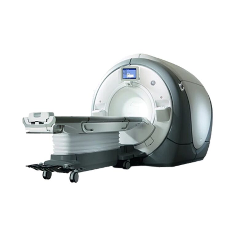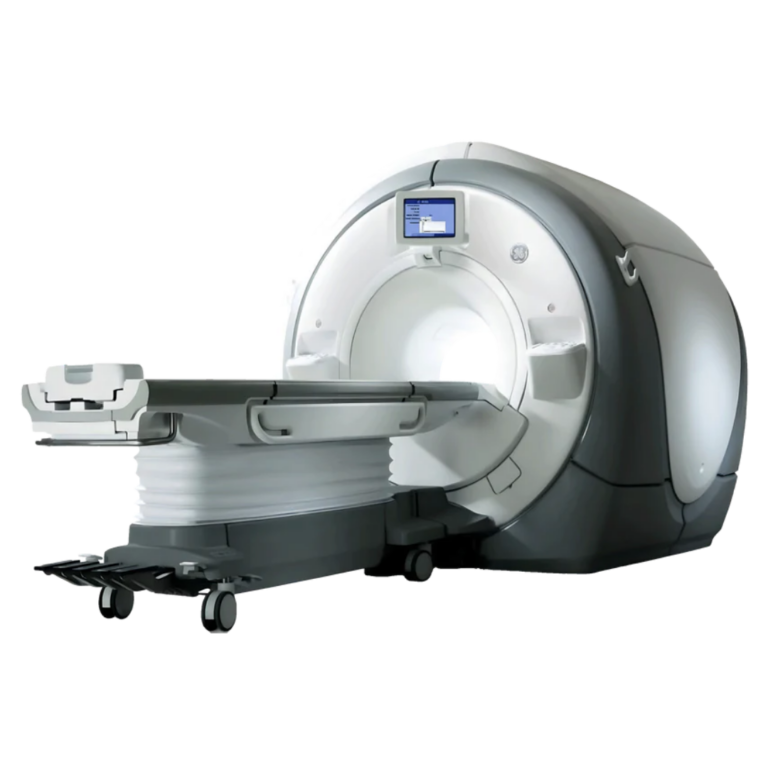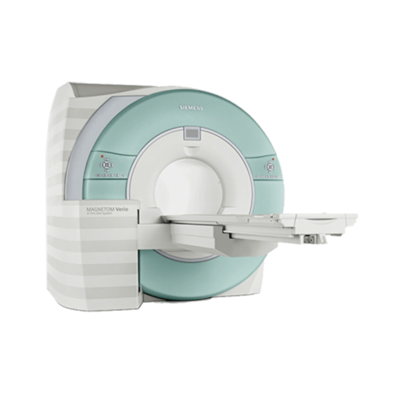
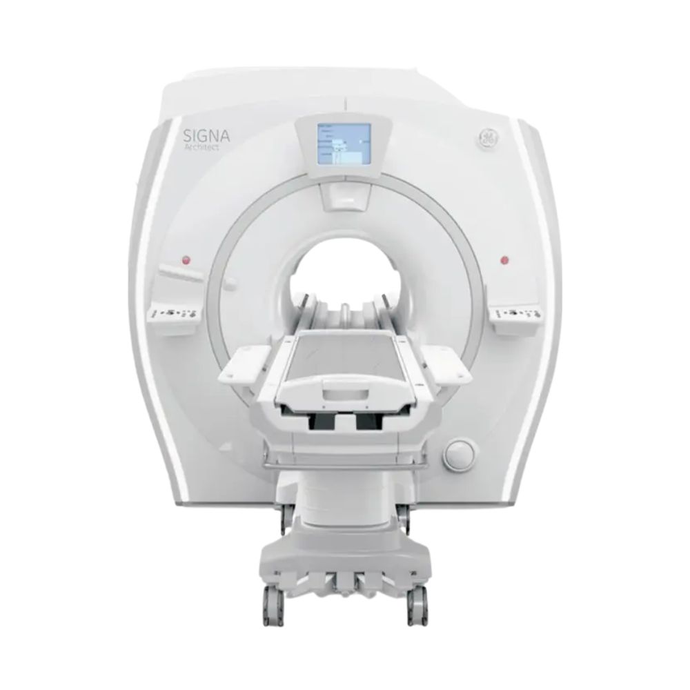
GE SIGNA Architect 3T MRI
- Category:
- MRI Systems
- Brand:
- GE
The GE SIGNA Architect 3T MRI is a wide-bore scanner well suited for hospitals, imaging centers, and specialty practices seeking advanced imaging capabilities. Its 70 cm bore, lightweight flexible coils, and feet-first imaging options combine detailed diagnostic imaging with greater patient comfort and positioning flexibility.
SIGNA Architect MRI Highlights & Benefits
The GE SIGNA Architect combines 3T imaging performance with wide-bore patient access and advanced imaging technology. Its digital signal architecture, flexible AIR Coils, and available software upgrades support detailed examinations across multiple specialties while helping facilities improve patient positioning and scanning efficiency.
Key Features & Benefits
-
- 3T High-Resolution Imaging: A superconducting magnet supports detailed brain, spine, musculoskeletal, and body imaging, including advanced clinical examinations.
- 70 cm Wide Bore: Provides additional patient space compared with conventional 60 cm MRI scanners, with flexible positioning and feet-first imaging capabilities.
- High-Performance Gradients: 44 mT/m gradient amplitude and 200 T/m/s slew rate support detailed anatomical imaging, diffusion studies, and advanced scan techniques.
- 128-Channel Digital RF Architecture: GE’s Total Digital Imaging (TDI) technology supports detailed signal acquisition through compatible multichannel coils.
- Flexible AIR Coil Technology: Lightweight coils conform to patient anatomy, helping improve comfort, positioning, and anatomical coverage.
- Advanced Imaging & Upgrade Options: Compatible AIR Recon DL, scan acceleration, and workflow automation technologies can improve image quality and examination efficiency.
MRI Performance & Image Quality
The SIGNA Architect combines a superconducting magnet, high-performance gradients, and digital signal acquisition for detailed anatomical imaging. These technologies support routine examinations and specialized clinical applications, including neurological, musculoskeletal, and body studies.
3T Magnet & High-Resolution Imaging
The scanner uses a 3.0 Tesla superconducting magnet designed for whole-body MRI.
Its high magnetic field strength supports detailed visualization of the brain, spine, joints, and soft tissue. It also provides the imaging performance needed for advanced diffusion, vascular, and neurological examinations.
The system offers a maximum field of view of 50 × 50 × 50 cm (19.7 × 19.7 × 19.7 in). This extended imaging coverage helps accommodate larger anatomical regions and off-center structures such as shoulders and hips.
Compatible image correction technologies also help maintain image uniformity and anatomical accuracy across the field of view.
Gradient Performance
The SIGNA Architect features gradients rated at 44 mT/m with a maximum slew rate of 200 T/m/s.
Gradient amplitude influences how precisely the scanner locates signals within the body, while slew rate measures how quickly those gradients can change.
Together, these capabilities support high-resolution anatomical imaging, diffusion studies, and advanced MRI sequences that require rapid gradient switching.
Digital RF System & Coil Performance
The scanner features GE’s Total Digital Imaging (TDI) technology with a 128-channel receive architecture.
The radiofrequency (RF) system collects signals from coils positioned around the patient and processes those signals into the data used to create MRI images.
Its digital architecture supports multichannel signal acquisition, detailed anatomical coverage, and compatible accelerated imaging techniques.
The number of receive channels represents the system’s signal-processing capacity rather than the number of elements in an individual coil. Architect configurations may also feature different RF hardware, including 96-channel variants.
Patient Access & Exam Experience
The SIGNA Architect’s 70 cm wide-bore design combines additional patient space with flexible coil technology and positioning options. These features help accommodate different body types, examination requirements, and mobility needs.
70 cm Wide Bore & Feet-First Imaging
The scanner features a 70 cm (27.6 in) bore, providing approximately 10 cm (3.9 in) more diameter than traditional 60 cm MRI scanners.
The additional space helps accommodate patients with broader body dimensions and examinations requiring specialized coil arrangements.
Compatible configurations also support feet-first imaging across a broad range of examinations, reducing the need for patients to enter the scanner head-first when clinically appropriate.
Dockable eXpress Patient Table
The SIGNA Architect supports the dockable eXpress Patient Table, allowing patients to be prepared and positioned outside the scanner before the table is docked for imaging.
The eXpress table supports a maximum patient weight of 500 lb (227 kg) in the applicable configuration.
The detachable design provides flexibility for patient transfers and specialized examinations. Compatible IntelliTouch controls also simplify patient positioning through touch-sensitive operation.
Alternative table configurations support specialized surgical-suite and radiation oncology workflows, with weight capacities and positioning specifications varying by configuration.
Flexible AIR Coils & Patient Comfort
GE AIR Coils use lightweight, flexible construction that conforms to patient anatomy rather than relying entirely on rigid coil housings.
This allows compatible coils to fit more naturally around the imaging area, improving positioning flexibility while reducing unnecessary bulk around the patient.
Additional patient-focused technologies include:
-
- 48-Channel Head Coil: Accommodates a range of patient head sizes while supporting detailed neurological imaging.
- Auto Navigator: Tracks respiratory motion without a separate external breathing sensor, supporting free-breathing examinations.
- Accelerated Imaging: Compatible acquisition and reconstruction technologies shorten selected sequences, reducing the time patients need to remain still.
Clinical Environments & Applications
The GE SIGNA Architect supports general diagnostic imaging and advanced clinical programs across multiple specialties. Its wide-bore design and available coil and software configurations accommodate a broad range of imaging requirements.
Clinical Environments
-
- Hospitals & Health Systems: General diagnostic MRI, inpatient and outpatient examinations, and advanced clinical programs.
- Outpatient Imaging Centers: Comprehensive MRI services with wide-bore patient access.
- Neurology & Neuroscience Centers: Brain, spine, diffusion, and specialized neurological imaging.
- Orthopedic & Musculoskeletal Imaging Centers: Joint, cartilage, extremity, and soft-tissue examinations.
- Oncology Programs: Tumor assessment, treatment monitoring, and specialized radiation therapy planning.
- Cardiac & Vascular Imaging Programs: Cardiovascular and vascular imaging with compatible coils and clinical software.
- Breast Imaging Facilities: Dedicated breast MRI with appropriate coil and software configurations.
Clinical Applications
The scanner supports the following applications, with specialized examinations dependent on installed coils, hardware, and clinical software:
-
- Neurological Imaging: Brain MRI, diffusion-weighted imaging, diffusion tensor imaging, and functional MRI.
- Spine Imaging: Cervical, thoracic, and lumbar spine examinations.
- Musculoskeletal Imaging: Shoulders, knees, hips, extremities, cartilage, joints, and soft tissue.
- Abdominal & Pelvic Imaging: Liver, pancreas, kidneys, prostate, pelvis, and general body examinations.
- Breast MRI: Contrast-enhanced breast examinations and specialized breast imaging.
- Cardiac MRI: Cardiac anatomy, function, and selected flow studies.
- Vascular Imaging: Magnetic resonance angiography (MRA) and vascular assessment.
- Oncology Imaging: Tumor characterization, disease monitoring, and treatment planning.
- Advanced Neuroimaging: Functional MRI, spectroscopy, and specialized diffusion techniques.
- Pediatric MRI: Appropriate brain, body, and musculoskeletal examinations with suitable protocols and coils.
MRI Workflow, Reconstruction & Imaging Technology
The SIGNA Architect supports examination planning, accelerated acquisition, image reconstruction, and digital image management. GE’s SIGNA Works platform provides tools for clinical workflows, while compatible AIR technologies extend automation and reconstruction capabilities.
Exam Setup & Scan Automation
SIGNA Works includes anatomy-specific imaging applications and protocol tools that help technologists prepare and perform MRI examinations.
Available workflow technologies include:
-
- AIR Touch: Automatically identifies appropriate coil elements based on patient positioning and the selected imaging area, reducing manual coil selection during supported examinations.
- AIR x: Uses automated anatomical identification to assist with scan planning and standardize image positioning for examinations such as brain and knee MRI.
- Auto Navigator: Tracks respiratory motion during scanning, supporting free-breathing abdominal imaging without requiring an external respiratory sensor.
Together, these technologies help simplify examination setup and reduce repetitive operator steps when included in the installed configuration.
Image Reconstruction & Scan Acceleration
The system supports advanced imaging technologies designed to improve image quality, reduce motion artifacts, and accelerate compatible examinations.
-
- AIR Recon DL: Uses deep-learning reconstruction to reduce image noise and improve sharpness. On compatible systems, it can support shorter acquisitions or higher-resolution imaging while maintaining useful image detail.
- HyperSense: Accelerates selected MRI sequences by acquiring less scan data and using advanced reconstruction methods to complete the image.
- HyperBand: Acquires information from multiple slices simultaneously to accelerate supported neurological examinations, including selected diffusion and functional MRI studies.
- PROPELLER: Uses a specialized data acquisition technique to reduce the effects of patient motion during suitable examinations.
- SONIC DL: Uses deep-learning acceleration for supported MRI acquisitions, including certain cardiac and three-dimensional imaging applications. Compatibility depends on the software generation, licensing, and available upgrade path.
These technologies can be incorporated into compatible Architect configurations, with installed functionality varying by software and hardware package.
Post-Processing & Connectivity
The scanner supports DICOM-based image transfer to compatible picture archiving and communication systems (PACS) for image storage, review, and distribution.
Available workstation and software configurations support multiplanar reconstruction, advanced visualization, and quantitative analysis.
Specialized post-processing capabilities depend on the installed clinical applications and workstation software.
Configurations, Coils, & Available Options
The GE SIGNA Architect is a configurable and upgradeable MRI platform. Hardware generations, coil packages, reconstruction software, and clinical applications can be matched to different imaging requirements.
System Configurations & Upgrades
The Architect platform combines a 3T superconducting magnet, 70 cm wide bore, high-performance gradients, and digital signal acquisition.
Configuration options include:
-
- RF receive architecture and hardware generation
- Coil connections and compatible coil families
- Software version and reconstruction capabilities
- AIR technology and workflow upgrades
- Patient table and positioning equipment
- Specialized clinical application packages
Compatible systems can receive supported technology enhancements through GE’s SIGNA Works AIR Edition and applicable upgrade programs.
This upgradeable architecture can help facilities extend the useful life of their MRI equipment while adapting to changing clinical requirements. Available upgrades depend on the scanner’s existing hardware and software configuration.
RF Coils
The SIGNA Architect supports dedicated radiofrequency coils for imaging different anatomical regions.
Compatible coil options include:
-
- Head Coils: Including a 48-channel head coil for neurological imaging.
- Head & Neck Coils: For brain and cervical anatomy coverage.
- Spine Coils: For cervical, thoracic, and lumbar examinations.
- Body & Anterior Array Coils: For abdominal, pelvic, and general body imaging.
- AIR Coils: Lightweight, flexible coil options for adaptable anatomical coverage.
- Extremity Coils: For joints and specialized musculoskeletal examinations.
- Breast Coils: For dedicated breast MRI.
- Cardiac Coil Arrangements: For supported cardiovascular imaging applications.
- Compatibility depends on the installed RF system, coil connections, and supported software.
The included coil package is matched to the available scanner configuration and may differ from the full range supported by the Architect platform. MXR can help identify appropriate coils for your intended examinations.
MXR also provides MRI coil repair services for supported equipment.
Software & Clinical Options
The system can be equipped with specialized software and accessories for additional clinical imaging capabilities.
Available options include:
-
- Advanced Neurology: Diffusion tensor imaging, functional MRI, and spectroscopy.
- Cardiac Imaging: Cardiac function, flow assessment, and compatible advanced cardiac applications.
- Breast Imaging: Dedicated acquisition and analysis tools.
- Vascular Imaging: Magnetic resonance angiography and specialized vascular techniques.
- Abdominal Imaging: Respiratory motion management and accelerated body imaging.
- Magnetic Resonance Elastography (MRE): Optional hardware and software for measuring tissue stiffness, including liver assessment applications.
- Multi-Nuclear Spectroscopy: Specialized optional equipment for advanced spectroscopy applications involving nuclei other than hydrogen.
- Oncology & Radiation Therapy: Specialized imaging and positioning workflows for treatment planning.
- Advanced Reconstruction & Automation: AIR Recon DL, AIR Touch, AIR x, and other compatible imaging enhancements.
Available software and clinical applications depend on the scanner’s hardware, software generation, licensing, and supported upgrade path. MXR can help match these capabilities to the facility’s intended clinical services.
MRI Magnet, Cryogen, & Site Requirements
The SIGNA Architect requires appropriate space, electrical infrastructure, cooling, and MRI safety provisions. Installation planning takes into account the equipment configuration, facility layout, and requirements for safe operation.
Superconducting Magnet & Helium System
The scanner uses a liquid-helium-cooled superconducting magnet.
Its mechanical refrigeration system maintains the extremely low temperatures required for superconducting operation and helps manage helium consumption during normal use.
Magnet condition, helium status, refrigeration performance, and service history are important considerations when planning equipment installation or relocation.
MXR provides MRI refrigeration and helium services for compatible systems.
MRI Room Size & Installation Requirements
Installation requires sufficient space for the magnet, patient table, operator workstation, and supporting equipment.
Minimum service-space requirements for a compatible platform configuration include approximately 224 sq ft (20.8 m²) for the magnet room, 81 sq ft (7.6 m²) for the equipment room, and 35 sq ft (3.2 m²) for the control room.
These are minimum planning dimensions for the specified configuration. Additional space may be needed to accommodate clinical workflow, specialized equipment, storage, safety requirements, and facility access.
Installation planning also includes:
-
- Magnet delivery and equipment access routes
- Structural support and floor loading
- RF shielding and magnetic field containment
- Patient and staff safety boundaries
- Cryogenic venting and emergency provisions
- Equipment placement and maintenance clearances
MXR supports MRI projects through custom MRI shielding solutions and turnkey imaging project management.
Electrical, Cooling, & Facility Requirements
The system requires dedicated electrical service and equipment cooling appropriate for a 3T MRI installation.
The installation must accommodate power and heat loads from the gradient electronics, RF hardware, refrigeration equipment, and supporting cabinets.
Facility planning includes electrical distribution, cooling capacity, HVAC, network connectivity, and equipment placement.
MXR can help coordinate facility requirements with the selected scanner configuration and installation conditions.
Available System Configuration
The GE SIGNA Architect can be supplied in different equipment configurations, allowing MXR to match scanner capabilities to a facility’s clinical, operational, and installation requirements.
System Hardware, Software, & Included Equipment
Configurations may differ in:
-
- Model year and hardware generation
- RF configuration and hardware upgrades
- Software version and reconstruction capabilities
- Included head, body, spine, and specialty coils
- Neuro, cardiac, breast, vascular, and body imaging software
- Patient table and positioning accessories
- Operator workstation and post-processing software
- Magnet, refrigeration, and helium service history
- Additional hardware, software, and accessories
MXR works with healthcare providers to identify a configuration suited to planned examinations, patient needs, clinical workflows, and facility infrastructure.
MRI Scanner Price & Availability
MXR Imaging offers GE SIGNA Architect systems with guidance on equipment selection, clinical configurations, installation planning, and ongoing support.
Pricing depends on the scanner’s configuration, installed coils and software, included accessories, and project requirements. MXR can help identify a system that aligns with your imaging needs, budget, and facility infrastructure.
Our team also supports equipment delivery, installation coordination, and relocation planning.
MXR provides MRI maintenance and repair services to support long-term equipment operation.
Contact MXR to discuss pricing, available configurations, and installation requirements.
GE SIGNA Architect Specifications
The specifications below cover the scanner’s core hardware, patient access, compatible equipment, and installation requirements. RF hardware, patient tables, installed software, and accessories may vary by configuration.
Magnet System
-
- Field Strength: 3.0T
- Magnet Type: Superconducting
- System Design: Whole-body, wide-bore MRI
- Bore Diameter: 70 cm (27.6 in)
- Maximum Field of View: 50 × 50 × 50 cm (19.7 × 19.7 × 19.7 in)
- Cryogenic Cooling: Liquid helium with mechanical refrigeration
- Magnet Homogeneity: Varies by measurement volume and magnet specification
Gradient System
-
- Maximum Gradient Amplitude: 44 mT/m
- Maximum Slew Rate: 200 T/m/s
- Gradient Architecture: High-performance whole-body gradient system
RF System
-
- RF Technology: Total Digital Imaging (TDI)
- Receive Architecture: 128-channel configuration
- Alternative Configurations: 96-channel Architect variants and upgrade packages
- Coil Architecture: Multichannel anatomical coils
- Flexible Coil Technology: Compatible GE AIR Coils
Bore & Patient Table
-
- Bore Diameter: 70 cm (27.6 in)
- Patient Table: Dockable eXpress table option
- Maximum Patient Weight: 500 lb (227 kg) for the applicable eXpress table
- Alternative Table: Optional surgical-suite transmobile configuration
- Patient Positioning: Feet-first imaging for compatible examinations
- Table Travel & Height: Vary by installed table configuration
Coils & Accessories
-
- Head Coil: Compatible 48-channel head coil
- Other Compatible Coils: Head/neck, spine, body, extremity, breast, and cardiac arrangements
- Flexible Coil Options: GE AIR Coils
- Included Coils: Based on selected system package
- Optional Equipment: Specialized clinical accessories, positioning supports, and advanced imaging hardware
Siting & Utilities
The following minimum room requirements apply to a compatible platform configuration with a 2050 mm scan range:
-
- Minimum Magnet Room: 224.1 sq ft (20.8 m²)
- Minimum Equipment Room: 81.4 sq ft (7.6 m²)
- Minimum Control Room: 35 sq ft (3.2 m²)
- Combined Minimum Room Area: 340.5 sq ft (31.6 m²)
- Minimum Finished Magnet Room Ceiling Height: 98.5 in (2500 mm)
- Preferred Magnet Room Ceiling Height: 105 in (2667 mm)
- Electrical Requirements: Based on installed system configuration
- Cooling: Dedicated equipment cooling and cryogenic refrigeration
- RF Shielding: Required for MRI installation
- Cryogen Safety: Appropriate venting and emergency safety provisions
Minimum room dimensions do not account for all clinical workflow, storage, accessibility, and facility-specific requirements. Some installations may require additional clearances or specialized provisions.
System Dimensions & Weight
For the applicable AR-series magnet assembly:
-
- Magnet Assembly Width: Approximately 97 in (2464 mm)
- Magnet Assembly Height: Approximately 93.7 in (2379 mm)
- Magnet Assembly Depth: Varies with rear assembly components
- eXpress Table Weight, Empty: 463 lb (210 kg)
- eXpress Table Weight at Maximum Patient Load: 963 lb (437 kg)
- Magnet Weight: Based on installed magnet configuration
- Installed System Weight: Varies by magnet, table, supporting cabinets, and accessories
Final equipment dimensions, component weights, and facility requirements are matched to the selected scanner configuration.