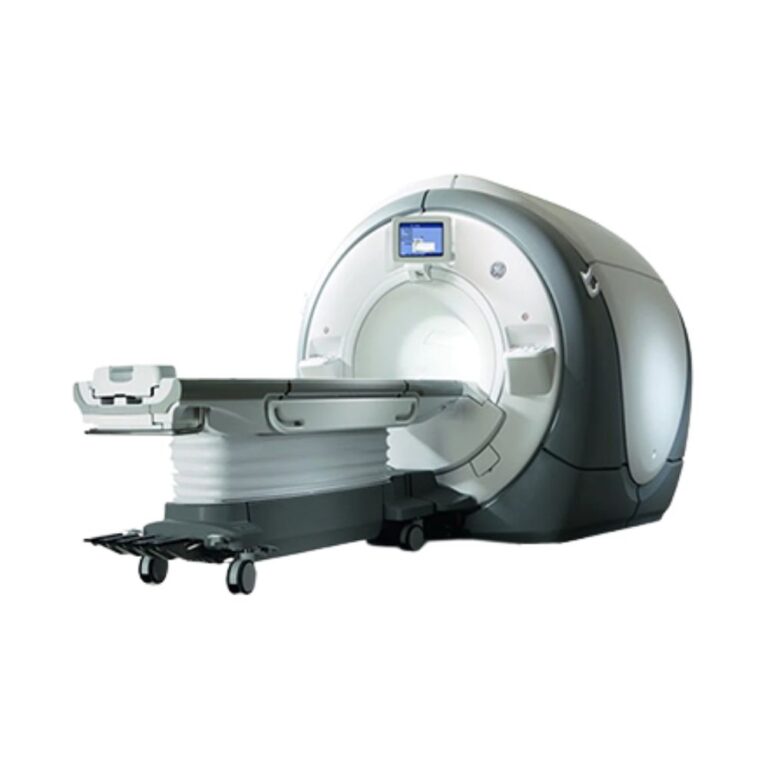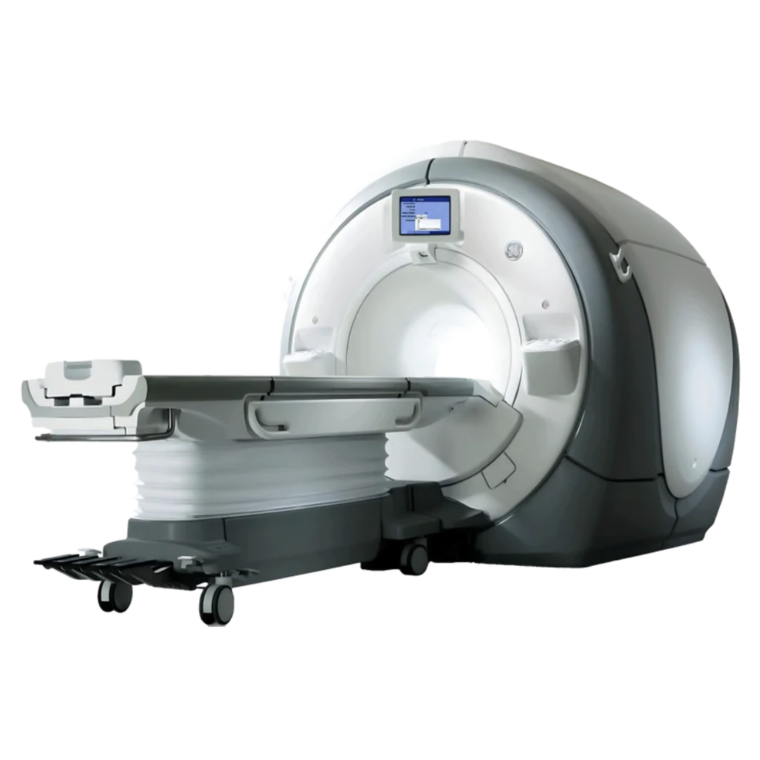
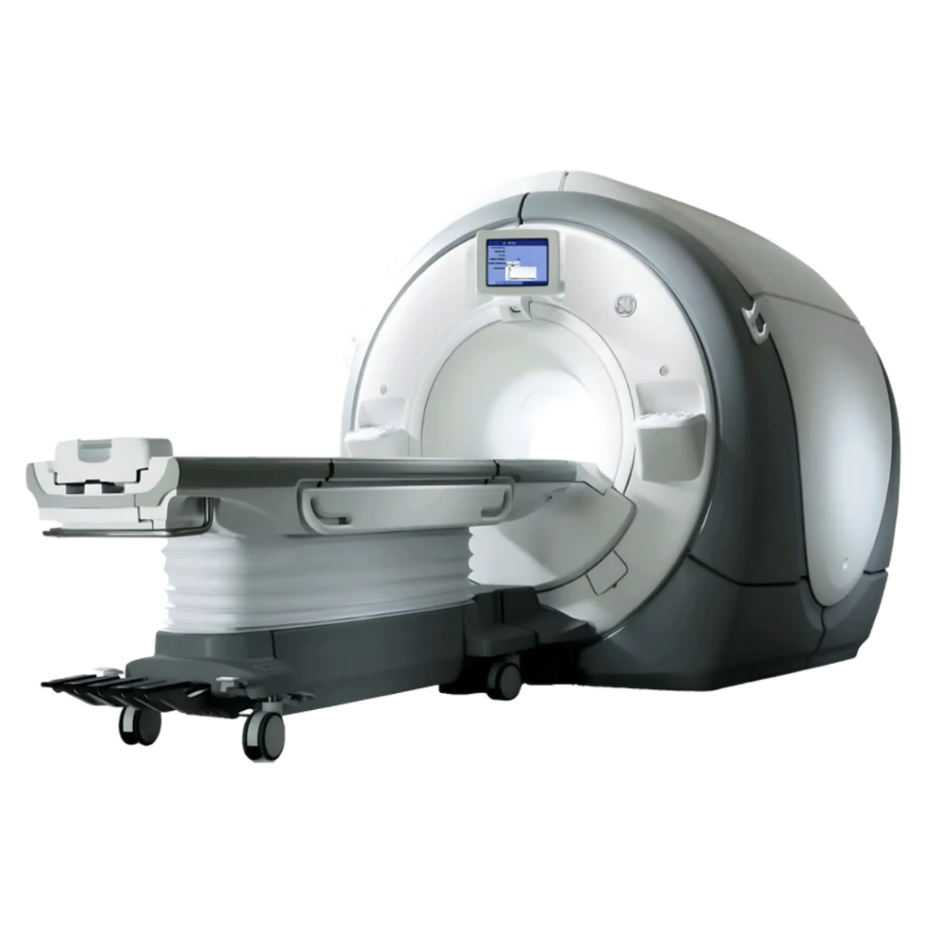
GE Discovery MR750W 3T MRI
- Category:
- MRI Systems
- Brand:
- GE
The GE Discovery MR750W 3T boasts exceptional, high definition image quality for a comprehensive range of advanced clinical applications, as well as a very friendly 70cm wide bore. Built for speed, and ultimately the highest return on investment.
GE Discovery MR750W 3T Highlights & Benefits
MR750W Key Features
-
- 3.0T superconducting magnet for high-resolution MRI
- 70 cm (27.6 in.) wide bore for greater patient access and positioning flexibility
- 50 × 50 × 50 cm (19.7 × 19.7 × 19.7 in.) usable field of view
- 44 mT/m gradient amplitude with up to 200 T/m/s slew rate
- MultiDrive RF Transmit for improved radiofrequency uniformity at 3T
- OpTix Optical RF architecture designed to preserve received signal quality
- In-room workflow tools including iROC and IntelliTouch patient positioning
- GEM coil and patient-table configurations available for streamlined multi-region imaging
- Maximum patient weight of 227 kg (500 lb) with the eXpress patient table
The GE Discovery MR750w combines the imaging performance of a 3T MRI scanner with the patient access of a 70 cm wide-bore system. Its large usable field of view also supports off-center anatomy such as the shoulder and hip, where positioning flexibility and image coverage are especially important.
3T Imaging Performance & Image Quality
Magnet Strength & Imaging Coverage
The 3.0T superconducting magnet provides the signal performance needed for detailed anatomical imaging, high-resolution studies, and advanced MRI applications.
A 50 × 50 × 50 cm (19.7 × 19.7 × 19.7 in.) usable field of view provides broad imaging coverage. This is particularly useful when anatomy cannot be positioned directly at the center of the magnet, including shoulder, hip, body, and larger-patient examinations.
Gradient Performance
The Discovery MR750w gradient system is rated at 44 mT/m peak amplitude and up to 200 T/m/s slew rate per axis.
Gradient amplitude determines the magnetic gradient strength available for spatial encoding, while slew rate describes how quickly those gradients can change. Together, they support demanding applications such as diffusion imaging, vascular studies, cardiac MRI, and high-resolution 3D acquisitions.
RF Uniformity & Signal Quality
At 3T, maintaining uniform radiofrequency excitation across different patient sizes and anatomies can be challenging. MultiDrive RF Transmit independently adjusts radiofrequency pulse amplitude and phase to improve transmit uniformity across the 70 cm bore.
OpTix Optical RF converts received MRI signals to digital optical data inside the scan room before sending them to the reconstruction system. This architecture is designed to reduce electrical noise and signal degradation between the receiver coils and system electronics.
Patient Access, Comfort, & Positioning
70cm Wide-Bore Patient Access
The 70 cm (27.6 in.) bore gives patients more lateral space than traditional 60 cm MRI designs while maintaining 3T field strength.
The wider, flared opening provides additional room for larger patients, receiver coils, padding, and specialized positioning. It also helps with off-center examinations where anatomy such as the shoulder or hip must be positioned away from the centerline.
Patient Table & Weight Capacity
Compatible eXpress patient-table configurations support patients up to 227 kg (500 lb) and provide powered vertical and longitudinal positioning.
On compatible GEM configurations, the table can integrate a posterior receiver array and allow portions of patient preparation to take place outside the scan room. The detachable design can also assist with patient transport when required.
A referenced legacy configuration provides an approximately 205 cm (80.7 in.) scan range for extended anatomical coverage.
GEM Positioning & Patient Comfort
GEM stands for Geometry Embracing Method, GE’s coil and patient-positioning approach designed to place receiver elements close to the anatomy while reducing unnecessary coil changes and patient repositioning.
Depending on the installed GEM components, configurations can support feet-first examinations, adjustable head and neck positioning, pressure-relieving padding, and broader multi-region coverage.
GE Discovery MR750w Clinical Environments & Applications
The GE Discovery MR750w supports routine and advanced 3T MRI across a broad range of human and veterinary imaging settings. Clinical capabilities depend on the coils, software, application packages, and supporting equipment installed on the individual system.
Clinical Environments
-
- Hospitals and health systems
- Outpatient diagnostic imaging centers
- Academic medical centers
- Specialty imaging centers
- Neurology and orthopedic imaging centers
- Cardiovascular imaging programs
- Oncology and radiation oncology programs when appropriately configured
- Veterinary hospitals and specialty referral centers
Clinical Specialties
-
- Diagnostic Radiology
- Neuroradiology
- Neurology & Neurosurgery
- Orthopedics & Musculoskeletal Imaging
- Cardiology
- Vascular Imaging
- Oncology
- Breast Imaging
- Abdominal & Pelvic Imaging
- Pediatric Radiology
- Radiation Oncology when configured for applicable MR treatment-planning workflows
- Veterinary Radiology & Advanced Veterinary Imaging
Common MRI Applications
-
- Brain and head MRI
- Spine and spinal cord imaging
- Diffusion, perfusion, and functional brain imaging when the required software is installed
- Shoulder, hip, knee, wrist, hand, ankle, foot, and other musculoskeletal MRI
- Abdominal and pelvic MRI
- Liver imaging and quantitative fat assessment on appropriately configured systems
- Prostate and other pelvic MRI
- Breast MRI with the appropriate dedicated coil
- Cardiac MRI for morphology, function, viability, and flow assessment
- MR angiography and peripheral vascular imaging
- Oncology imaging and treatment follow-up
- Imaging around orthopedic implants with compatible metal artifact reduction software
- Pediatric MRI using appropriate coils and examination protocols
- Veterinary MRI, including neurological and musculoskeletal/oncology applications, when appropriate coils, positioning, anesthesia, and monitoring are available
- MR elastography, spectroscopy, and other advanced applications when installed
- MR simulation and image acquisition for applicable radiation treatment-planning workflows
MRI Workflow & Image Processing
In-Room Setup & Operator Controls
High-Resolution In-Room Operator Control, or iROC, provides scan and patient information on a display at the front of the magnet. This allows technologists to complete supported setup tasks while remaining near the patient.
Dual-sided scanner controls provide access from either side of the patient table, which can simplify positioning when working around intravenous lines, monitoring equipment, cardiac gating leads, or other accessories.
IntelliTouch patient positioning reduces manual landmarking steps by automatically establishing patient position. Together, these features help streamline the transition from patient setup to scanning.
Acquisition & Reconstruction
The GE Discovery MR750w supports a broad range of GE acquisition and reconstruction technologies. Exact capabilities depend on software release and installed licenses.
Available configurations can include parallel imaging, accelerated 3D acquisition, motion-management tools, multi-contrast imaging, and specialized reconstruction technologies.
MAGiC, for example, can create multiple brain image contrasts from a compatible acquisition. Other available software can target faster body imaging, metal artifact reduction, fat and water separation, cardiac imaging, or advanced neurological examinations.
Post-Processing & DICOM Connectivity
The system supports Digital Imaging and Communications in Medicine (DICOM) connectivity for integration with picture archiving and communication systems (PACS), modality worklists, and other compatible clinical imaging systems.
Post-processing capabilities vary with the workstation and installed software package. Available applications can extend into neurological, cardiovascular, body, vascular, quantitative, and 3D image analysis.
Two GE Discovery MR750w systems can therefore provide different workflows and clinical capabilities even when the core MRI scanner model is the same.
MR750w Configurations, Coils & Available Options
System Configuration Differences
GE documentation identifies both Discovery MR750w and Discovery MR750w GEM configurations.
An MR750w GEM configuration incorporates components from the Geometry Embracing Method platform, which can include the integrated posterior receiver array, GEM Express patient table, and compatible GEM coils. The complete GEM package should not be assumed to be included with every MR750w.
The Discovery MR750w should also not be confused with the Discovery MR750. The key physical difference is bore diameter: the MR750w is the 70 cm wide-bore MRI platform, while the Discovery MR750 uses a 60 cm bore.
Individual MR750w systems can also differ by model year, software release, RF hardware, patient table, coil package, workstation, application licenses, and later upgrades.
RF Coil Options
GEM-equipped configurations can combine an integrated posterior receiver array with compatible anterior and specialty coils to support multi-region imaging with fewer coil changes.
Available coil configurations can include:
-
- GEM Posterior Array
- GEM Anterior Array
- GEM Head and Neck Unit
- Peripheral and lower-extremity vascular arrays
- GEM Flex coils
- Dedicated breast and musculoskeletal coils
Specific knee, shoulder, hand/wrist, foot/ankle, and other specialty coils can also be available depending on system configuration.
The exact coil inventory should be verified for each available MRI system based on the examinations the facility intends to perform.
GE MRI Software & Clinical Packages
Software availability can vary substantially between GE Discovery MR750w configurations.
Depending on software generation, licensing, and subsequent upgrades, available technologies can include MAGiC for multi-contrast brain imaging, MAVRIC SL for reducing metal-related artifacts, IDEAL IQ for fat and water separation and quantitative applications, DISCO and Turbo LAVA for dynamic or accelerated body imaging, and SilentSuite for reduced acoustic noise during supported sequences.
Later software configurations can also support SIGNA Works technologies such as HyperSense, HyperBand, SIGNA Flow, and additional reconstruction, acceleration, cardiac, vascular, and neurological applications.
GE continues to list the Discovery MR750w and MR750w GEM within its SIGNA Continuum upgrade program. Depending on starting configuration and eligibility, upgrade pathways can include SIGNA Architect Lift and SIGNA Premier Lift.
Magnet, Cryogen, & Site Requirements
Magnet & Cryogen Considerations
The Discovery MR750w uses a liquid-helium-cooled superconducting 3T magnet with a dedicated cryocooler system.
Magnet configuration, helium status, cryocooler condition, and service history are important considerations when evaluating an individual system. GE documentation covers more than one magnet configuration associated with the MR750w, so magnet-specific dimensions, weight, and installation requirements should be confirmed for the system being considered.
MRI Room Planning & Shielding
Installing a 3T wide-bore MRI scanner requires planning for the magnet room, equipment room, and control room along with RF shielding, magnetic-field containment, cryogen venting, equipment access, structural support, electrical service, cooling, and service clearances.
GE’s published dimensions represent minimum service-space references rather than recommended final room sizes. Actual space requirements can increase based on magnetic-field containment, workflow, local codes, shielding, accessories, storage, and facility layout.
Electrical & Cooling Requirements
Legacy site-planning documentation supports 380, 400, 415, or 480 VAC three-phase electrical configurations depending on the installation.
The scanner also requires continuous facility cooling for the heat exchanger and cryocooler equipment. Final electrical, chilled-water, HVAC, emergency-power, and construction requirements should be established using documentation for the specific system and installation site.
Available GE Discovery MR750w Configurations
Available GE Discovery MR750w MRI scanners can differ substantially even when they share the same model designation.
Configuration Details to Verify
Key configuration details include:
-
- Model year and system history
- Installed software release
- MR750w or MR750w GEM configuration
- RF and receiver hardware
- Included head, body, musculoskeletal, breast, vascular, and specialty coils
- Patient-table configuration
- Installed clinical application licenses
- Workstation and post-processing package
- Magnet, helium, and cryocooler status
- Hardware and software upgrade history
- Included accessories
MXR can help match the available configuration to the examinations and clinical applications a facility plans to perform, allowing systems to be evaluated based on actual capability rather than model name alone.
MRI Scanner Price & Availability
GE Discovery MR750w pricing varies based on system year, hardware configuration, software level, included coils, clinical applications, magnet and cryogen condition, accessories, installation scope, and service requirements.
An MXR representative can review current availability and help identify a 3T MRI configuration that fits the facility’s clinical needs, site requirements, workflow, and budget. Service options can also be reviewed for the specific system under consideration.
GE Discovery MR750W 3T Specifications
Magnet System
-
- System type: Whole-body MRI scanner
- Magnetic field strength: 3.0T
- Magnet type: Superconducting
- Bore diameter: 70 cm (27.6 in.)
- Usable field of view: 50 × 50 × 50 cm (19.7 × 19.7 × 19.7 in.)
- Cooling: Liquid helium with cryocooler system
- Magnet configuration and weight: Vary by installed magnet and system configuration
Gradient System
-
- Peak gradient amplitude: 44 mT/m per axis
- Maximum slew rate: Up to 200 T/m/s per axis
- Maximum usable field of view: 50 cm (19.7 in.)
- Gradient cooling: Water-cooled
RF System
-
- MultiDrive RF Transmit
- OpTix Optical RF receive architecture
- Digital optical signal transmission from the scan room
- RF receiver configuration: Varies by hardware generation and installed configuration
Bore & Patient Table
-
- Bore diameter: 70 cm (27.6 in.)
- Maximum patient weight with eXpress table: 227 kg (500 lb)
- Referenced scan range: Approximately 205 cm (80.7 in.) on the applicable legacy configuration
- Powered vertical and longitudinal positioning
- GEM Express table available on compatible GEM configurations
- Integrated posterior receiver array available with GEM Express configuration
Workflow
-
- High-Resolution In-Room Operator Control (iROC)
- IntelliTouch patient positioning
- Dual-sided scanner controls
- Feet-first imaging supported with compatible GEM configurations
- Patient setup outside the scan room supported with compatible GEM Express configurations
Coils & Accessories
Available depending on configuration:
-
- GEM Posterior Array
- GEM Anterior Array
- GEM Head and Neck Unit
- GEM Flex coils
- Peripheral and lower-extremity vascular arrays
- Dedicated breast and musculoskeletal coils
- Cardiac and physiological monitoring accessories
- MR elastography equipment
- Spectroscopy equipment
- Radiation oncology positioning accessories
Electrical & Cooling Requirements
-
- Facility input power: 380 / 400 / 415 / 480 VAC depending on installation
- Electrical service: Three-phase
- Referenced continuous system demand: 99 kVA
- Referenced five-second peak system demand: 123 kVA
- Facility coolant flow: 114 to 132 L/min (30 to 35 GPM)
- Minimum referenced chiller capacity: 70 kW
- Final utilities depend on system and site configuration
Minimum Room Planning Reference
For the referenced 205 cm (80.7 in.) scan-range legacy configuration:
-
- Equipment room: 2,774 × 2,860 mm (109.2 × 112.6 in.)
- Equipment room area: 7.9 m² (85.4 ft²)
- Magnet room: 3,645 × 6,331 mm (143.5 × 253 in.)
- Magnet room area: 20.8 m² (224.1 ft²)
- Minimum magnet-room ceiling height: 2,500 mm (98.5 in.)
- Control room: 1,524 × 2,134 mm (60 × 84 in.)
- Control room area: 3.2 m² (35 ft²)
- Minimum referenced total system area: 32.1 m² (346.1 ft²)
These dimensions represent GE’s minimum service-space reference. Final room size must account for workflow, shielding, magnetic-field containment, cryogen venting, codes, accessories, storage, and facility-specific requirements.