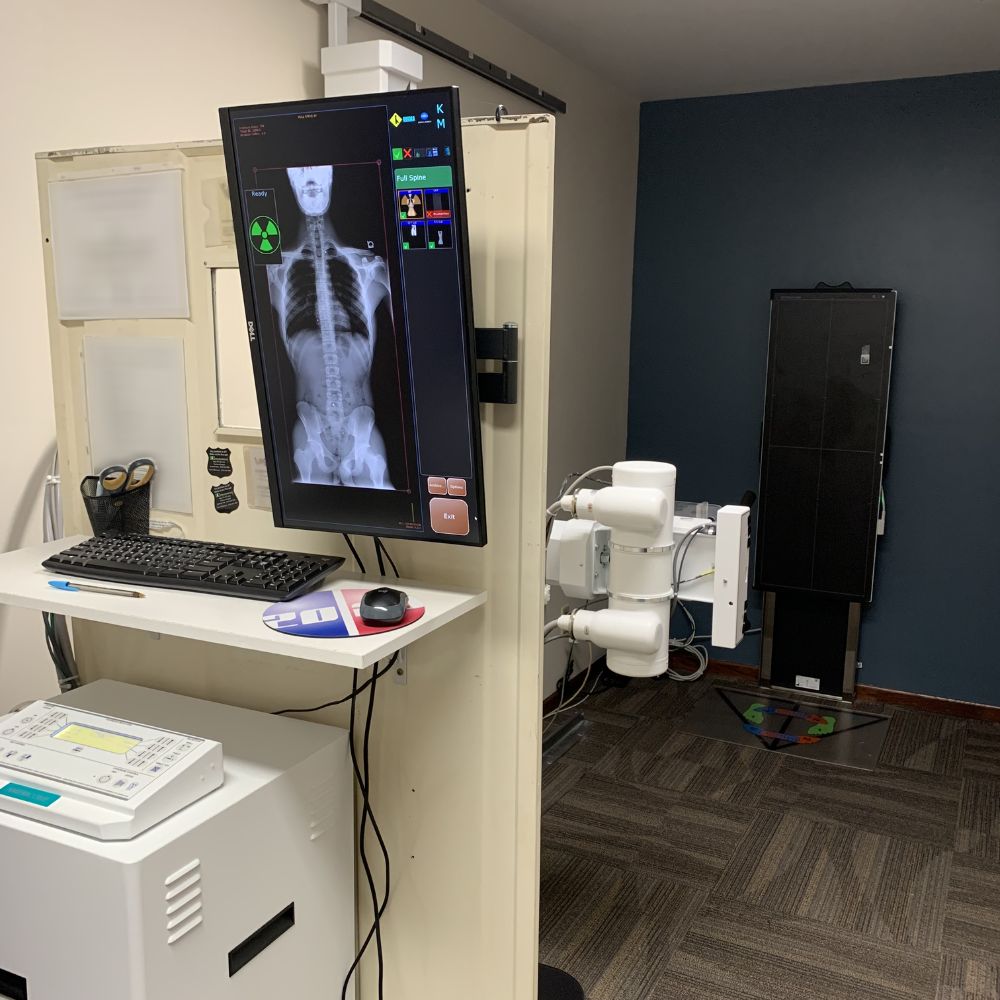
20/20 Imaging 17x48 Long-Length DR Panel
- Category:
- X-Ray Systems
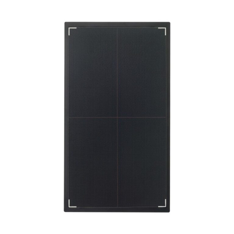
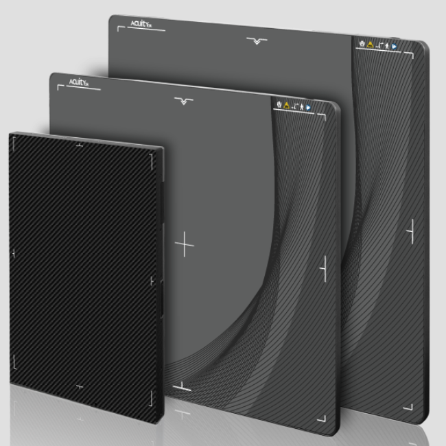
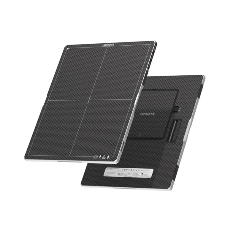
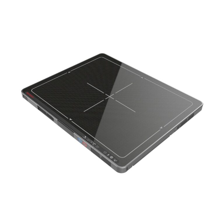
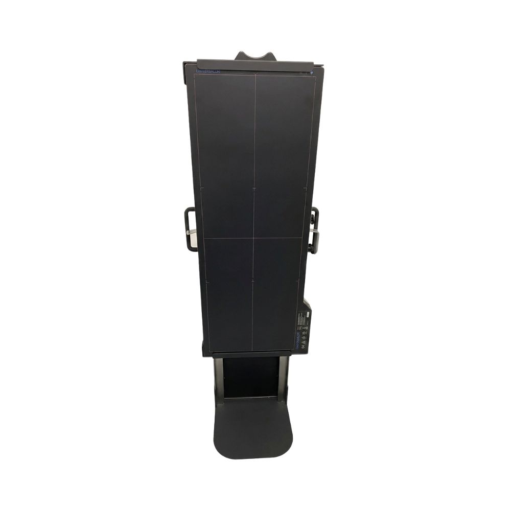
The 20/20 Imaging 17×48 DR Panel, also referred to in some product literature as the 17 × 48 Full Spine DR Panel, is a long-length digital radiography detector designed to capture extended anatomy in a single exposure. Its 17 × 48-inch active imaging area, Cesium (CsI) scintillator, and high-resolution detector technology support full-spine imaging, long-leg alignment studies, scoliosis evaluations, and other examinations that extend beyond the field of view of a conventional DR detector.
Capture Long-Length Anatomy in a Single Exposure
The 20/20 Imaging 17×48 is designed for imaging applications that extend beyond the field of view of a standard DR detector. By capturing extended anatomy in a single exposure, the detector helps reduce the need for stitched images while simplifying patient positioning and improving workflow for compatible long-length digital radiography systems.
Key Benefits
- 17 × 48-inch active imaging area
- Single-exposure long-length imaging
- Cesium (CsI) scintillator
- Amorphous silicon detector technology
- 139 μm pixel pitch
- 3.6 lp/mm spatial resolution
- Designed for compatible long-length digital radiography systems
The 20/20 Imaging 17×48 combines a long active imaging area with single-exposure image acquisition to streamline examinations that traditionally require multiple exposures and software image stitching.
When a 17 × 48 Long-Length Detector Fits Your Workflow
Capture Extended Anatomy Without Image Stitching
Traditional DR detectors often require multiple exposures to image anatomy that extends beyond the detector’s field of view.
The 17×48 detector captures extended anatomy in a single exposure, making it well suited for full-spine imaging, long-leg alignment studies, scoliosis evaluations, and other long-length examinations while helping reduce workflow associated with stitched images.
Improve Workflow for Standing Long-Length Imaging
Single-exposure imaging reduces repositioning and repeat acquisitions, helping improve workflow while creating a more efficient imaging experience for both patients and technologists during standing radiographic examinations.
Reduce Patient Repositioning
Because the detector captures more anatomy in a single exposure, patients typically require fewer position changes than workflows that depend on multiple stitched images. This helps streamline long-length digital radiography while maintaining consistent anatomical positioning throughout the examination.
Maintain Anatomical Continuity Across Extended Anatomy
Maintaining the anatomical relationship between the spine, pelvis, hips, knees, ankles, or other extended anatomy is often important during long-length radiographic examinations. Capturing the anatomy in a single exposure helps preserve this continuity without relying on stitched acquisitions.
Where Long-Length Digital Radiography Makes the Greatest Impact
The 20/20 Imaging 17×48 is designed for facilities that routinely perform long-length radiographic examinations requiring anatomical coverage beyond the field of view of a standard detector.
Common Imaging Environments
- Hospitals and health systems
- Orthopedic practices
- Spine centers
- Chiropractic practices
- Outpatient imaging centers
- Scoliosis imaging programs
- Veterinary facilities performing compatible long-bone imaging
Common Radiographic Examinations
The detector is particularly well suited for:
- Full-spine standing radiographs
- Long-leg alignment studies
- Scoliosis evaluations
- Bilateral lower-extremity imaging
- Standing orthopedic alignment studies
- Long-bone veterinary examinations using compatible imaging systems
How the 17×48 Supports Long-Length Imaging Workflows
Upgrade Compatible Long-Length Imaging Rooms
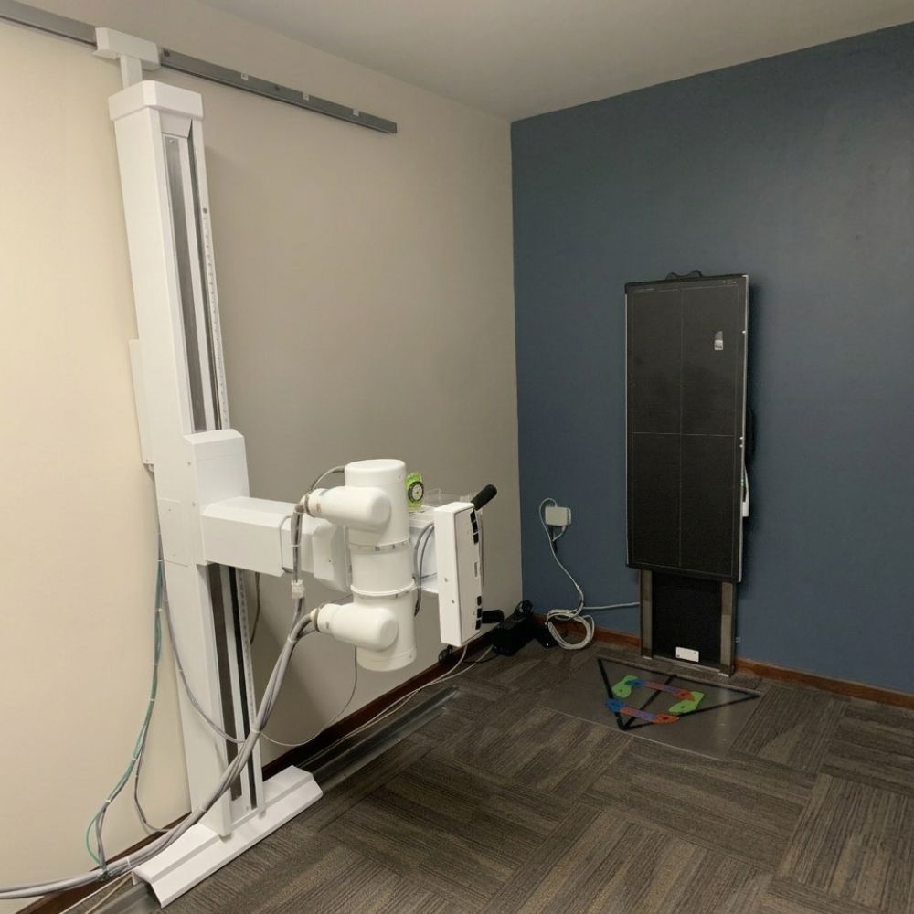
The detector can be incorporated into compatible DR retrofit projects, allowing facilities to modernize existing long-length imaging rooms while preserving compatible X-ray equipment.
Integrate Into New Digital Radiography Installations
The detector can also be integrated into new digital radiography installations where single-exposure long-length imaging is an important part of the clinical workflow.
See the 20/20 Imaging 17×48 DR Panel in Action
Seeing the detector in use provides additional context that specifications alone cannot. The video below demonstrates the 20/20 Imaging 17×48 DR Panel, highlighting its long-length detector design, single-exposure imaging workflow, and clinical applications for extended anatomy imaging. It offers a closer look at how the detector helps simplify imaging while reducing reliance on stitched acquisitions.
Technologies That Support Long-Length Imaging
Cesium (CsI) Scintillator
The 20/20 Imaging 17×48 uses a Cesium (CsI) scintillator to efficiently convert X-ray energy into digital image data while supporting excellent detector efficiency and image quality.
Amorphous Silicon Detector Technology
The detector incorporates amorphous silicon technology with a 139 μm pixel pitch and 3.6 lp/mm spatial resolution to support detailed long-length digital radiography.
Single-Exposure Long-Length Detector Design
The detector’s 17 × 48-inch active imaging area allows compatible imaging systems to capture extended anatomy in a single exposure rather than combining multiple images through software stitching. This helps simplify long-length digital radiography workflows while preserving anatomical continuity across the entire image.
Compatible Imaging Platforms
The 20/20 Imaging 17×48 is designed for compatible digital radiography systems that support long-length detector integration. Compatibility depends on detector configuration, acquisition software, and the overall imaging system design.
Acquisition Software Designed for the 17×48
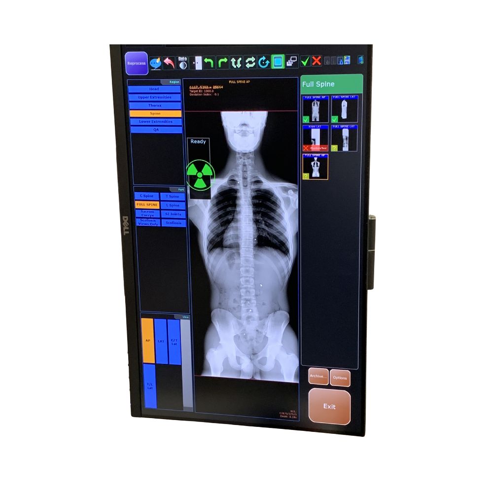
The 20/20 Imaging 17×48 integrates with compatible acquisition software to support long-length image acquisition, image enhancement, DICOM communication, image review, and routine digital radiography workflow. Available software capabilities vary depending on the complete imaging system configuration.
Compare 20/20 Imaging DR Detector Options
Each 20/20 Imaging detector is designed to support a different imaging workflow.
- Need a compact detector for smaller anatomy and extremity imaging? Explore the 20/20 Imaging 1012V Tethered DR Detector.
- Need a full-size detector for everyday digital radiography? Explore the 20/20 Imaging 17 × 17 DR Detector.
Built for Single-Exposure Long-Length Imaging
Whether performing full-spine imaging, long-leg alignment studies, scoliosis evaluations, or other examinations involving extended anatomy, the 20/20 Imaging 17×48 is designed to capture more anatomy in a single exposure than a conventional DR detector.
By reducing reliance on stitched acquisitions, the detector helps simplify workflow, improve positioning efficiency, and support consistent long-length digital radiography across compatible imaging systems.
20/20 Imaging 17×48 Long-Length DR Panel Specifications
Detector Specifications
-
- Detector size: 17 × 48 in.
- Active imaging area: 17 × 48 in.
- Amorphous silicon detector technology
- Cesium (CsI) scintillator
- Pixel pitch: 139 μm
- Spatial resolution: 3.6 lp/mm
- Single-exposure long-length detector
Workflow & Connectivity
-
- Designed for long-length digital radiography
- Single-exposure image acquisition
- Compatible with supported DR systems
- Designed for standing imaging workflows