Vet-Ray by Sedecal Digital DX-V Veterinary X-ray
- Category:
- Veterinary X-ray Products
- Brand:
- Vet-Ray by Sedecal
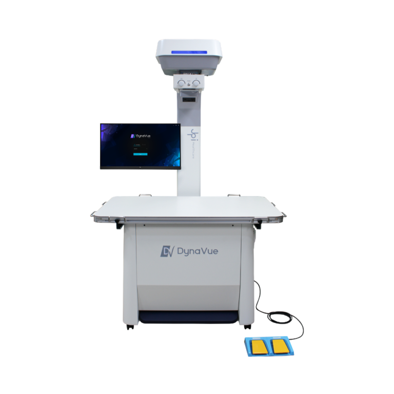
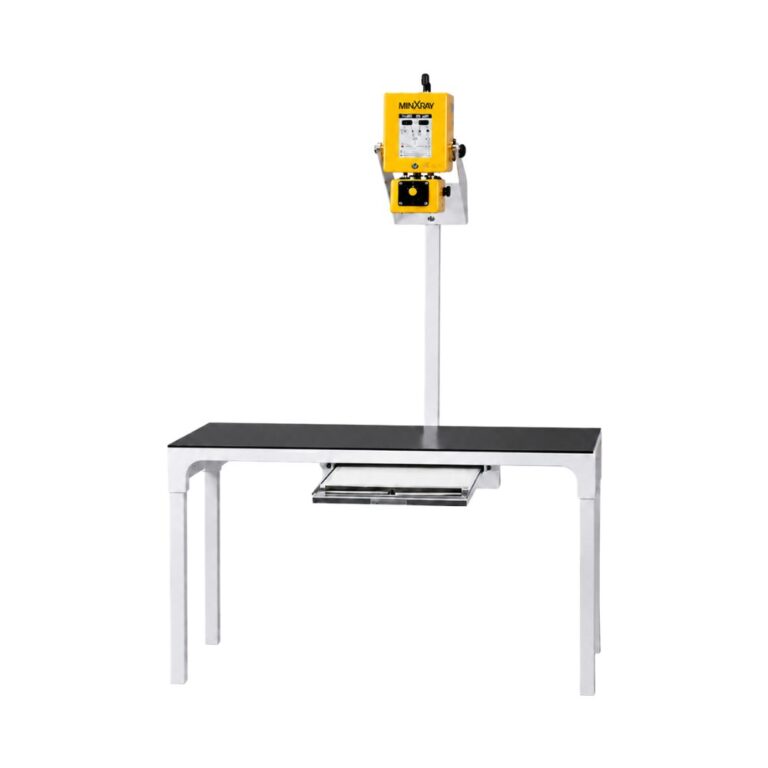
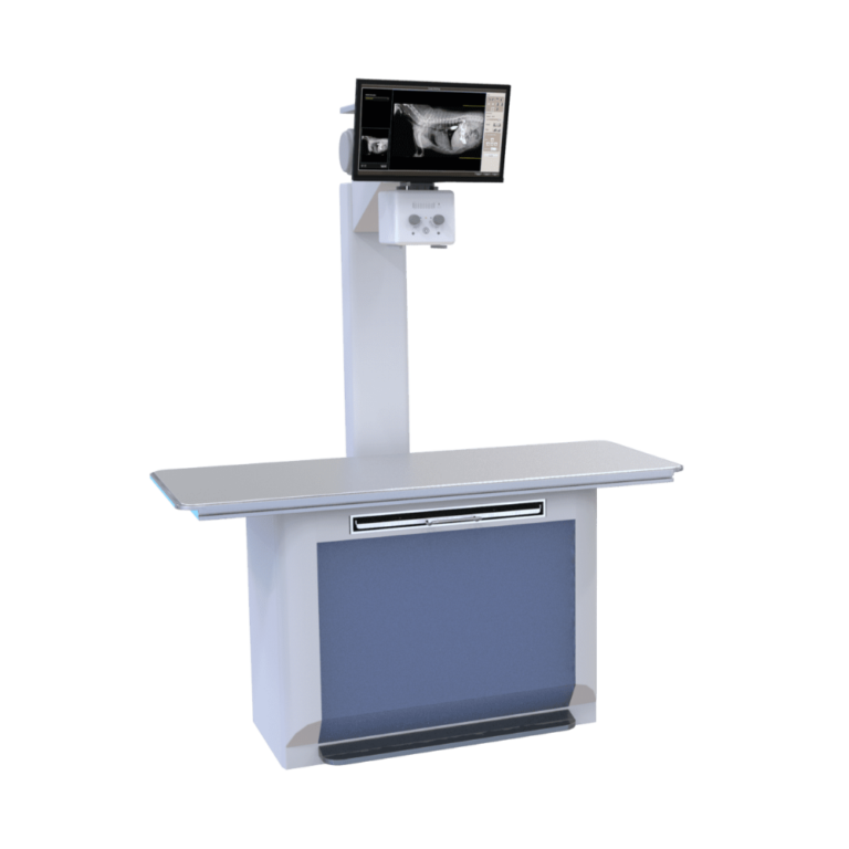
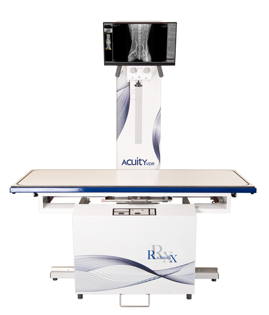
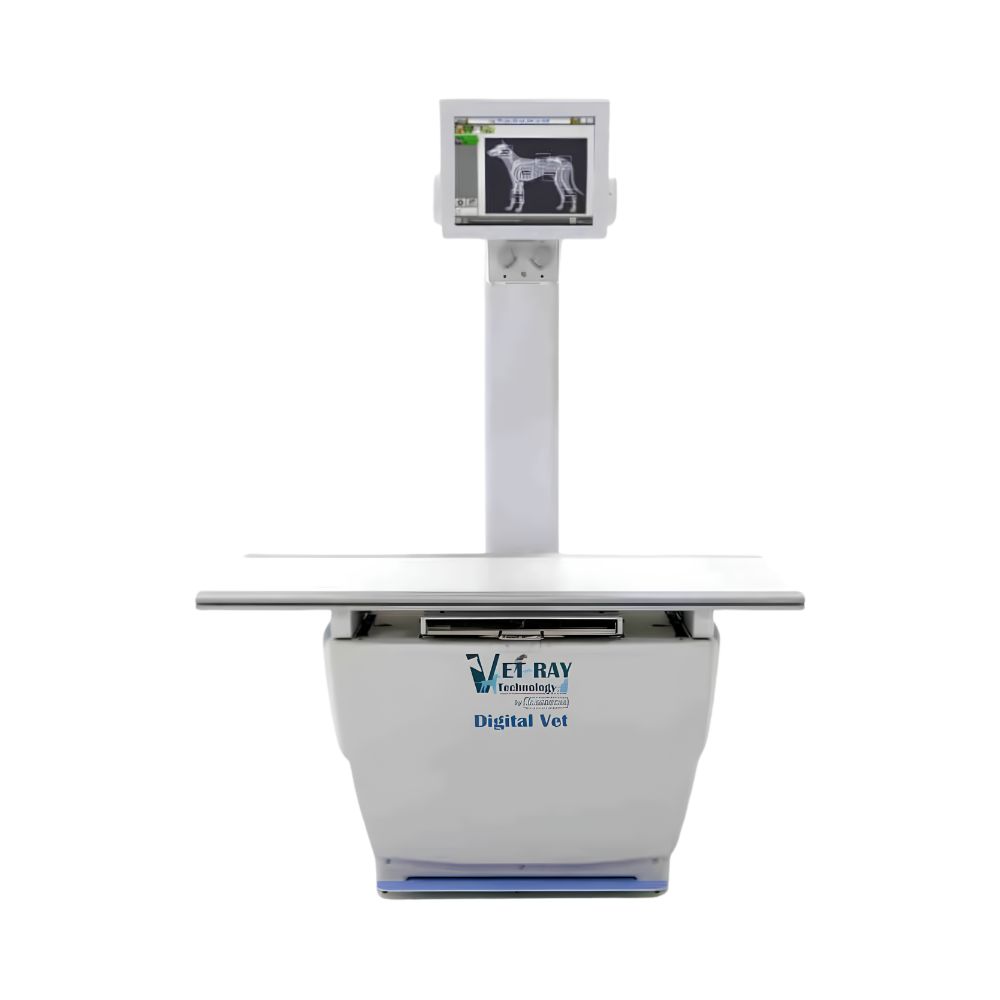
The Vet-Ray by Sedecal Digital DX-V is a complete veterinary radiography system for clinics imaging animals from felines to large breeds and exotics. Its choice of VIVIX detector sizes, floating tabletop, touch-screen controls, and rapid image processing supports flexible positioning and efficient digital exams.
Complete Digital Veterinary X-Ray for Efficient Exams
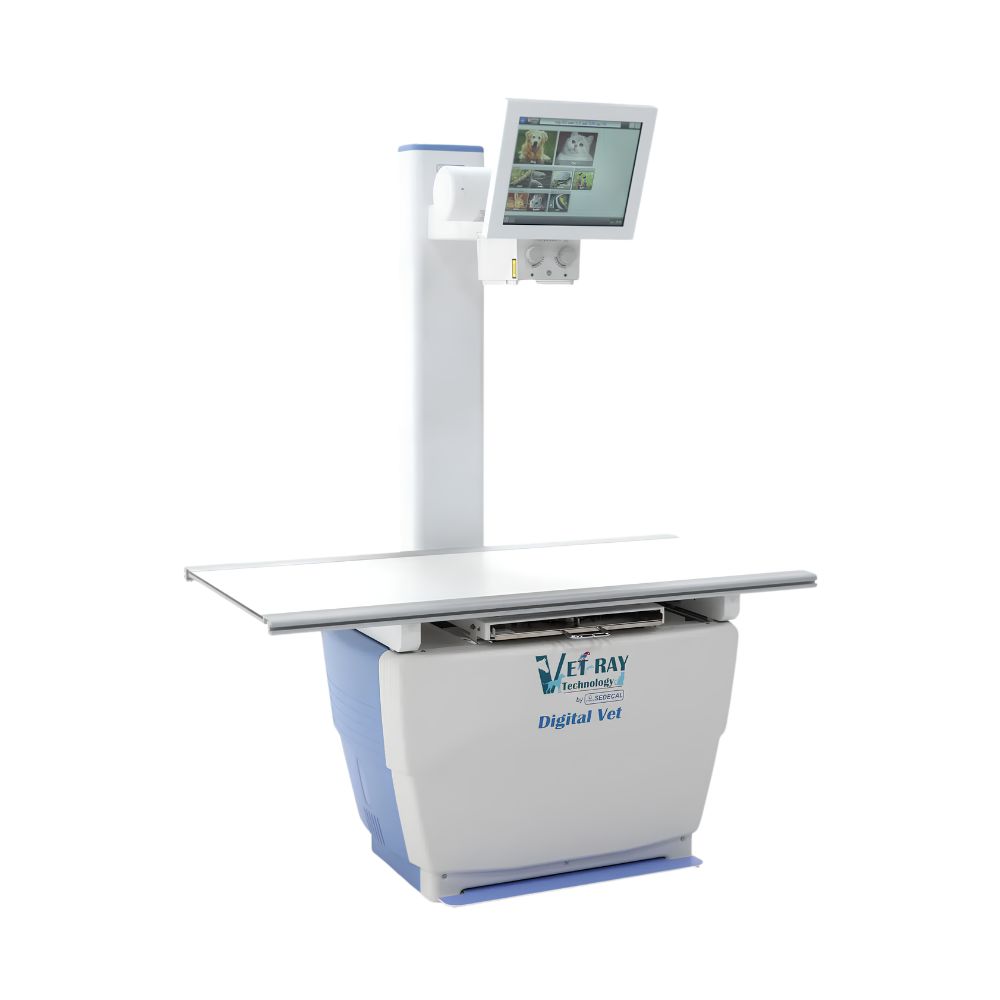
The Vet-Ray by Sedecal Digital DX-V combines a veterinary X-ray table, integrated high-frequency generator, digital detector, acquisition workstation, and image-sharing tools in one system. Practices can select a 17 x 17 in. (43.2 x 43.2 cm) detector for broad anatomical coverage or a removable 14 x 17 in. (35.6 x 43.2 cm) detector for more varied positioning.
Key benefits include:
-
- Choice of 17 x 17 in. (43.2 x 43.2 cm) or 14 x 17 in. (35.6 x 43.2 cm) VIVIX HD detectors
- Four-way floating tabletop for controlled patient positioning
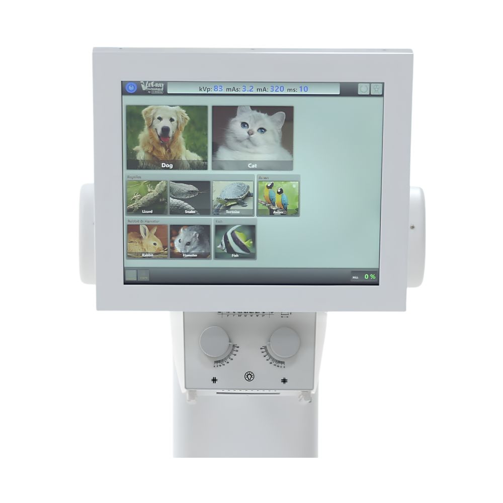
- Single Touch II touch-screen monitor for patient, body-part, and image selection
- Four-second image cycle time for rapid image availability
- Hands-free collimator light control and table lock release
- DICOM-compatible transfer, PACS workflow, and remote image access options
The complete configuration is intended to reduce movement between exposure, review, and distribution. The generator fits inside the table base, while the touch-screen monitor keeps acquisition controls and image review accessible within the radiology area.
Tour the Complete Veterinary X-Ray System
Watch an overview of the Vet-Ray Digital DX-V, including the floating tabletop, integrated generator, touch-screen controls, digital detector, and image acquisition workflow. The video provides a closer look at how the system is configured for routine veterinary radiography.
Veterinary Clinical Environments & Applications
The Vet-Ray Digital DX-V supports routine and specialty veterinary radiography in practices that need a fixed digital room with flexible patient positioning. Detector selection, table length, and positioning accessories can be matched to the animal sizes and examination types handled by the facility.
Veterinary Care Environments
Appropriate environments include:
-
- Companion animal veterinary practices
- Feline-focused practices
- Emergency and specialty hospitals
- Exotic animal practices
- Practices that routinely image large dog breeds
Supported Radiographic Applications
The system supports common digital veterinary X-ray examinations, including:
-
- Thoracic and abdominal radiography
- Orthopedic and extremity studies
- Spinal imaging
- Skull and dental-region overview imaging
- Horizontal-beam and cross-table studies with the appropriate detector and tube-stand configuration
- Weight-bearing and oblique views with the appropriate detector and tube-stand configuration
How Does the Vet-Ray Digital DX-V Support Daily Imaging Workflow?
Four-Way Floating Tabletop for Patient Positioning
The standard 5 ft (1.52 m) tabletop floats in four directions to help position the anatomy beneath the X-ray beam while limiting unnecessary patient movement. Adjustable tie-down rails support secure positioning. A 6 ft (1.83 m) tabletop is available for larger breeds, while a shorter 3 ft (0.91 m) tabletop is available for feline-focused applications.
Touch-Screen Control at the Radiology Table
The Single Touch II monitor system places patient selection, body-part selection, image review, and acquired-image display on a heads-up touch screen. This keeps the primary exam workflow in the imaging area rather than requiring repeated movement to a separate workstation.
Hands-Free Positioning Controls
Hands-free collimator light control and table lock release allow the operator to keep both hands available for patient positioning. This is especially useful when maintaining alignment or restraint immediately before exposure.
Fast Image Availability
The VIVIX HD detector acquires and displays images in approximately four seconds. Advanced Veterinary Image Processing software applies exam-specific image processing, helping the operator move from positioning to review without manually adjusting every image.
Which VIVIX Detector Best Fits the Practice?
17 x 17 in. Detector for Broad Anatomical Coverage
The 17 x 17 in. (43.2 x 43.2 cm) VIVIX detector provides the largest available image field in the DX-V lineup. Its square format is especially useful when imaging large breeds because the detector does not need to be rotated between portrait and landscape orientation.
14 x 17 in. Detector for Advanced Positioning
The 14 x 17 in. (35.6 x 43.2 cm) detector is removable and rotatable. When configured with the variable-SID tube stand used in the Premium Vet system, it supports horizontal-beam, weight-bearing, oblique, and cross-table examinations.
Digital Imaging, Processing & Connectivity Technologies
Tethered or Wireless Digital Acquisition
VIVIX HD detector configurations are available with tethered or wireless connectivity. This gives the practice a choice between a fixed wired workflow and greater detector-placement flexibility, depending on room design and examination needs.
VIVIX HD Image Quality
The detector provides 3.5 line pairs per millimeter spatial resolution, 14-bit analog-to-digital conversion, and 16-bit digital output. Depending on the selected detector, scintillator options include cesium or gadolinium.
Advanced Veterinary Image Processing
Advanced Veterinary Image Processing software applies exam-specific processing based on the selected anatomy and study. This helps produce review-ready images quickly while reducing the need for repetitive manual adjustment.
Acquisition Workstation & Display
The digital system includes an acquisition computer and wall mount, a 24 in. (61 cm) high-resolution 2-megapixel monitor, and the Single Touch II monitor interface. These components provide a dedicated environment for acquisition, review, and study management.
PACS, DICOM & Remote Collaboration
The system includes QXLINK PACS and a PACS workstation with a PC and 24 in. (61 cm) monitor. DICOM transfer supports specialist collaboration, while studies can also be distributed by CD, USB drive, or compatible cloud storage for viewing on web-enabled computers and mobile devices.
Compact High-Frequency Generator Options
The solid-state high-frequency generator is installed inside the table base to minimize the system footprint. The standard configuration is rated at 32 kW, 400 mA, 125 kVp, and 500 mAs with 220 VAC single-phase input. Optional generator configurations are available for three-phase 208 VAC or 480 VAC installations.
Vet-Ray by Sedecal Digital DX-V Specifications
Digital Detector
-
- Detector type: VIVIX HD tethered or wireless flat-panel detector
- Available image fields: 17 x 17 in. (43.2 x 43.2 cm) or 14 x 17 in. (35.6 x 43.2 cm)
- Spatial resolution: 3.5 lp/mm
- Image cycle time: approximately 4.0 seconds
- Scintillator options: cesium or gadolinium
- Analog-to-digital conversion: 14 bit
- Digital output: 16 bit
- Grayscale: 4,096 levels from 14-bit acquisition; up to 16,384 levels in 16-bit output
X-Ray Table & Positioning
-
- Tabletop movement: four-way floating
- Standard tabletop length: 5 ft (1.52 m)
- Optional large-breed tabletop length: 6 ft (1.83 m)
- Optional feline tabletop length: 3 ft (0.91 m)
- Patient positioning: adjustable tie-down rail track
- Controls: hands-free collimator light control and table lock release
High-Frequency Generator
-
- Generator type: compact solid-state high-frequency
- Installation: integrated inside the table base
- Standard generator rating: 32 kW
- Maximum tube current: 400 mA
- Maximum tube voltage: 125 kVp
- Maximum mAs: 500 mAs
- Standard electrical input: 220 VAC, single phase
- Optional electrical configurations: three-phase 208 VAC or 480 VAC
Monitor, Workstation & Connectivity
-
- Operator interface: Single Touch II touch-screen monitor system
- Acquisition display: 24 in. (61 cm), 2-megapixel high-resolution monitor
- Acquisition hardware: computer and wall mount
- Image processing: Advanced Veterinary Image Processing software
- PACS: QXLINK PACS and PACS workstation
- PACS display: PC with 24 in. (61 cm) monitor
- Study distribution: DICOM transfer, CD, USB drive, and compatible cloud storage
- Included viewing licenses: 10
Warranty
-
- X-ray system warranty: 5 years
- Digital detector warranty: 3 years
Warranty terms, detector configuration, electrical requirements, and included accessories should be confirmed for the quoted system.