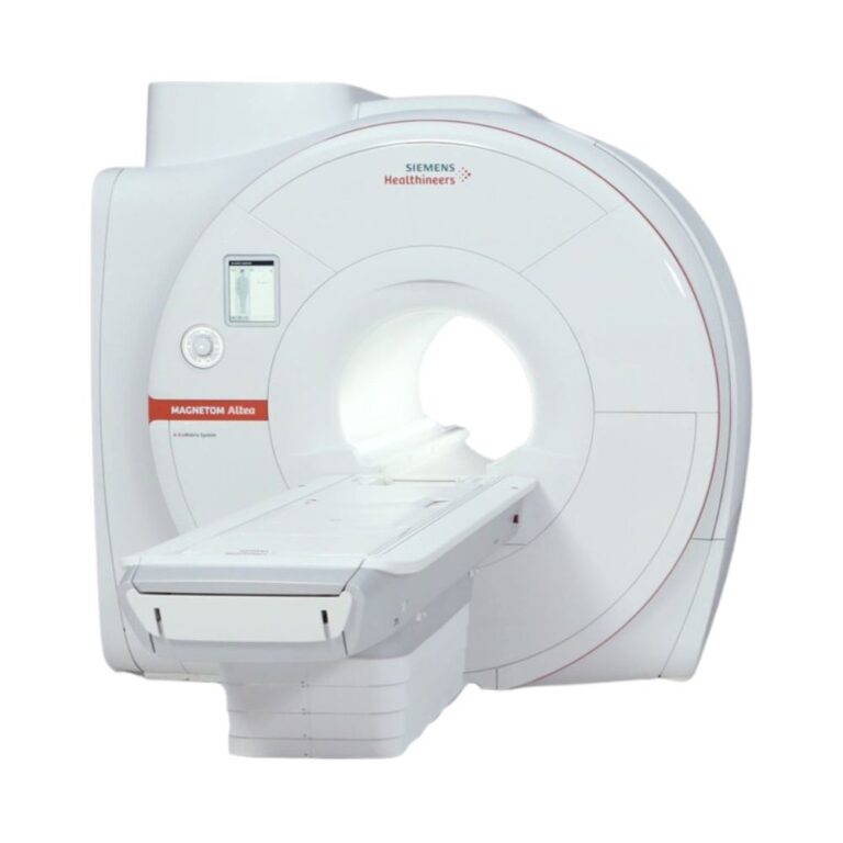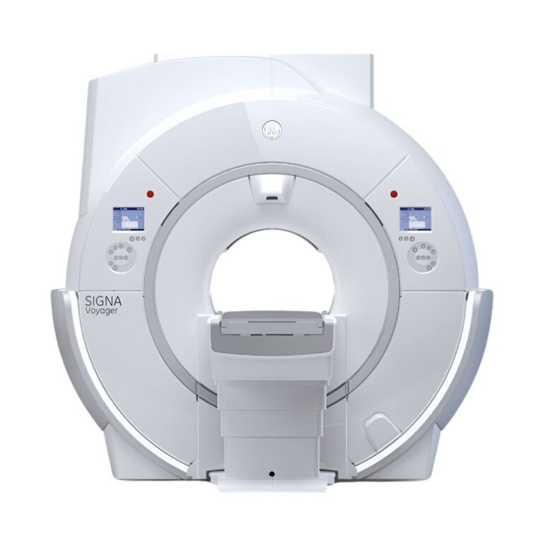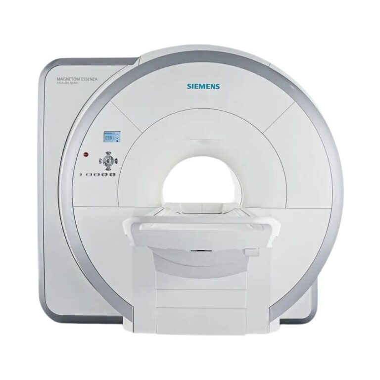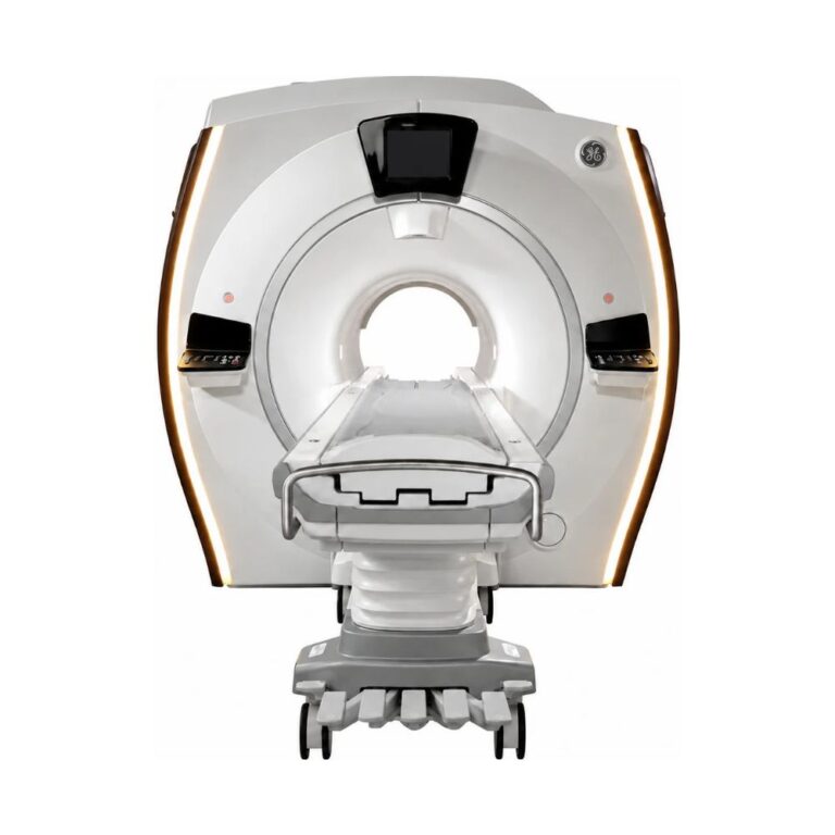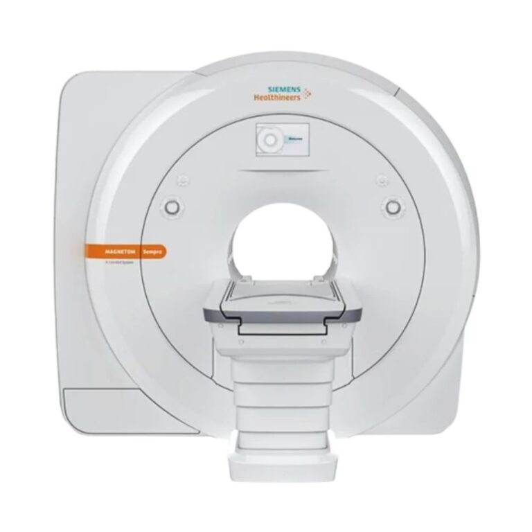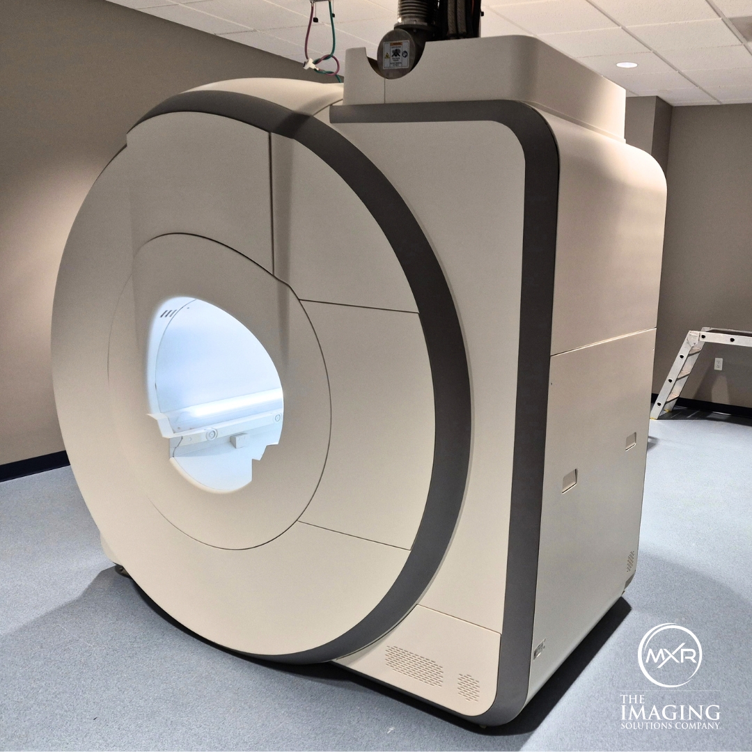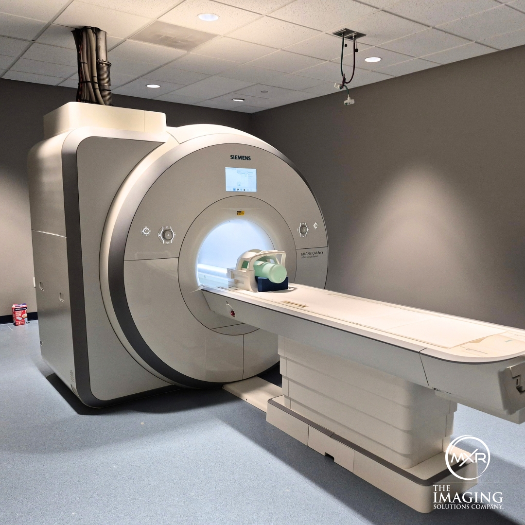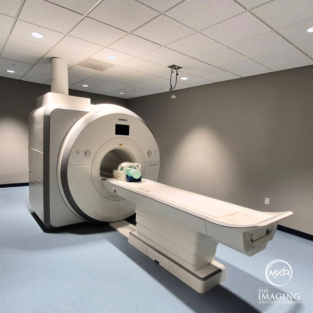
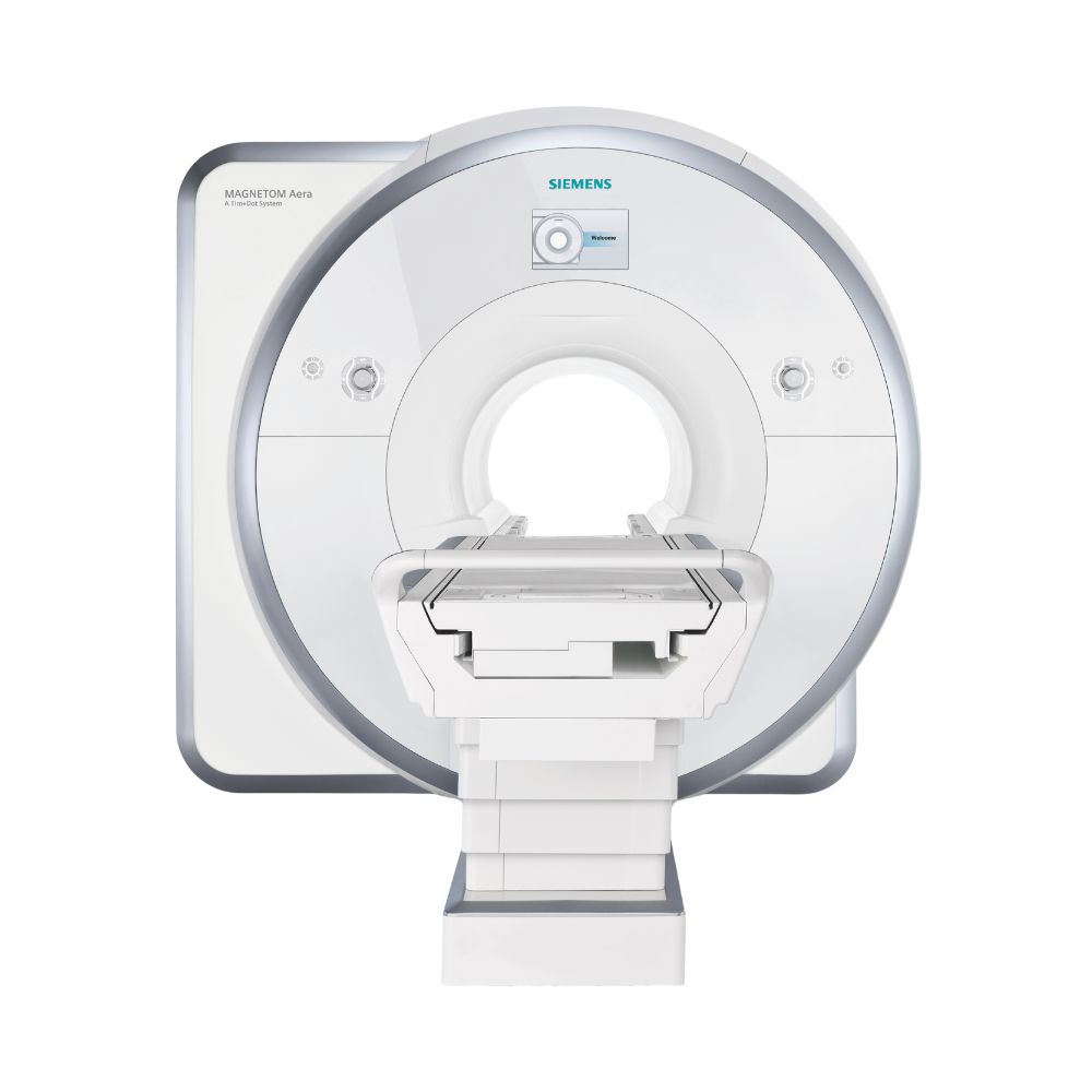
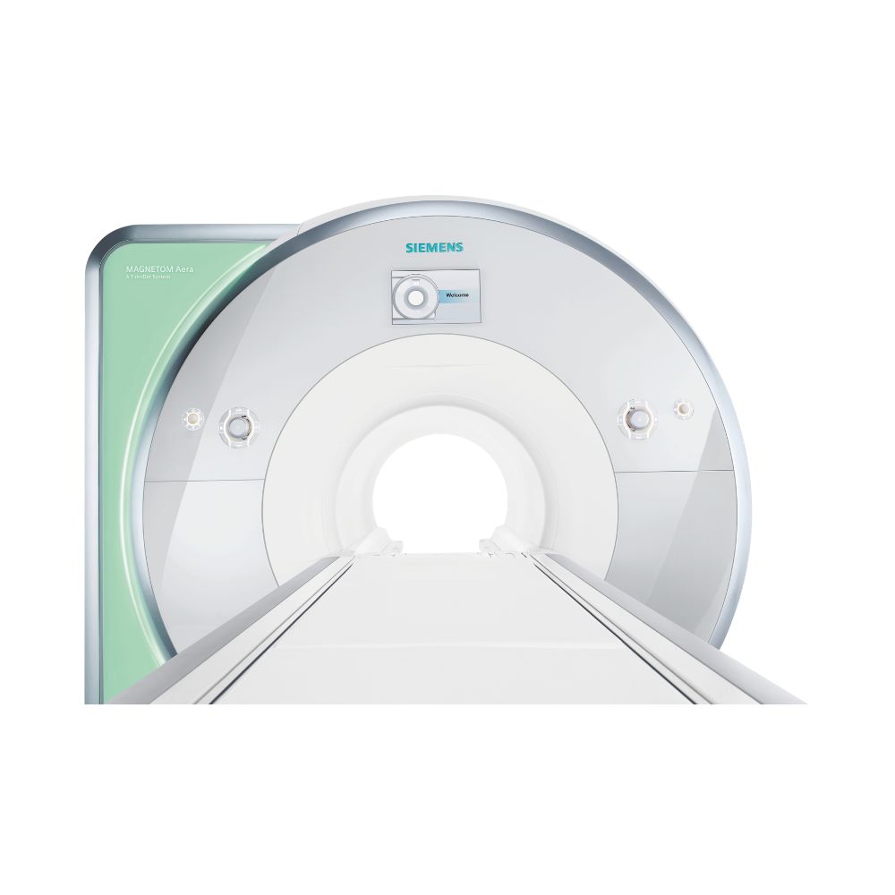
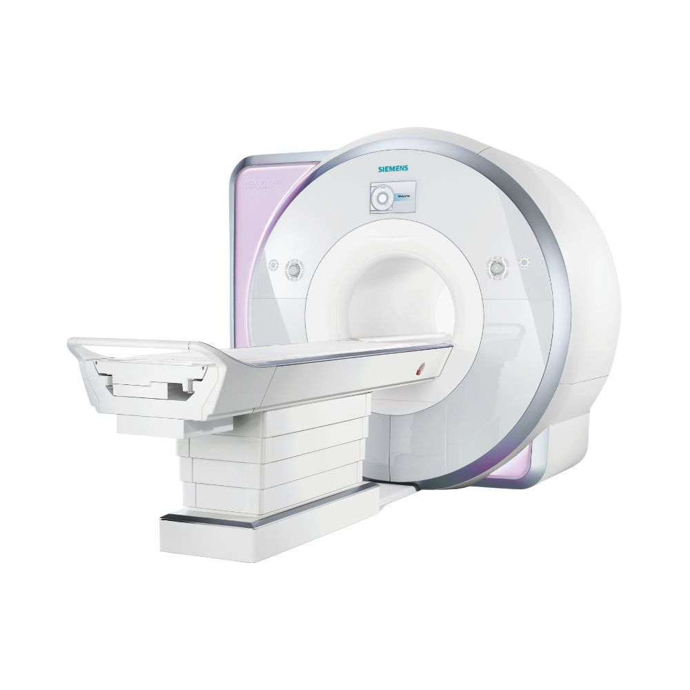
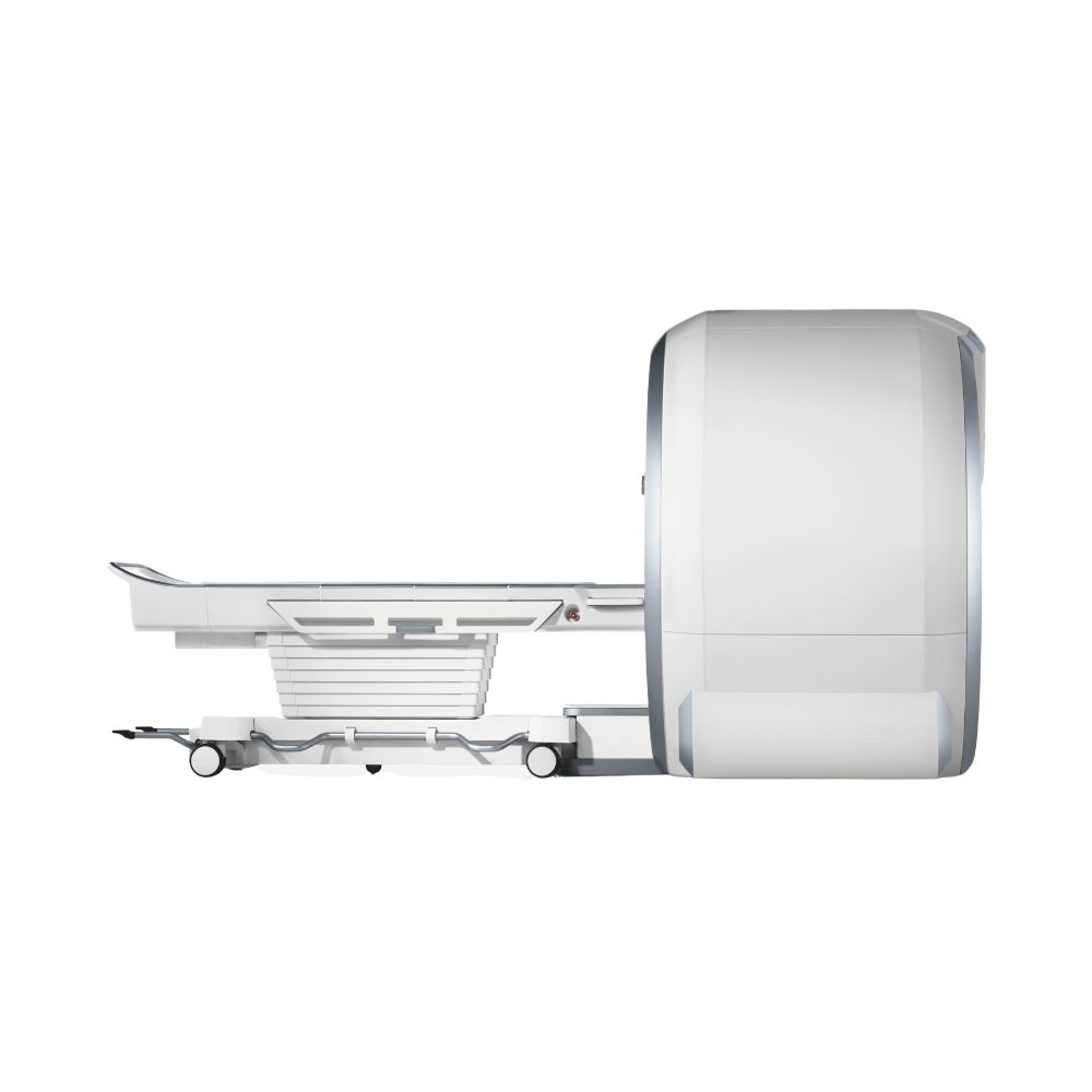
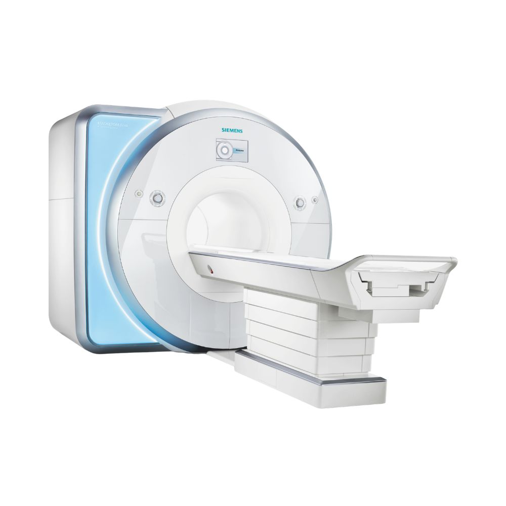
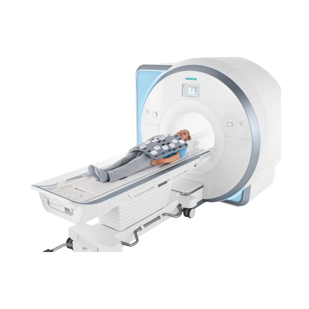
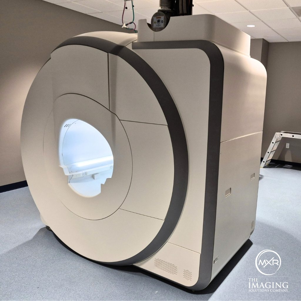
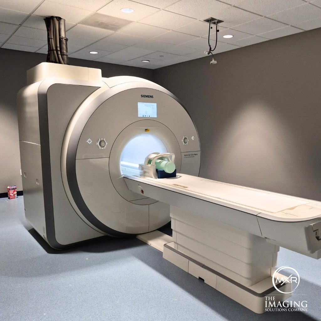
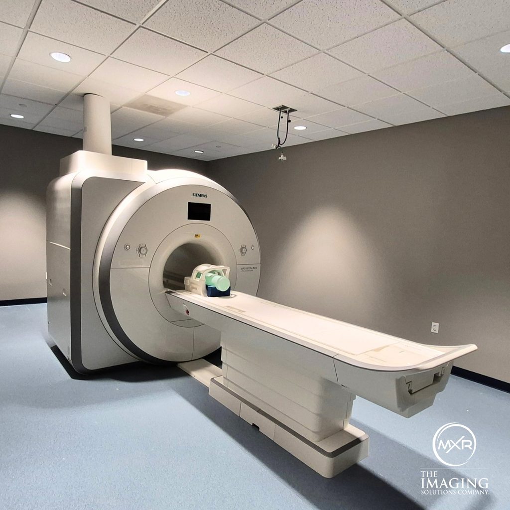
Siemens MAGNETOM Aera 1.5T MRI
- Category:
- MRI Systems
- Brand:
- Siemens
The Siemens MAGNETOM Aera 1.5T MRI is a 70 cm wide-bore, whole-body scanner for independent practices, imaging centers, hospitals, and health systems. Flexible receiver configurations, guided exam workflows, and available high-performance gradients support efficient routine and advanced imaging.
Siemens Aera MRI Highlights & Benefits
The Siemens Aera MRI combines a 1.5T superconducting magnet with wide-bore patient access, flexible imaging hardware, and workflow automation. XJ and XQ gradient options plus 24-, 48-, and 64-channel receiver configurations allow facilities to match the scanner to their clinical workload instead of treating every Aera configuration as equivalent.
Key Benefits
-
- 1.5T Whole-Body MRI: Supports routine and advanced neuro, spine, musculoskeletal, body, cardiac, vascular, breast, prostate, pediatric, and oncology imaging with the appropriate coils and software.
- 70 cm Wide Bore: Provides more room around the patient than conventional 60 cm systems while maintaining whole-body imaging capability.
- 250 kg (550 lb) Patient Capacity: Supports a broad patient population with an appropriately configured patient table.
- XJ or XQ Gradients: Available performance ranges from 33 mT/m at 125 T/m/s to 45 mT/m at 200 T/m/s for different routine and advanced imaging needs.
- 24-, 48-, or 64-Channel Configurations: Tim 4G supports high-density MRI coils and scan acceleration, with receiver configurations available for different clinical requirements.
- Automated Exam Workflows: Siemens Dot and DotGO technology guides exam setup, planning, and selected acquisition steps to improve consistency between patients and technologists.
Aera MRI Performance & Image Quality
The magnet, gradients, radiofrequency system, receiver architecture, and coils determine how the MAGNETOM Aera acquires imaging data. Because these components vary between configurations, buyers should evaluate the installed hardware rather than relying on the Aera model name alone.
1.5T Magnet & 50 cm Imaging Field
The MAGNETOM Aera uses a 1.5T superconducting magnet with a maximum imaging field of view of approximately 50 × 50 × 45 cm (19.7 × 19.7 × 17.7 in).
Siemens TrueForm magnet and gradient design helps maintain consistent imaging performance across larger and off-center anatomy. With an appropriate Tim whole-body configuration, imaging coverage can extend to approximately 205 cm (80.7 in) without repeatedly repositioning the patient.
XJ & XQ Gradient Performance
The Aera was offered with XJ and XQ gradient packages, which provide different levels of performance for routine and advanced imaging.
The Aera was offered with two primary gradient configurations:
-
- XJ Gradients: Up to 33 mT/m gradient amplitude with a maximum slew rate of 125 T/m/s per axis.
- XQ Gradients: Up to 45 mT/m gradient amplitude with a maximum slew rate of 200 T/m/s per axis.
These specifications describe gradient strength and switching speed. XJ can support a broad routine MRI workload and many specialty applications, while XQ provides additional performance for demanding diffusion, neurological, cardiovascular, accelerated, and other advanced protocols.
The installed gradient package should be confirmed for the individual system.
MAGNETOM Aera Receiver Channels & Tim 4G RF System
The MAGNETOM Aera uses Siemens Tim 4G radiofrequency (RF) and coil architecture and was offered with 24, 48, or 64 independent receiver channels.
The platform supports up to 204 connected coil elements, while the receiver-channel count determines how many independent coil signals the system can process simultaneously. For example, a Tim [204 × 48] configuration supports up to 204 connected coil elements with 48 independent receiver channels.
Higher channel configurations provide more flexibility for high-density coils and accelerated imaging, but receiver count is only one part of the system. Gradient package, coil inventory, software, and clinical licenses also affect overall capability.
DirectRF digitizes the MRI signal close to the magnet to help preserve signal quality before image reconstruction.
Aera Patient Access & Exam Experience
The Aera combines whole-body 1.5T performance with greater physical access than traditional 60 cm MRI systems. Its wide opening, short magnet, flexible table options, and patient-comfort features can benefit facilities serving a varied patient population.
70 cm Aera Bore Size & Patient Access
The Siemens Aera bore size is 70 cm (27.6 in).
Siemens describes this as a 70 cm Open Bore design. In practical equipment terms, the Aera is a wide-bore cylindrical MRI system rather than an open-sided MRI scanner.
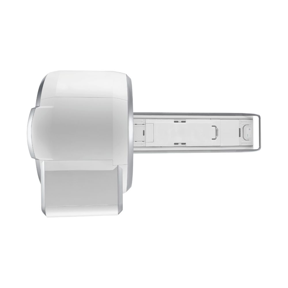
The magnet is approximately 137 cm (53.9 in) long, while the complete scanner measures approximately 145 cm (57.1 in) from cover to cover.
The combination of the 70 cm opening and short system can make positioning easier for larger patients and patients dealing with limited mobility, pain, kyphosis, or difficulty tolerating confined spaces.
Many examinations can also be performed feet first when the protocol and coil arrangement allow, leaving more of the patient’s body outside the scanner.
Patient Table, Weight Capacity, & Positioning
The Aera patient table supports up to 250 kg (550 lb) with the applicable configuration.
The standard table provides approximately 140 cm (55.1 in) of scan range. With an appropriate Tim whole-body configuration, imaging coverage can extend to approximately 205 cm (80.7 in).
The table can lower to approximately 52 cm (20.5 in) from the floor, helping with patient transfers and accessibility.
Fixed and mobile or dockable table configurations are available.
The Tim Dockable Table can be separated from the scanner for patient preparation and transport. This can simplify transfers and positioning for patients with limited mobility while allowing compatible coils to remain integrated with the table.
Patient Comfort & Communication
The Aera includes several product-specific features designed to improve the exam experience:
-
- Adjustable Bore Ventilation: Provides airflow inside the scanner for patient comfort.
- Adjustable Bore Lighting: Provides illumination inside the scanner opening.
- Illumination MoodLight: Adds ambient lighting around appropriately configured systems.
- Quiet Suite: Available specialized sequences reduce acoustic noise during supported neurological and orthopedic examinations.
- Patient Intercom: Allows two-way communication between the technologist and patient.
- Patient Call Device: Gives the patient a handheld control to alert the technologist.
- Automated Voice Commands: Provides standardized breathing and timing instructions during supported examinations.
- Lightweight Tim 4G Coils: Reduce the amount of heavy equipment positioned directly over the patient.
- Feet-First Imaging: Available for many supported examinations to reduce how much of the patient enters the bore.
Comfort features and software vary by configuration.
Aera Clinical Environments & Applications
The MAGNETOM Aera is a versatile whole-body 1.5T MRI platform. Its fit for a specific facility depends on the clinical workload, installed coils, software packages, receiver configuration, and gradient package.
Clinical Environments
The Aera is appropriate for:
-
- Independent outpatient imaging practices
- Multi-site diagnostic imaging centers
- Hospitals and health systems
- Orthopedic and musculoskeletal imaging centers
- Neurology and neuroscience programs
- Oncology and cancer imaging programs
- Cardiac and vascular imaging programs
- Breast and women’s imaging centers with MRI capability
- Pediatric imaging programs
- Academic medical centers and teaching hospitals
- Radiation oncology programs using appropriately configured MRI planning applications
Clinical Applications
Depending on configuration, the Aera can support:
-
- Brain & Neurological MRI: Routine brain imaging, diffusion, perfusion, and other advanced neurological examinations.
- Functional MRI: Functional MRI, or fMRI, maps changes associated with brain activity when the appropriate software and protocol are installed.
- Spine MRI: Cervical, thoracic, lumbar, and extended spine examinations.
- Musculoskeletal MRI: Shoulder, knee, hip, wrist, hand, foot, ankle, and other joint imaging.
- Imaging Near Eligible Implants: WARP software can reduce image distortion around certain MR-conditional implants. MR-conditional means the implant can be scanned only under its specified MRI safety conditions.
- Abdominal MRI: Liver, pancreas, kidney, bowel, and other abdominal examinations.
- Pelvic MRI: Pelvic anatomy and disease evaluation using appropriate protocols and coils.
- MRCP: Magnetic resonance cholangiopancreatography is an MRI technique used to visualize the bile and pancreatic ducts.
- MR Urography: MRI evaluation of the kidneys, ureters, and urinary tract.
- Prostate MRI: Prostate imaging with appropriate clinical software and coil configuration.
- Cardiac MRI: Cardiac anatomy, ventricular function, tissue characterization, and other supported cardiovascular applications.
- Vascular Imaging: Magnetic resonance angiography, or MRA, images arteries and veins with or without contrast depending on the examination.
- Breast MRI: Diagnostic breast imaging and compatible biopsy-guidance workflows with the appropriate breast coil and software.
- Oncology MRI: Regional and extended whole-body examinations for supported cancer imaging applications.
- Pediatric MRI: Age-appropriate MRI protocols across supported anatomical regions.
- MR Spectroscopy: Optional spectroscopy software evaluates chemical information within selected tissue in addition to anatomical images.
- Radiation Therapy Planning: Appropriately configured systems can support MRI examinations used in radiation-treatment planning workflows.
Advanced clinical applications depend on installed hardware, coils, software, and licenses.
Aera Exam Setup & Workflow Automation
The Aera uses Siemens Dot workflow technology to reduce repetitive setup steps and improve consistency across examinations.
Dot and DotGO are workflow-automation technologies, not diagnostic artificial intelligence. They help the technologist prepare and perform the examination but do not interpret MRI images or make diagnoses.
DotGO & Dot Engines
-
- DotGO: Organizes protocols, user guidance, and automated exam steps within a common workflow so facilities can standardize scanning while retaining protocol flexibility.
- Brain Dot Engine: Guides planning and selected workflow steps for supported brain examinations.
- Abdomen Dot Engine: Helps standardize abdominal imaging and coordinate supported breath-hold examinations.
- Cardiac Dot Engine: Automates portions of cardiac localization, planning, and workflow.
- Angio Dot Engine: Coordinates supported magnetic resonance angiography examinations, including scan and contrast timing.
- Breast Dot Engine: Provides structured workflow guidance for supported breast MRI examinations.
- Large Joint & Knee Dot Engines: Automate portions of planning and positioning during supported orthopedic examinations.
- TimCT Onco Dot Engine: Coordinates extended anatomical coverage for supported whole-body oncology imaging.
Installed Dot Engines vary by software generation and clinical package.
Automated Planning & Patient Guidance
-
- AutoAlign: Assists with consistent slice orientation for supported examinations.
- AutoCoverage: Helps select appropriate anatomical coverage automatically.
- AutoAlign Heart: Assists with cardiac localization and planning on appropriately configured systems.
- AutoVoice Commands: Provides standardized patient breathing and timing instructions.
These tools reduce repetitive manual tasks while keeping the technologist in control of the examination.
Aera Scan Acceleration & Motion Management
The Aera can use information collected from multiple coil elements to acquire supported examinations more efficiently. Several software technologies can also help when breathing, movement, or metal would otherwise reduce image quality.
Faster Imaging with GRAPPA, CAIPIRINHA, & SMS
-
- GRAPPA: A parallel imaging technique that uses signals from multiple coil elements to reconstruct images from less acquired data, reducing scan time on supported protocols.
- CAIPIRINHA: Accelerates supported multi-slice and volumetric imaging and can shorten breath-holds during body examinations.
- Simultaneous Multi-Slice (SMS): Acquires multiple image slices during the same acquisition step to accelerate supported two-dimensional imaging.
- GOBrain: Combines optimized protocols, workflow automation, and accelerated acquisition to support clinically validated routine brain examinations in under six minutes on appropriately configured systems.
The achievable acceleration depends on the receiver configuration, coils, software, and protocol.
Motion & Metal Artifact Reduction
-
- FREEZEit: Combines body-imaging techniques designed to reduce problems caused by respiratory motion and scan-timing variability.
- StarVIBE: Supports motion-resistant volumetric imaging and can allow selected body examinations to be performed during free breathing.
- BLADE: Acquires MRI data in a rotating pattern that makes supported sequences less sensitive to certain types of patient motion.
- WARP & Advanced WARP: Reduce distortion and signal loss around eligible metal implants during supported orthopedic imaging.
Aera Reconstruction, Post-Processing, & Connectivity
After an acquisition, the Aera can automatically reconstruct and process selected images before they move into the facility’s reading and storage workflow.
Inline Reconstruction & Extended Imaging
-
- Inline 3D: Automatically generates supported three-dimensional image processing as part of the exam workflow.
- Multiplanar Reconstruction (MPR): Creates views in different anatomical planes from volumetric imaging data without requiring another scan.
- Inline Composing: Combines data from multiple imaging stations into a larger anatomical view, such as an extended spine study.
- TimCT: Coordinates imaging over an extended anatomical range with compatible coils and table movement, reducing the need to reposition the patient.
- MyoMaps: Available cardiac software provides quantitative information about heart tissue during supported cardiac examinations.
Availability depends on software generation and installed clinical packages.
DICOM, PACS, & Workstation Integration
The Aera operates within the Siemens syngo MR environment and supports standard DICOM connectivity and PACS integration for transferring, storing, and reviewing MRI studies within the facility’s imaging network.
Available post-processing, advanced visualization, quantitative analysis, and image-review capabilities depend on the installed workstation and software configuration.
Aera Configurations, Coils, & Available Options
Two Siemens Aera MRI systems can have the same model name while offering different imaging performance and clinical capability. Gradient hardware, receiver channels, coils, software, and clinical licenses should therefore be evaluated together.
Choosing XJ, XQ, & Receiver Configurations
For facilities evaluating an Aera, the main hardware differences include the gradient package and receiver-channel configuration.
-
- XJ gradients provide 33 mT/m amplitude and 125 T/m/s slew rate. They can support a broad routine MRI workload, including common brain, spine, musculoskeletal, and body examinations.
- XQ gradients increase performance to 45 mT/m and 200 T/m/s. They can be valuable when advanced diffusion, cardiovascular, neurological, or accelerated imaging is a greater part of the expected workload.
Receiver options include 24, 48, or 64 independent channels.
An independent imaging practice focused primarily on routine MRI may have different hardware needs from a tertiary hospital performing advanced neuro, cardiac, oncology, and other specialty examinations.
More receiver channels do not automatically make one system the better choice. Coil inventory, software licenses, gradient hardware, clinical applications, and expected workload should all be considered.
Tim 4G RF Coils
The Tim 4G architecture supports a broad selection of anatomical and specialty coils, including:
-
- Head and head/neck coils
- Integrated spine coils
- Body array coils
- Cardiac coil combinations
- Breast coils
- Shoulder coils
- Knee coils
- Hand and wrist coils
- Foot and ankle coils
- Flexible multi-purpose coils
- Peripheral vascular coil combinations
- Other specialty coils
DirectConnect and SlideConnect are designed to simplify coil setup on compatible Aera configurations. DirectConnect allows supported coils to connect directly through table interfaces, reducing loose cabling, while SlideConnect provides a guided connection point for compatible removable coils.
The specific coil package varies by system, so available coils should be matched to the facility’s planned examinations rather than assumed from platform compatibility alone. MXR Imaging can also support ongoing coil needs with MRI coil repair for supported MRI coils.
Software & Clinical Options
Depending on software generation and installed licenses, available applications can include:
-
- Advanced neurological imaging
- Diffusion and perfusion imaging
- Functional MRI
- MR spectroscopy
- Orthopedic imaging
- Metal artifact reduction
- Abdominal and pelvic imaging
- Cardiac MRI
- MR angiography
- Breast MRI
- Prostate MRI
- Oncology and whole-body imaging
- Pediatric protocols
- Scan acceleration
- Quantitative cardiac tools
- Radiation therapy planning applications
- Advanced reconstruction and post-processing
Platform compatibility does not mean every option is installed. MXR Imaging can verify the software and clinical licenses included with an available system.
Aera Magnet, Cryogen, & Site Requirements
An Aera installation requires appropriate room dimensions, structural support, magnetic-field planning, radiofrequency shielding, cooling, electrical service, and helium infrastructure.
Superconducting Magnet & Helium System
The MAGNETOM Aera uses a superconducting 1.5T magnet with liquid helium and Zero Helium Boil-Off technology designed to limit routine helium loss during normal operation.
The system still requires magnet refrigeration, helium service capability, and a properly designed quench pipe as part of the MRI installation.
MXR Imaging provides MRI refrigeration and helium services for supported MRI systems.
Room Planning, RF Shielding, & Quench Venting
Siemens lists approximately 30 m² (323 sq ft) of minimum total space for the magnet, equipment, and console areas combined. This should not be interpreted as a universal 323 sq ft examination-room requirement.
Representative planning requirements include:
-
- Examination Room Minimum Finished Height: Approximately 240 cm (94.5 in)
- Equipment Room Minimum Finished Height: Approximately 240 cm (94.5 in)
- Control Room Minimum Finished Height: Approximately 210 cm (82.7 in)
- Whole-Body Room Length with Mobile Table: Minimum approximately 642 cm (252.8 in), with approximately 662 cm (260.6 in) recommended in the referenced layout
- Whole-Body Room Length with Fixed Table: Minimum approximately 612 cm (240.9 in)
The Aera requires an RF-shielded scan room, appropriate magnetic-field planning, quench venting, and adequate service clearances. Final room dimensions, shielding, structural support, transport access, and installation requirements should be verified for the specific system and facility.
MXR Imaging provides custom MRI shielding solutions for new MRI rooms, replacements, and facility upgrades.
Power, Cooling, & Facility Requirements
The Aera requires water cooling for the gradient system and electronics. Facilities can use a compatible central cooling-water supply or an appropriate local chiller.
Representative requirements include:
-
- XJ Cooling Load: Approximately 45 kW
- XQ Cooling Load: Approximately 60 kW
- XJ Electrical Connection: Approximately 63 kVA
- XQ Electrical Connection: Approximately 88 kVA
- Optional Local Chiller: Approximately 48 kVA
- Referenced Electrical Supply: 400 V three-phase
Final electrical, cooling, HVAC, structural, and utility requirements should be confirmed for the specific system and installation.
Available Siemens Aera MRI Configuration
An available Siemens Aera MRI can differ substantially from another Aera of the same model family. MXR Imaging evaluates the complete configuration so the system can be matched to the buyer’s exam mix, patient population, workflow, and facility.
Important configuration factors include:
- Model year
- XJ or XQ gradient package
- 24-, 48-, or 64-channel receiver configuration
- Tim 4G coil inventory
- Software generation
- Dot and DotGO capabilities
- Installed clinical licenses
- Scan-acceleration options
- Fixed or dockable patient table
- Operator console
- Post-processing workstation
- Patient-comfort options
- Hardware and software upgrades
- Magnet refrigeration and helium status
- Accessories
For an independent practice or single-site imaging center, configuration matching can help avoid purchasing capabilities that do not support the expected exam mix.
For hospitals and health systems, the same review can help align clinical capability, workflow, service needs, and equipment standards across departments or locations.
MXR Imaging can verify the installed hardware, software, coils, and accessories of an available system before purchase.
Siemens MAGNETOM Aera Price & Availability
The price of a Siemens MAGNETOM Aera depends on the actual system configuration, including XJ or XQ gradients, receiver channels, coil inventory, software level, clinical licenses, patient table, workstation, system condition, and project scope.
MXR Imaging offers Aera systems in configurations matched to the facility’s clinical and technical requirements, subject to availability. Qualifying systems may also be available through the Platinum Certified process.
Project support can include equipment planning, deinstallation, relocation, installation, shielding, coil support, and facility coordination.
MXR Imaging also supports the system after installation with MRI maintenance and repair, helium and refrigeration services, parts support, and turnkey imaging project management.
This gives independent practices access to comprehensive equipment and service support while also accommodating larger hospital and health-system projects.
Siemens MAGNETOM Aera 1.5T Specifications
The specifications below describe the MAGNETOM Aera platform. Gradient, receiver, coil, software, table, and clinical configurations vary by individual system.
Magnet System
-
- Field Strength: 1.5T
- Magnet Type: Superconducting
- MRI Type: Whole-body, wide-bore cylindrical MRI
- Bore Diameter: 70 cm (27.6 in)
- Magnet Length: Approximately 137 cm (53.9 in)
- System Length: Approximately 145 cm (57.1 in), cover to cover
- Maximum Field of View: Approximately 50 × 50 × 45 cm (19.7 × 19.7 × 17.7 in)
- Maximum Imaging Coverage: Approximately 205 cm (80.7 in) with an appropriate whole-body configuration
- Magnet Design: Siemens TrueForm
- Cryogen: Liquid helium
- Helium Management: Zero Helium Boil-Off under normal operating conditions
- Magnet Shielding: Actively shielded
Gradient System
-
- Available Gradient Packages: XJ and XQ
- XJ Maximum Gradient Amplitude: 33 mT/m per axis
- XJ Maximum Slew Rate: 125 T/m/s per axis
- XQ Maximum Gradient Amplitude: 45 mT/m per axis
- XQ Maximum Slew Rate: 200 T/m/s per axis
- Installed Gradient Package: Configuration dependent
RF System
-
- RF Architecture: Siemens Tim 4G
- Signal Architecture: DirectRF digital transmit and receive design
- Maximum Connected Coil Elements: Up to 204
- Receiver Configurations: 24, 48, or 64 independent channels
- Maximum Simultaneous Independent Receiver Channels: 64
- Parallel Imaging: Supported with compatible coils and software
- Installed RF Configuration: System specific
Bore & Patient Table
-
- Bore Diameter: 70 cm (27.6 in)
- Maximum Patient Weight: 250 kg (550 lb) with the applicable table
- Standard Scan Range: Approximately 140 cm (55.1 in)
- Optional Whole-Body Imaging Coverage: Approximately 205 cm (80.7 in)
- Minimum Table Height: Approximately 52 cm (20.5 in)
- Patient Orientation: Head-first and feet-first for supported examinations
- Patient Table Options: Fixed and mobile or dockable
- Integrated Spine Coil: Available with compatible table configurations
Coils & Accessories
-
- Coil Architecture: Siemens Tim 4G
- Compatible Coil Categories: Head, head/neck, spine, body, cardiac, breast, shoulder, knee, hand/wrist, foot/ankle, flexible, vascular, and other specialty coils
- DirectConnect: Available with compatible coil designs
- SlideConnect: Available with compatible coil designs
- Included Coils: System specific
- Accessories: Depend on equipment package and clinical configuration
Siting & Utilities
-
- Minimum Total Space: Approximately 30 m² (323 sq ft) for magnet, electronics, and console areas combined
- Minimum Examination Room Height: Approximately 240 cm (94.5 in)
- Minimum Equipment Room Height: Approximately 240 cm (94.5 in)
- Minimum Control Room Height: Approximately 210 cm (82.7 in)
- RF Shielding: Required
- Quench Pipe: Required
- Cooling: Water cooled
- XJ Heat Dissipation to Cooling Water: Approximately 45 kW
- XQ Heat Dissipation to Cooling Water: Approximately 60 kW
- Representative XJ Electrical Connection: Approximately 63 kVA
- Representative XQ Electrical Connection: Approximately 88 kVA
- Optional Local Chiller Connection: Approximately 48 kVA
- Referenced Electrical Supply: 400 V ±10%, three-phase
- Final Facility Requirements: Must be verified for the specific system and installation
System Dimensions & Weight
-
- Magnet Length: Approximately 137 cm (53.9 in)
- System Length: Approximately 145 cm (57.1 in), cover to cover
- System Weight in Operation: Approximately 4.8 metric tons (10,580 lb)
- Facility Access: Delivery route, structural loading, openings, and installation clearances must be evaluated for the individual site