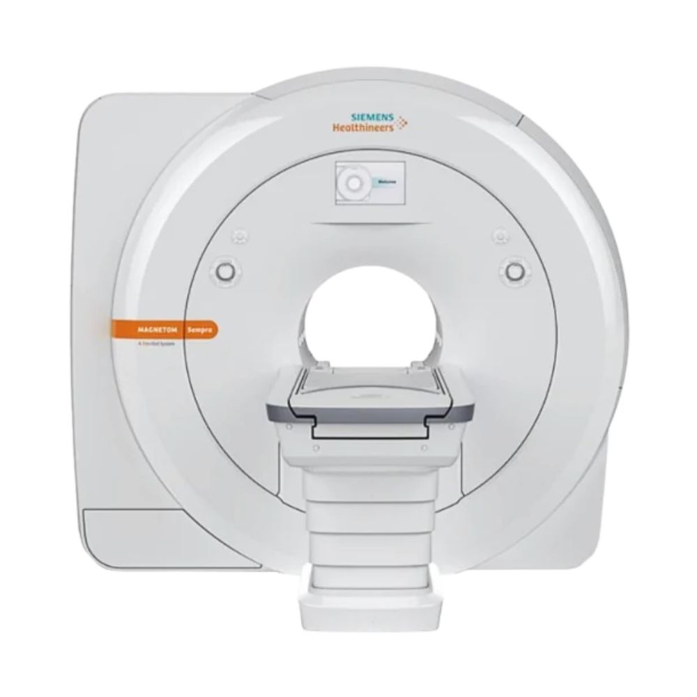
Siemens MAGNETOM Sempra 1.5T MRI
The Siemens MAGNETOM Sempra is a compact 1.5T MRI scanner well suited to facilities seeking efficient routine imaging with options for specialized examinations. Its 60 cm patient opening, guided exam setup, and lightweight imaging coils support consistent scanning across a broad range of clinical needs.
Siemens MAGNETOM Sempra MRI Highlights & Benefits
The MAGNETOM Sempra combines 1.5T imaging with guided examination workflows and a compact installation footprint. Its lightweight coils, automated scanning features, and energy-efficient design make it practical for facilities performing routine neurological, spinal, musculoskeletal, and body MRI examinations.
Key Features & Benefits
-
- 1.5T Superconducting Magnet: Provides the stable magnetic field needed for detailed brain, spine, joint, and body examinations.
- 60 cm Bore & Compact Design: Combines a 60 cm (23.6 in) patient opening with a 171 cm (67.3 in) system length for space-conscious MRI installations.
- Tim 4G Imaging Coils: Siemens’ lightweight signal-receiving coils support detailed anatomical imaging and efficient patient preparation, with 8- or 16-channel receiver configurations.
- Guided MRI Workflows: Siemens DotGO or newer myExam Companion software guides technologists through examination setup and scanning, helping maintain consistent results.
- Advanced Imaging Options: Compatible technologies reduce image distortion near certain metal implants, support shorter examinations, and improve image clarity through advanced reconstruction, including optional AI-assisted processing.
- Energy-Efficient Operation: Zero helium boil-off refrigeration limits helium loss during normal operation, while Eco-Power technology helps reduce electricity consumption.
MAGNETOM Sempra 1.5T Imaging Performance
The Sempra combines a superconducting 1.5T magnet, Siemens XF gradient technology, and multichannel signal reception to produce detailed diagnostic images. The magnet provides the main magnetic field, gradients identify where imaging signals originate, and the receiving system collects the information used to create each image.
1.5T TrueForm Magnet for Whole-Body Imaging
The Sempra uses a 1.5 Tesla superconducting magnet with Siemens TrueForm technology, a magnet design that supports consistent magnetic field conditions across the imaging area.
Passive and active shimming help maintain field uniformity. Shimming adjusts the magnetic field to reduce variations that could interfere with image quality, particularly during examinations of larger or anatomically complex regions.
The scanner provides a maximum field of view of approximately 50 × 50 × 45 cm (19.7 × 19.7 × 17.7 in), describing the anatomical coverage available within the specified imaging field.
The 1.5T platform supports routine brain, spine, joint, abdominal, pelvic, and vascular imaging, with specialized techniques available through compatible clinical software.
XF Gradients for Detailed MRI Acquisition
The Sempra uses Siemens XF gradient technology to identify where MRI signals originate within the patient.
Its gradient system provides:
-
- Maximum Gradient Amplitude: 30 mT/m per axis
- Maximum Slew Rate: 100 T/m/s per axis
Gradient amplitude measures the strength of the additional magnetic fields used to locate anatomical information. Slew rate measures how quickly those fields can change during scanning.
Together, these capabilities support detailed anatomical imaging, diffusion-weighted examinations that evaluate water movement within tissue, and other compatible advanced imaging techniques.
For routine brain, spine, and joint examinations, the gradient system helps balance anatomical detail, scan time, and image quality.
Tim 4G RF Technology for Flexible Coil Coverage
Siemens Tim 4G uses multiple imaging coil elements positioned near the patient to collect radiofrequency (RF) signals. These signals provide the information used to reconstruct MRI images.
The Sempra supports two principal configurations:
-
- Tim [96 × 8]: Supports up to 96 connected coil elements with eight independent receiver channels operating during an acquisition.
- Tim [96 × 16]: Supports up to 96 connected coil elements with 16 independent receiver channels operating during an acquisition.
Coil elements collect signals from the anatomy, while receiver channels process those signals during scanning. The number 96 refers to the maximum connected coil elements, not the number of independent receiver channels.
Siemens DirectRX processes signals received from compatible coils, helping preserve imaging information for reconstruction.
The 16-channel configuration can process more coil signals simultaneously, supporting additional compatible coil arrangements and certain advanced scanning techniques.
Sempra Patient Access & Exam Comfort
The MAGNETOM Sempra combines a conventional 60 cm patient opening with a short magnet, powered patient positioning, and quieter scanning options. These features support efficient preparation and help accommodate patients requiring additional positioning assistance.
60 cm Bore in a Compact 171 cm System
The Sempra features a 60 cm (23.6 in) bore, a magnet length of approximately 155 cm (61 in), and an overall system length of 171 cm (67.3 in).
Its relatively short design limits the length of the scanner surrounding the patient compared with longer conventional MRI systems.
Head-first and feet-first positioning are available for appropriate examinations, allowing technologists to select the orientation suited to the anatomy being studied.
The compact design also reduces the space required to accommodate the scanner within an appropriately planned MRI suite.
440 lb Patient Capacity with Powered Positioning
The Sempra supports a maximum patient weight of 440 lb (200 kg) and a continuous scanning range of up to 140 cm (55.1 in).
Compatible patient-table configurations include:
-
- Powered Horizontal Movement: Moves the patient into and out of the scanner during examination setup.
- Optional Vertical Movement: Adjusts table height to assist patient access, with a representative adjustable range of 58–89 cm (22.8–35 in).
The height-adjustable configuration is particularly useful when patients need assistance getting onto or off the table.
Controls on the front of the scanner and the Siemens Dot Display, a screen showing positioning and examination information, provide convenient access to patient-positioning functions.
Quiet Suite for Less MRI Scanning Noise
The Sempra supports Siemens Quiet Suite, a collection of scanning techniques designed to reduce the noise created by rapidly changing magnetic gradients.
Compatible options include:
-
- QuietX: Adjusts the operation of the magnetic gradients during supported examinations to reduce scanning noise while preserving useful image quality.
- PETRA: Uses a specialized three-dimensional scanning technique that produces particularly quiet images during compatible examinations.
Quiet Suite can make neurological and musculoskeletal examinations more manageable for patients sensitive to noise.
In-bore ventilation, lighting, patient communication, and lightweight imaging coils provide additional comfort during scanning.
Clinical Environments & Applications
The Siemens MAGNETOM Sempra is well suited to routine brain, spine, and joint MRI, with additional capabilities for specialized examinations when appropriately equipped.
Clinical Environments
-
- Hospitals and radiology departments
- Outpatient diagnostic imaging centers
- Orthopedic and sports medicine imaging centers
- Specialty diagnostic imaging facilities
- Pediatric imaging departments
- Veterinary hospitals and specialty referral centers
Clinical Specialties & Applications
-
- Neurology & Neurosurgery: Brain and spine MRI, including advanced studies of tissue changes, blood flow, and brain activity. The Brain Dot Engine guides examination setup for consistent brain imaging.
- Orthopedics & Sports Medicine: Joint, ligament, cartilage, tendon, and soft-tissue MRI, with Advanced WARP software to reduce image distortion near compatible metal implants.
- Abdominal Imaging & Hepatology: Abdominal and liver MRI, with FREEZEit to reduce breathing-related blur and LiverLab to help measure liver tissue characteristics.
- Urology: Detailed prostate MRI using the Sempra’s compatible spine and body imaging coils to capture multiple types of images for tissue evaluation.
- Cardiology: Heart MRI to evaluate anatomy and pumping function, with optional MyoMaps software for measuring heart muscle tissue characteristics.
- Vascular Medicine: Imaging of arteries and veins, with or without contrast dye, including guided timing for contrast-enhanced examinations.
- Breast Radiology: Diagnostic breast MRI, implant evaluation, and supported MRI-guided biopsy procedures using dedicated breast coils.
- Oncology: Tumor assessment and follow-up MRI using Onco Suite, a collection of specialized scanning protocols and image-review tools for cancer-related examinations.
- Pediatric Radiology: Brain, spine, joint, and body examinations using protocols adapted for younger patients.
- Veterinary Medicine: Brain, spine, joint, and selected body imaging for companion animals using the Sempra Veterinary Edition.
Specialized examinations depend on installed software and compatible imaging coils. Pediatric examinations require appropriate patient-specific safety assessment, particularly for children under two. Veterinary applications require the dedicated equipment configuration and appropriate clinical protocols.
Sempra MRI Exam Planning & Workflow Automation
The Sempra uses guided scanning software to simplify MRI examination preparation and maintain consistency across operators. Earlier systems use Siemens DotGO, while compatible newer systems offer updated guidance and AI-assisted workflow features through myExam Companion.
Guided Scanning with DotGO or myExam Companion
Siemens DotGO organizes examination preparation and protocol selection into guided workflows called Dot Engines. These provide structured steps for planning and completing specific types of MRI examinations.
The original standard Sempra workflow package includes:
-
- Brain Dot Engine: Guides neurological scanning and helps standardize anatomical positioning.
- Spine Dot Engine: Supports consistent spine examination planning, including automatic alignment and labeling.
- Large Joint Dot Engine: Guides knee and other supported large-joint examinations with standardized imaging planes and anatomical coverage.
Compatible Sempra systems running Siemens syngo MR XA50M software use myExam Companion, which updates the guided scanning interface and incorporates automation and AI-assisted features.
These tools help technologists select examination strategies, position imaging slices, and organize scanning steps while allowing adjustments for individual patients.
Compatible features include:
-
- myExam Assist: Guides technologists through supported examinations using predefined scanning strategies that can be adjusted to the patient’s anatomy and clinical requirements.
- myExam Cockpit: Provides a central interface for creating, organizing, and customizing scanning protocols so facilities can maintain consistent examination procedures.
- myExam Brain Autopilot: Automates supported brain examination steps, including positioning, scan planning, and acquisition. It reduces repetitive operator actions during routine neurological imaging.
myExam Companion combines conventional automation with AI-assisted functions. Individual capabilities depend on the installed software and supported workflow applications.
AutoAlign for Consistent Scan Positioning
Siemens AutoAlign uses anatomical landmarks identified during initial imaging to establish appropriate scan orientations.
Instead of requiring technologists to position every imaging plane manually, compatible workflows automatically align subsequent acquisitions to those landmarks.
Related positioning technologies include:
-
- AutoAlign: Establishes consistent scan orientations for supported neurological and other examinations.
- AutoAlign Heart: Assists with positioning standard views of the heart during compatible cardiac MRI examinations.
- AutoPosition: Helps move the selected anatomy toward the center of the scanner’s imaging area during supported examinations.
These tools reduce repetitive adjustments and help maintain consistent positioning across patients and follow-up studies.
Specialty Exam Workflows for Body, Cardiac & Breast MRI
The Sempra supports additional guided workflows for specialized examinations.
Compatible applications include:
-
- Abdomen Dot Engine: Organizes abdominal scanning protocols, including steps needed to accommodate breathing-related movement.
- Cardiac Dot Engine: Guides heart examination planning and helps establish standard imaging views.
- Angio Dot Engine: Coordinates blood vessel imaging, including timing scans to capture an injected contrast agent at the appropriate moment.
- Breast Dot Engine: Standardizes breast imaging protocols and supports consistent examination preparation.
These applications extend guided scanning beyond routine brain, spine, and joint imaging. On newer software configurations, corresponding workflows are available under myExam Assist naming, such as myExam Abdomen Assist.
Sempra Technologies for Faster Scans & Clearer Images
The Sempra supports scanning and image-reconstruction technologies that help shorten compatible examinations and improve image clarity. Turbo Suite reduces acquisition time, while Deep Resolve provides advanced noise reduction and AI-based image reconstruction on appropriately equipped systems.
Turbo Suite for Faster MRI Examinations
Siemens Turbo Suite combines scanning and image-processing technologies that reduce the amount of information the scanner needs to collect, helping shorten supported MRI examinations.
Compatible Sempra configurations support different acceleration technologies:
-
- GRAPPA: Uses signals collected from multiple imaging coils to reconstruct images from fewer measurements. This allows compatible examinations to finish sooner.
- CAIPIRINHA: Uses a coordinated scanning pattern and multiple coil signals to accelerate three-dimensional imaging. During abdominal examinations, this can reduce the amount of time patients need to hold their breath.
- Simultaneous Multi-Slice (SMS): Collects information from several imaging slices during the same acquisition and separates those signals when producing the images. This can shorten compatible examinations.
- Compressed Sensing: Uses fewer measurements and specialized mathematical processing to reconstruct images. This can reduce scanning time for supported three-dimensional and other examinations.
Turbo Suite Essential and Turbo Suite Excelerate provide different combinations of these technologies.
SMS applications are not supported on the Sempra’s Tim [96 × 8] receiver configuration. Compatible SMS capabilities require appropriate RF hardware, software, and licenses.
Deep Resolve AI for Improved Image Clarity
Siemens Deep Resolve combines advanced image processing with artificial intelligence (AI) to improve MRI image quality and support shorter examinations.
Its AI-powered technologies use trained computer models to process information collected during scanning. These models help reconstruct images with less visible noise, improve fine anatomical detail, and produce useful images from shorter acquisitions.
The Deep Resolve family includes three technologies with different functions:
-
- Deep Resolve Gain: Identifies where image noise is strongest and reduces it using information collected during the scan. This helps improve image clarity without requiring additional measurements. Unlike Sharp and Boost, Gain does not use neural-network AI.
- Deep Resolve Sharp: Uses AI trained on lower- and higher-resolution MRI data to reconstruct sharper images. It combines the trained model with the scanner’s actual measurements to improve the visibility of smaller anatomical structures.
- Deep Resolve Boost: Uses AI to reconstruct images directly from the MRI measurements collected during scanning. Its noise-reduction capabilities help support faster acquisitions while maintaining useful image quality. The strength of the noise reduction can be adjusted for different examinations.
These technologies help balance examination time and image detail, supporting shorter compatible scans and more efficient use of scanner time.
Deep Resolve Gain, Sharp, and Boost are available as purchasable options for compatible Sempra systems with syngo MR XA50M software. Deep Resolve Sharp requires compatible Gain or Boost functionality, while Boost has additional hardware requirements. Individual availability depends on the installed software, equipment configuration, and licenses.
Sempra MRI Techniques to Reduce Image Distortion
Different imaging challenges require different solutions. The Sempra supports specialized techniques that help reduce distortion in diffusion images, improve visibility near certain metal implants, and limit blurring caused by breathing during abdominal examinations.
RESOLVE for Reduced Diffusion Image Distortion
RESOLVE is a specialized diffusion-weighted imaging technique. Diffusion imaging examines how water moves within tissue and can provide information beyond what conventional anatomical images show.
Traditional diffusion imaging can produce distortion or blurring, particularly in areas where magnetic-field conditions vary.
RESOLVE divides image acquisition into several shorter segments, helping reduce these effects and improve the visibility of anatomical structures.
It is particularly useful for supported brain, prostate, breast, and other diffusion examinations where distortion may obscure relevant anatomy.
Compatible newer Sempra software also supports BLADE Diffusion, which uses a different scanning pattern to help reduce distortion in challenging anatomical regions.
Advanced WARP for Imaging Near Metal Implants
Metal implants can interfere with MRI imaging by distorting the surrounding magnetic field. This can produce dark areas, displaced anatomy, or other visual artifacts that make nearby tissue harder to evaluate.
Siemens Advanced WARP combines techniques that help correct these distortions.
Its methods include:
-
- View Angle Tilting: Helps correct displacement of anatomy caused by magnetic-field variations around metal.
- Slice Encoding for Metal Artifact Correction (SEMAC): Collects additional information to correct distortion extending between imaging slices.
These techniques improve the visibility of tissue near suitable joint replacements and other MR-conditional implants.
An MR-conditional implant is a device approved for MRI only under specified scanning conditions. Every examination must follow the implant’s applicable safety requirements.
Advanced WARP is particularly relevant for orthopedic and postoperative imaging.
FREEZEit for Motion-Resistant Abdominal MRI
Breathing can cause images to blur during abdominal MRI because the organs move while the scanner is collecting information.
Siemens FREEZEit combines scanning techniques designed to reduce the effects of this movement and improve abdominal image consistency.
Compatible techniques help acquire imaging information more efficiently or manage respiratory motion during examinations.
For liver and other abdominal imaging, this can preserve anatomical detail and reduce the need to repeat scans affected by breathing.
Sempra Image Processing & Hospital Connectivity
The Sempra uses Siemens syngo software to reconstruct, review, and process MRI images. The available tools depend on the installed software version.
Compatible functions include:
- Recon&GO: Automatically completes selected image-reconstruction and processing tasks, reducing manual work after scanning.
- View&GO: Provides tools for reviewing images, creating additional viewing angles, and managing examination results directly at the scanner.
- DICOM Connectivity: Uses Digital Imaging and Communications in Medicine (DICOM), the standard format for exchanging medical images, to transfer scans and examination information to compatible systems.
- Clinical Worklist Integration: Exchanges patient and scheduled examination information with compatible hospital or radiology systems, reducing repetitive data entry.
These capabilities support integration with a picture archiving and communication system (PACS), which stores medical images and makes them available for clinical review.
Additional processing and quantitative analysis functions depend on the installed software and clinical applications.
Sempra System Configurations
The MAGNETOM Sempra uses the same core 1.5T magnet and gradient architecture across its principal configurations. Receiver channels, patient tables, imaging coils, and software options can differ, affecting which examinations the system supports and how efficiently they can be performed.
8-Channel vs. 16-Channel Sempra Configurations
The Sempra is available with two Siemens Tim 4G receiver configurations. Receiver channels process signals from the scanner’s imaging coils, making their number an important consideration when selecting equipment.
-
- Tim [96 × 8]: Provides eight independent receiver channels for routine brain, spine, joint, and body MRI. It supports compatible multichannel coils and imaging techniques for general diagnostic examinations.
- Tim [96 × 16]: Provides 16 independent receiver channels, allowing more coil signals to be processed simultaneously. This supports additional coil configurations and certain advanced imaging and scan-acceleration techniques.
In both configurations, 96 refers to the maximum connected coil elements, not the number of independent receiver channels.
The 16-channel version provides additional flexibility for facilities planning more specialized examinations, although scanning performance also depends on compatible coils and software.
Patient Table Options: The Sempra offers powered horizontal positioning, with an available height-adjustable table that makes access easier for patients with limited mobility.
Both receiver configurations belong to the same MAGNETOM Sempra model. The appropriate combination depends on the facility’s planned examinations and patient-access needs.
Tim 4G Coils for Brain, Spine, Body & Joint Imaging
The Sempra uses Siemens Tim 4G imaging coils to collect detailed anatomical information while simplifying patient preparation. Their lightweight construction makes them easier to handle, while compatible DirectConnect and SlideConnect connections help technologists attach coils with fewer setup steps.
The available coil portfolio includes:
- Head/Neck 10 & Head/Neck 16: Dedicated coils for brain and head/neck examinations. Selection depends on receiver configuration and imaging requirements.
- Spine 18 & Body 6: Support spine, abdominal, pelvic, and prostate examinations. The spine coil is positioned within the patient table, while body coils provide additional coverage.
- Shoulder Small/Large 6, Wrist 8, Foot/Ankle 10 & Extremity 12: Dedicated options for detailed orthopedic and musculoskeletal imaging, including shoulders, wrists, knees, ankles, and other joints.
- Flex Small 4 & Flex Large 4: Flexible coils positioned around different anatomical regions, useful when a dedicated rigid coil is less practical.
- Breast 4 & 4-Channel BI Breast: Specialized coils for diagnostic breast imaging and compatible MRI-guided breast biopsy procedures.
- Compatible newer software configurations also support additional flexible coil options, including Flex Small 8 and Flex Large 8.
The coil portfolio supports facilities offering a combination of routine brain and spine examinations with orthopedic and body imaging.
Some Sempra coils can also be shared with compatible Siemens MAGNETOM Aera and Amira systems, potentially benefiting facilities that already operate those scanners. Compatibility depends on the individual coil and system configuration.
MXR Imaging can help select the appropriate coil package for planned examinations. Compatible coils are not necessarily included with every system, and MRI coil repair is available for supported equipment.
Sempra Software for Routine & Advanced MRI
The Sempra’s clinical capabilities extend beyond its core hardware through Siemens imaging and workflow software.
Installed software determines which specialized examinations, automated scanning functions, AI-assisted capabilities, and image-enhancement technologies a system can support.
Routine MRI & Guided Scanning
Earlier Sempra systems use DotGO to guide brain, spine, and large-joint examinations. Compatible newer systems use myExam Companion, which combines guided scanning with AI-assisted features that help organize examinations, automate selected setup steps, and maintain consistent scanning protocols.
Orthopedic & Neurological Imaging
Advanced WARP reduces image distortion around compatible metal implants, while RESOLVE improves clarity during specialized diffusion examinations. Quiet Suite provides quieter scanning techniques for supported examinations.
Body, Cardiac & Specialty Imaging
Compatible software supports specialized abdominal, liver, prostate, cardiac, vascular, and breast examinations. Applications include tools that reduce breathing-related blur, evaluate liver tissue, and assess heart function.
MyoMaps provides additional measurements of heart muscle tissue characteristics for compatible cardiac studies. Onco Suite brings together dedicated scanning protocols and image-review tools for cancer-related examinations.
Scan Acceleration & Image Enhancement
Turbo Suite supports faster examinations by reducing the amount of imaging information that must be collected. Compatible Deep Resolve applications add advanced noise reduction and AI-powered reconstruction, helping improve image detail and shorten supported scans.
Software capabilities vary by generation. Earlier Sempra systems use Siemens syngo MR E11S or XA12-family software, while appropriately updated systems can use syngo MR XA50M. Access to individual technologies depends on the installed release, receiver hardware, and licenses.
MXR Imaging works with customers to identify a Sempra configuration that supports their clinical services, including appropriate coils, workflow software, and advanced imaging applications.
Sempra Magnet Cooling & Site Requirements
The Sempra combines a compact superconducting magnet with helium-conservation and energy-management technology. Its relatively small footprint can simplify facility planning, although the scanner still requires dedicated MRI infrastructure.
Zero Helium Boil-Off for Reduced Cryogen Consumption
The Sempra uses liquid helium to keep its superconducting magnet at the extremely low temperature required for MRI operation.
Its zero helium boil-off technology uses refrigeration to limit helium evaporation during normal operation, reducing routine helium replenishment requirements.
The scanner also incorporates Eco-Power technology, which manages electricity consumption during supported standby and low-demand operating periods.
Together, these features help control ongoing operating costs.
The magnet still requires refrigeration maintenance, monitoring, and appropriate emergency helium venting. A quench vent provides a controlled route for helium to escape outside the building if an emergency causes the magnet to lose its superconducting state.
MXR Imaging provides MRI refrigeration and helium services for compatible systems.
Compact MRI Installation with RF Shielding
The Sempra’s 171 cm (67.3 in) system length supports relatively compact MRI suite layouts.
Its representative minimum total installation-space requirement is approximately 28 m² (302 sq ft), including the magnet, equipment, and control areas.
For one fixed-table configuration, the minimum finished examination room measures approximately 595 × 354 cm (234.3 × 139.2 in).
These are representative planning dimensions rather than universal room requirements.
Installation planning must account for:
-
- Magnet Delivery & Access: Adequate routes and clearances for moving the equipment into place.
- Structural Support: Floors capable of supporting the scanner and related equipment.
- RF Shielding: Specialized room shielding that blocks outside radiofrequency interference from affecting image quality.
- Magnetic Field Boundaries: Safe placement of equipment and controlled areas around the magnet.
- Emergency Quench Venting: A route for safely directing helium outside if an emergency magnet shutdown releases gas.
- Service Access: Space for inspection, maintenance, and equipment repairs.
MXR Imaging provides custom MRI shielding solutions and turnkey imaging project management to coordinate new installations, replacement systems, and equipment relocations.
Power & Cooling Requirements for MRI Installation
The Sempra requires dedicated three-phase electrical service, equipment cooling, and appropriate environmental controls.
Representative configurations use 400 V ±10%, three-phase, 50/60 Hz electrical service.
Published planning arrangements identify connection values of 29 kVA and 39 kVA, depending on the cooling configuration. Kilovolt-amperes (kVA) describe the electrical power capacity a facility must provide.
The system supports passive and active equipment-cooling arrangements, depending on the installation infrastructure.
Representative minimum finished ceiling heights include:
-
- Examination Room: 240 cm (94.5 in)
- Control Room: 210 cm (82.7 in)
- Equipment Room: 240 cm (94.5 in)
Electrical distribution, cooling-water requirements, heating and air-conditioning systems, and equipment placement must be planned for the selected scanner and facility.
Available MAGNETOM Sempra System Configuration
MXR Imaging helps hospitals, imaging centers, and specialty practices select a Siemens 1.5T MRI scanner suited to their clinical applications, patient volume, facility requirements, and budget.
Available MAGNETOM Sempra equipment packages may differ in receiver configuration, patient table, software generation, installed clinical applications, imaging coils, workstations, accessories, and magnet-service history.
For facilities requiring advanced neurological, orthopedic, abdominal, cardiac, or other specialty examinations, MXR Imaging can help match the appropriate coils, software, and system capabilities to those requirements.
MXR Imaging also provides MRI maintenance and repair, equipment relocation, and installation planning to support the scanner throughout its operating life.
MAGNETOM Sempra MRI Price & Availability
MAGNETOM Sempra MRI scanner pricing depends on the selected system’s hardware, installed software, imaging coils, equipment condition, and installation requirements.
MXR Imaging provides configuration matching and equipment planning for facilities considering a Siemens MAGNETOM Sempra 1.5T MRI scanner, with pre-owned and Platinum Certified options where applicable, subject to availability.
Our team supports equipment evaluation, service planning, and turnkey project management for new installations and replacement systems.
Contact MXR Imaging to request a Siemens MAGNETOM Sempra quote and discuss system configurations, pricing, and installation requirements.
Siemens MAGNETOM Sempra 1.5T Specifications
Magnet System
-
- Field Strength: 1.5T
- Magnet Type: Superconducting, actively shielded
- Magnet Technology: Siemens TrueForm
- Bore Diameter: 60 cm (23.6 in)
- Maximum Imaging Field of View: Approximately 50 × 50 × 45 cm (19.7 × 19.7 × 17.7 in)
- Magnetic Field Adjustment: Passive and active shimming
- Cooling System: Liquid helium with zero helium boil-off under normal operating conditions
Gradient System
-
- Gradient Technology: Siemens XF Gradients
- Maximum Gradient Amplitude: 30 mT/m per axis
- Maximum Slew Rate: 100 T/m/s per axis
- Advanced Acquisition Compatibility: Configuration-dependent diffusion imaging, parallel imaging, and scan acceleration
RF System
-
- RF Technology: Siemens Tim 4G with DirectRX signal processing
- Receiver Configurations: Tim [96 × 8] or Tim [96 × 16]
- Maximum Connected Coil Elements: 96
- Independent Receiver Channels: 8 or 16, depending on configuration
- Coil Connections: Compatible Siemens DirectConnect and SlideConnect arrangements
Bore & Patient Table
-
- Bore Diameter: 60 cm (23.6 in)
- Maximum Patient Weight: 200 kg (440 lb)
- Maximum Continuous Scan Range: 140 cm (55.1 in)
- Patient Table: Powered horizontal movement
- Vertical Table Movement: Available on compatible configurations
- Representative Adjustable Table Height: 58–89 cm (22.8–35 in) with applicable vertical-drive hardware
- Positioning Features: Controls on the scanner, Dot Display, and compatible automated positioning
Coils & Accessories
-
- Coil Architecture: Siemens Tim 4G
- Head/Neck Coils: Head/Neck 10 and Head/Neck 16
- Body and Spine Coils: Body 6 and Spine 18
- Extremity Coils: Shoulder, wrist, foot/ankle, and extremity options
- Flexible Coils: Flex Small 4, Flex Large 4, and compatible 8-channel flexible options
- Specialty Coils: Compatible breast and application-specific coils
- Included Equipment: Determined by the selected system configuration
Siting & Utilities
-
- Representative Minimum Total Installation Space: 28 m² (302 sq ft), including magnet, equipment, and control areas
- Example Minimum Examination Room: 595 × 354 cm (234.3 × 139.2 in) for a specified fixed-table arrangement
- Minimum Examination Room Height: 240 cm (94.5 in)
- Minimum Control Room Height: 210 cm (82.7 in)
- Minimum Equipment Room Height: 240 cm (94.5 in)
- Representative Electrical Service: 400 V ±10%, three-phase, 50/60 Hz
- Representative Connection Values: 29 kVA or 39 kVA, depending on cooling configuration
- Equipment Cooling: Passive or active cooling arrangements, depending on installation
- RF Shielding: Required
- Quench Venting: Required
- Final Facility Specifications: Dependent on the selected system and site requirements
System Dimensions & Weight
-
- Magnet Length: 155 cm (61 in)
- System Length Including Covers: 171 cm (67.3 in)
- Bore Diameter: 60 cm (23.6 in)
- Representative Operating System Weight: 4,550 kg (10,035 lb)
- Magnet Installation Weight: Approximately 4,380 kg (9,656 lb)
- Magnet Transport Weight: Approximately 4,600 kg (10,141 lb)
- Overall Installation Layout: Depends on patient table, equipment cabinets, shielding, and service clearances