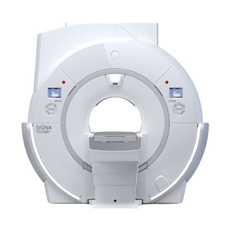
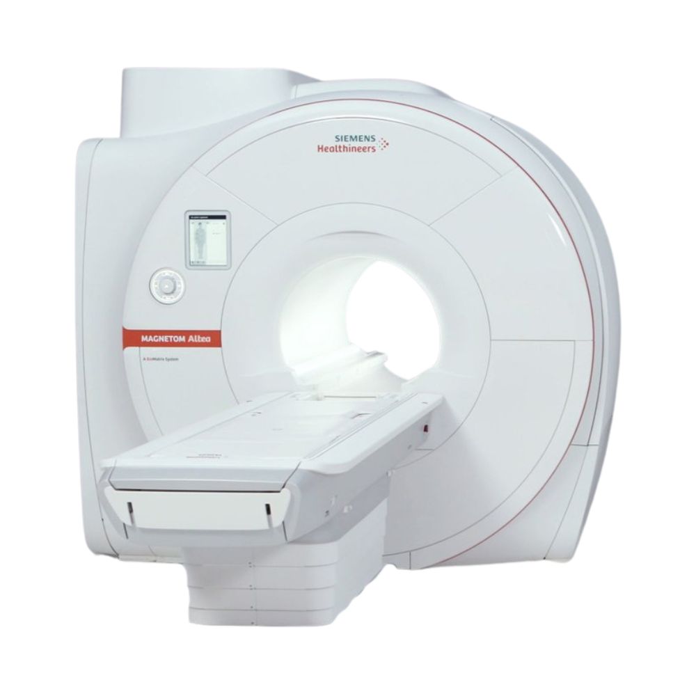
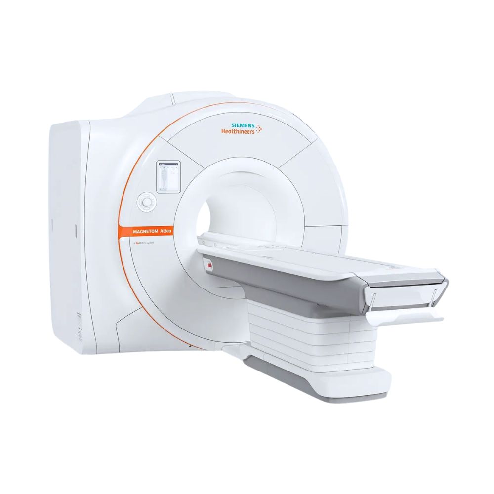
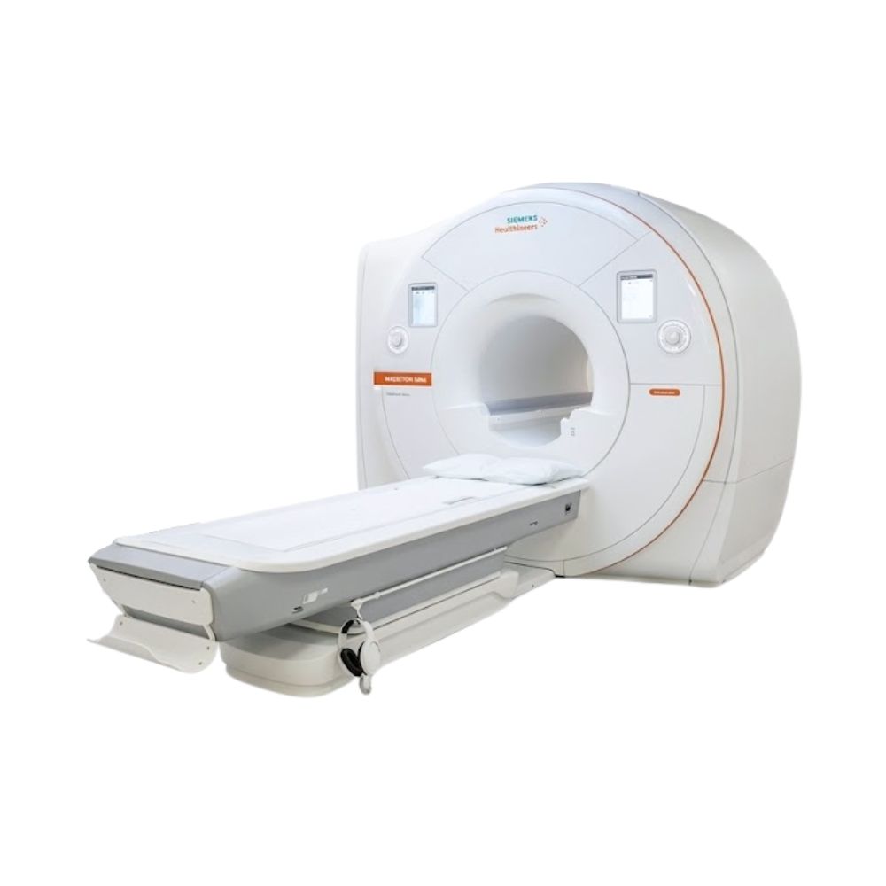
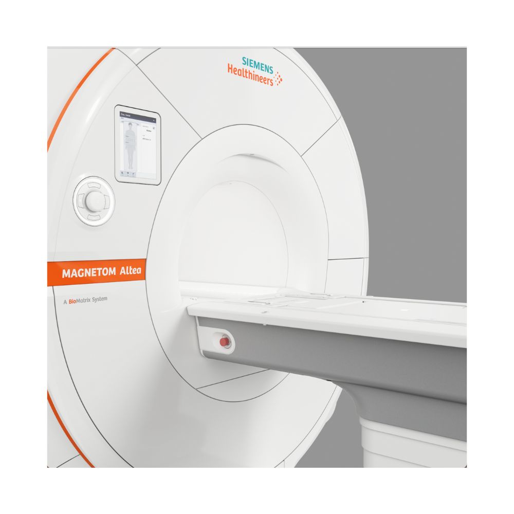
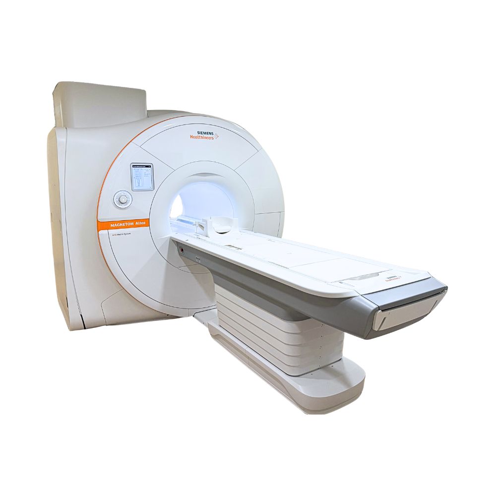
Siemens MAGNETOM Altea 1.5T MRI
- Category:
- MRI Systems
- Brand:
- Siemens
The Siemens MAGNETOM Altea is a 1.5T MRI scanner well suited to facilities seeking wide-bore patient access and advanced imaging. Its 70 cm opening, 32-channel receiver system, patient-adaptive technology, and guided scanning workflows support consistent routine and specialty examinations across a broad range of clinical needs.
Siemens MAGNETOM Altea MRI Highlights & Benefits
The Siemens MAGNETOM Altea combines wide-bore access with Siemens BioMatrix technology, advanced imaging coils, and automated workflows. Its 1.5T platform supports routine and specialty MRI while helping facilities improve patient comfort, examination consistency, and efficiency.
Key Features & Benefits
-
- 70 cm Wide Bore: Provides a larger patient opening within a compact 157 cm (61.8 in) scanner for greater positioning flexibility.
- 32-Channel Imaging System: Siemens Tim 4G technology supports up to 180 connected coil elements and 32 independent receiver channels for detailed imaging and accelerated examinations.
- BioMatrix Patient-Adaptive Technology: Compatible sensors, imaging coils, and automated positioning tools adapt scanning to patient anatomy and movement for more consistent examinations.
- Guided MRI Workflows: Siemens Dot Engines and compatible myExam Companion software simplify examination setup, protocol selection, and scan planning.
- Faster Scanning & Advanced Reconstruction: Turbo Suite acceleration and compatible Deep Resolve technologies support shorter examinations and improved image clarity.
- Patient-Friendly Table Options: Supports patients weighing up to 550 lb (250 kg), with an optional detachable patient table that lowers to approximately 20.5 in (52 cm).
- Energy-Efficient Operation: Zero helium boil-off refrigeration and Eco-Power technology help control helium consumption and electricity use.
MAGNETOM Altea 1.5T Imaging Performance
The Altea combines a superconducting 1.5T magnet, Siemens XJ gradients, and a 32-channel Tim 4G receiving system to support detailed anatomical imaging, specialized examinations, and faster scanning with compatible software.
1.5T Magnet with 50 cm Whole-Body Imaging Coverage
The Altea uses a 1.5 Tesla superconducting magnet designed to maintain consistent magnetic-field conditions during routine and advanced examinations.
Its maximum imaging field of view measures approximately 50 × 50 × 50 cm (19.7 × 19.7 × 19.7 in), providing anatomical coverage for brain, spine, joint, abdominal, pelvic, and other supported examinations.
Passive and active shimming adjust small variations in the magnetic field that could otherwise reduce image quality.
Compatible BioMatrix technologies provide additional adjustments for specific anatomical regions, improving image consistency where magnetic-field variations are challenging.
XJ Gradients for Detailed Anatomical Imaging
The Altea uses Siemens XJ gradients to identify the location of signals collected during scanning.
The gradient system provides:
-
- Maximum Gradient Amplitude: 33 mT/m per axis, supporting detailed anatomical imaging and compatible advanced acquisitions.
- Maximum Slew Rate: 125 T/m/s per axis, allowing rapid changes in the magnetic fields used to collect imaging information.
Gradient amplitude describes the strength of the additional magnetic fields used to locate anatomical information. Slew rate measures how quickly those fields can change.
These capabilities support routine imaging, diffusion-weighted MRI that evaluates water movement within tissue, and compatible advanced scanning techniques.
The system also has a combined three-axis gradient amplitude of 57 mT/m and a combined slew rate of 216 T/m/s. These represent the combined performance of all three gradient axes, not an individual axis.
Tim 4G Technology with 32 Independent Receiver Channels
The Siemens MAGNETOM Altea uses Siemens Tim 4G technology to collect radiofrequency (RF) signals from imaging coils positioned near the patient. These signals provide the information needed to reconstruct MRI images.
Its Tim [180 × 32] architecture includes:
-
- Up to 180 Connected Coil Elements: Supports extensive anatomical coverage through compatible coil combinations, reducing coil changes between certain imaging regions.
- 32 Independent Receiver Channels: Processes 32 independent coil signals simultaneously during an acquisition, supporting detailed imaging and compatible scan acceleration.
- DirectRF Signal Processing: Processes MRI signals close to the magnet to preserve signal quality and reduce interference before reconstruction.
The Altea can connect up to 180 coil elements, but 32 independent receiver channels operate simultaneously during a single acquisition.
This combination supports detailed imaging, flexible coil arrangements, and extended examinations without repeatedly repositioning the patient.
For facilities evaluating a 32-channel MRI scanner, coil compatibility and installed software also determine which examinations and acceleration techniques are supported.
Altea Patient Access & Exam Comfort
The Siemens MAGNETOM Altea combines a 70 cm wide bore with a short magnet, flexible imaging coils, and patient-positioning options to accommodate different patient sizes and mobility needs.
70 cm Wide Bore in a Compact 157 cm System
The Altea features a 70 cm (27.6 in) patient opening and an overall system length of approximately 157 cm (61.8 in).
The wider opening provides more space than conventional 60 cm MRI scanners, helping accommodate larger patients, those with limited mobility, and individuals uncomfortable in enclosed spaces.
Its short magnet also limits the length of the scanner surrounding the patient.
This combination makes the Altea suitable for facilities seeking wider patient access without requiring a substantially longer MRI platform.
550 lb Patient Capacity with Dockable Table Options
The Altea supports patients weighing up to 550 lb (250 kg), depending on the installed table configuration.
Available positioning features include:
-
- Powered Patient Positioning: Moves the patient into the scanner during examination setup, reducing manual table movement.
- BioMatrix Select&GO: Uses a patient-side touchscreen and automated positioning to place the selected anatomy near the center of the scanner.
- Optional BioMatrix Dockable Table: Separates from the scanner for patient preparation and transport, reducing transfers for patients requiring assistance.
- Low Table Entry: The dockable table lowers to approximately 52 cm (20.5 in), making it easier for patients with limited mobility to get onto the scanning surface.
- AutoDock: Guides a compatible detachable table into position when reconnecting it to the scanner.
A detachable table is particularly useful for facilities imaging patients who use wheelchairs, require assisted transfers, or have difficulty walking.
Table selection also affects installation requirements. Mobile-table configurations require additional room length and clearance for docking, patient movement, and equipment access.
Lightweight Coils & Quieter MRI Examinations
Siemens Tim 4G and BioMatrix coils use lightweight, flexible designs that adapt to different anatomical regions, reducing the need for heavy or restrictive equipment.
Siemens Quiet Suite uses specialized scanning techniques to reduce MRI noise during compatible brain, spine, and large-joint examinations. Quieter scanning can improve comfort for patients sensitive to loud sounds.
Additional patient-experience options may include ambient lighting and compatible audiovisual comfort features.
Available comfort technologies, coils, and patient-table equipment depend on the selected scanner configuration.
Clinical Environments & Applications
The Siemens MAGNETOM Altea supports routine brain, spine, and joint MRI, with advanced capabilities for body, cardiac, vascular, and whole-body examinations. Its wide bore, 32-channel receiving system, and BioMatrix technology support a broad range of diagnostic imaging services.
Clinical Environments
-
- Hospitals and radiology departments
- Outpatient diagnostic imaging centers
- Orthopedic and sports medicine imaging centers
- Neurology and neuroscience imaging departments
- Multispecialty diagnostic imaging facilities
- Cardiac and vascular imaging centers
- Oncology imaging departments
- Pediatric imaging facilities
- Veterinary teaching hospitals and specialty imaging centers
Clinical Specialties & Applications
-
- Neurology & Neurosurgery: Brain and spine MRI, with guided examination planning and BioMatrix CoilShim technology to improve image consistency in challenging head and neck regions.
- Orthopedics & Sports Medicine: Joint, ligament, tendon, cartilage, and soft-tissue imaging using dedicated orthopedic coils, with compatible techniques for reducing distortion around certain metal implants.
- Abdominal Imaging & Hepatology: Abdominal and liver MRI, with BioMatrix Respiratory Sensors to detect breathing patterns and optional software for patients who have difficulty holding their breath.
- Urology: Detailed prostate and pelvic imaging using compatible body and spine coils, including diffusion imaging to evaluate tissue characteristics.
- Cardiology: Heart MRI to evaluate anatomy, pumping function, and tissue characteristics. The optional BioMatrix Beat Sensor supports cardiac scanning without conventional ECG lead placement.
- Vascular Medicine: Artery and vein imaging using compatible contrast-enhanced and non-contrast techniques, with guided scanning workflows.
- Breast Radiology: Diagnostic breast MRI and compatible MRI-guided procedures using dedicated breast coils and guided protocols.
- Oncology: Tumor assessment and follow-up imaging, with the Whole-Body Dot Engine supporting guided examinations for investigating bone and lymph node metastases.
- Pediatric Radiology: Brain, spine, joint, and body examinations using appropriately selected coils and patient-specific protocols.
- Veterinary Medicine: Neurological, musculoskeletal, and other clinically appropriate animal MRI examinations using suitably configured Altea equipment.
Specialized examinations require compatible coils, software, and clinical protocols. Veterinary use is established in practice, but the standard Altea is not a dedicated veterinary-edition scanner. Pediatric and veterinary examinations require appropriate patient-specific safety and equipment assessments.
Altea BioMatrix Technology for Consistent MRI Exams
Siemens BioMatrix technology helps the Altea accommodate differences in patient anatomy, breathing, and positioning.
It combines motion-detection sensors, magnetic-field adjustments, and patient-positioning interfaces. Individual capabilities depend on the installed sensors, coils, and options.
BioMatrix Sensors for Breathing & Heart Motion
Breathing and heart movement can interfere with MRI images, particularly during abdominal and cardiac examinations.
Compatible BioMatrix sensors detect these movements to help coordinate scanning.
-
- BioMatrix Respiratory Sensors: Integrated into compatible spine coils, including BioMatrix Spine 24. They detect breathing patterns without a separate breathing belt, simplifying respiratory-triggered abdominal examinations.
- Optional BioMatrix Beat Sensor: Integrated into compatible BioMatrix Body 12 or Body 18 coil configurations, this sensor detects heart movement to coordinate cardiac image acquisition. It can eliminate conventional ECG electrodes during supported examinations, reducing preparation time.
Respiratory triggering coordinates scanning with breathing, while cardiac triggering synchronizes image acquisition with heart motion.
The BioMatrix Beat Sensor requires a compatible coil, software, and the appropriate installed option.
CoilShim & SliceAdjust for Improved Image Consistency
Patient anatomy can cause magnetic-field variations that affect image clarity.
The Altea supports BioMatrix technologies designed to correct these variations during compatible examinations.
-
- CoilShim: Uses magnetic-field adjustment elements integrated into the compatible BioMatrix Head/Neck 20 coil. These improve image consistency and reduce uneven tissue appearance during specialized head, neck, and diffusion examinations.
- SliceAdjust: Automatically adjusts magnetic-field conditions for different anatomical regions during supported examinations, reducing uneven image quality and visible inconsistencies between sections of a whole-body scan.
These technologies are particularly useful for challenging head and neck, spine, abdominal, and whole-body examinations.
They help maintain image consistency without requiring technologists to perform every adjustment manually.
Select&GO for Simplified Patient Positioning
BioMatrix Select&GO uses a patient-side touchscreen to select the anatomy being examined.
Information from connected coils and Siemens’ computerized positioning model helps identify the selected anatomical region and move it into the appropriate scanning position.
This reduces manual positioning steps during routine examinations.
Compatible interfaces can also display breathing patterns detected by installed sensors, helping technologists monitor patients during preparation.
Altea Guided Scanning & Workflow Automation
The Altea supports guided scanning technologies that simplify examination preparation, protocol selection, and standardized imaging across operators.
Dot Engines & myExam Companion for Guided Scanning
Siemens Dot Engines provide guided workflows for different anatomical regions.
Compatible workflows include:
-
- Brain & Spine: Guide positioning and protocols for consistent neurological and spinal imaging.
- Large Joints: Standardize planning for knees and other supported joints, reducing repetitive setup.
- Abdomen: Organize abdominal scans and account for breathing-related movement.
- Cardiac & Vascular: Support heart and blood vessel examinations, including compatible contrast-timing workflows.
- Breast: Guide breast examination setup and maintain consistent imaging protocols.
- Whole-Body Imaging: Coordinate examinations across multiple anatomical regions, including selected oncology applications.
Depending on the installed software generation, Altea systems use Siemens Dot Engines or newer myExam Companion workflows.
myExam Companion helps technologists select scanning strategies and adjust protocols without planning every step manually.
Available workflows and assistance features depend on the installed software and licenses.
Automated Brain & Knee Exam Planning
Compatible Altea software reduces repetitive setup through Siemens AutoAlign and myExam automation.
-
- AutoAlign: Identifies anatomical landmarks and automatically positions imaging slices for supported examinations.
- myExam Assist: Guides examinations using predefined scanning strategies that can be adjusted for individual patients.
- myExam Brain Autopilot: Automates supported brain examination steps, including scan planning, image positioning, and acquisition.
- myExam Knee Autopilot: Automates supported knee examination preparation and scanning to maintain consistent positioning and protocols.
Brain and Knee Autopilot are standard features of the compatible syngo MR XA51A software release, although individual scanners may run earlier versions.
myExam Implant Suite for Patients with Compatible Implants
The Altea supports optional myExam Implant Suite software to assist technologists in planning examinations around scanning restrictions for MR-conditional implants.
An MR-conditional implant may be scanned only under specific conditions established by its manufacturer.
The software helps organize supported implant-related scanning parameters and workflows. It does not replace implant identification, safety screening, or compliance with the manufacturer’s MRI conditions.
Altea Turbo Suite for Faster MRI Examinations
Siemens Turbo Suite uses accelerated scanning techniques to shorten compatible MRI examinations. The original Altea platform includes Turbo Suite Essential, while optional Excelerate expands the available acceleration capabilities.
Turbo Suite Essential vs. Excelerate
Turbo Suite Essential
Turbo Suite Essential is the Altea’s original standard acceleration package. It uses two core technologies to shorten routine brain, joint, and body examinations:
-
-
- GRAPPA: Uses signals from multiple imaging coils to reconstruct images from fewer measurements, reducing scanning time while preserving anatomical detail.
- CAIPIRINHA: Uses coordinated scanning patterns and coil information to accelerate three-dimensional imaging, including selected abdominal and orthopedic examinations.
-
Siemens demonstrates selected routine examinations completed in approximately 10–15 minutes using supported protocols.
Turbo Suite Excelerate
Optional Turbo Suite Excelerate expands acceleration capabilities with technologies designed to collect imaging information more efficiently:
-
-
- Simultaneous Multi-Slice (SMS): Collects signals from multiple imaging slices during a single acquisition, shortening supported two-dimensional and diffusion examinations.
- Compressed Sensing: Collects fewer measurements and uses advanced mathematical reconstruction to produce images more efficiently during supported examinations.
-
These technologies can reduce acquisition times by up to 50% for selected neurological, orthopedic, and body MRI protocols. Actual savings depend on the examination and installed options.
Optional Compressed Sensing GRASP-VIBE
Compressed Sensing GRASP-VIBE supports dynamic abdominal and liver MRI during free breathing, helping examine patients who cannot reliably hold their breath or follow breathing instructions.
It also supports automatic identification of relevant imaging phases during compatible contrast-enhanced liver examinations, reducing manual processing.
Turbo Suite Essential was part of the original standard Altea offering. Excelerate and GRASP-VIBE provide additional capabilities that depend on installed software, licenses, and compatible hardware.
MXR Imaging helps customers identify new or pre-owned configurations that support their clinical scanning requirements.
Altea Deep Resolve for Clearer MRI Images
Siemens Deep Resolve combines advanced noise reduction with artificial intelligence (AI) to improve MRI image clarity and support shorter examinations. Each technology addresses a different aspect of image reconstruction.
- Deep Resolve Gain: Reduces uneven image noise using information collected during scanning, improving clarity without additional measurements. Gain does not rely on neural-network AI.
- Deep Resolve Sharp: Uses AI trained on lower- and higher-resolution MRI images to reconstruct sharper anatomical detail, improving the visibility of smaller structures.
- Deep Resolve Boost: Uses AI to reconstruct images directly from original scanner measurements, reducing noise and supporting faster compatible acquisitions.
Gain focuses on noise reduction, Sharp improves visible detail, and Boost helps balance scan speed with image quality.
Deep Resolve Gain and Sharp are supported on compatible Altea configurations beginning with appropriate syngo MR XA31 software. Boost requires syngo MR XA50 or a later supported release and additional reconstruction hardware.
A compatible software release does not automatically mean a feature is installed or licensed. MXR Imaging can help identify a configuration with the image-quality and scan-speed capabilities suited to the facility’s examinations.
Altea Imaging Techniques for Challenging Examinations
The Altea supports specialized imaging methods for examinations involving compatible metal implants or extensive anatomical coverage.
Imaging Near Metal Implants
Orthopedic implants can distort the magnetic field and obscure nearby anatomy.
Compatible metal-artifact-reduction techniques, including Advanced WARP and SEMAC-based imaging, help improve visibility around certain MR-conditional implants.
Advanced WARP uses specialized scanning and correction methods to reduce distortion. SEMAC collects additional information to correct anatomy displaced between imaging slices.
These techniques support postoperative joint and musculoskeletal examinations. Each scan must still follow the implant’s MRI safety conditions.
Whole-Body Imaging with Extended Coil Coverage
The Altea’s Tim 4G system supports a maximum scanning range of approximately 205 cm (80.7 in) with compatible coil combinations, enabling broad anatomical coverage without repeatedly repositioning the patient.
The optional Whole-Body Dot Engine guides multiregion examinations, including supported oncology protocols used to investigate bone and lymph node metastases.
BioMatrix SliceAdjust can improve image consistency across anatomical regions during these examinations.
Altea Image Processing & Hospital Connectivity
The Altea uses Siemens syngo MR software for image reconstruction, examination management, and clinical system connectivity.
Compatible capabilities include:
-
- Recon&GO: Automates selected image-reconstruction and post-processing tasks after scanning, reducing repetitive workstation activity.
- View&GO: Provides image review and processing tools at the MRI workstation, helping technologists prepare examination results for clinical interpretation.
- DICOM Connectivity: Supports Digital Imaging and Communications in Medicine (DICOM), the standard format used to transfer medical images to compatible hospital and radiology systems.
- Clinical Worklist Integration: Exchanges patient and scheduled examination information with compatible systems, reducing manual data entry and helping prevent identification errors.
These capabilities support integration with a picture archiving and communication system (PACS), which stores medical images for clinical review.
Compatible facilities may also use syngo Virtual Cockpit, a remote assistance platform that allows qualified MRI personnel to support examinations from another location.
Remote assistance requires appropriate software, connectivity, equipment, and operational arrangements.
Altea System Configurations
The MAGNETOM Altea uses a common 1.5T magnet, XJ gradient system, and Tim [180 × 32] RF architecture.
Individual systems primarily differ in patient tables, BioMatrix features, imaging coils, software, and installed clinical options.
Patient Table & BioMatrix Configurations
Available configurations include:
-
- Fixed Patient Table: Provides powered positioning with a permanent table arrangement.
- BioMatrix Dockable Table: Allows patient preparation and transport away from the scanner, supporting assisted transfers.
- AutoDock: Simplifies reconnection and alignment of a compatible detachable table.
- BioMatrix Respiratory Sensors: Detect breathing through compatible spine coils for respiratory-triggered examinations.
- BioMatrix Beat Sensor: Provides optional cardiac triggering without conventional ECG electrodes when a compatible Body 12 or Body 18 coil and required software are installed.
- BioMatrix CoilShim: Improves magnetic-field conditions around the head and neck through the compatible BioMatrix Head/Neck 20 coil.
- BioMatrix Select&GO: Provides guided positioning through a patient-side control interface.
The appropriate configuration depends on patient needs, planned examinations, workflow, and installation space.
Software, Coils & Equipment Packages
Individual Altea systems may also differ in:
-
- Software Generation: Determines compatibility with guided workflows, AI reconstruction, and newer imaging technologies.
- Clinical Applications: Specialized neurological, cardiac, orthopedic, oncology, and other applications may require additional licenses.
- Imaging Coils: Determine which examinations the system can perform with its installed hardware.
- Reconstruction Hardware: Certain advanced processing applications require compatible computing components.
- Accessories: Patient comfort equipment, positioning aids, and additional workstations vary by system.
MXR Imaging helps customers select equipment suited to their clinical requirements rather than assuming every Altea has identical features.
Tim 4G & BioMatrix Coils for Specialized MRI
The MAGNETOM Altea supports Siemens Tim 4G and BioMatrix coils for brain, spine, body, joint, breast, and other specialized imaging.
Available coil combinations depend on the selected system and compatible hardware and software.
The supported coil portfolio includes:
- BioMatrix Head/Neck 20: Supports brain, cervical spine, and neck imaging. Integrated CoilShim improves image consistency in challenging anatomical regions. The coil tilts from 0° to 18° to accommodate patients who cannot comfortably keep their head flat.
- BioMatrix Spine 24: Provides spine coverage and can incorporate respiratory sensors for breathing-coordinated abdominal imaging.
- BioMatrix Body 12 & Body 18: Support abdominal, pelvic, and other body examinations. Compatible configurations support the optional BioMatrix Beat Sensor for cardiac triggering without conventional ECG electrodes.
- UltraFlex Small 18 & Large 18: Lightweight, breathable coils with a flexible, blanket-like design that conforms to different anatomical regions. They accommodate different patient sizes and support joint and soft-tissue imaging without rigid coil positioning.
- Shoulder Shape 16: Uses flexible wings to fit different shoulder sizes for detailed tendon, cartilage, and shoulder imaging.
- Hand/Wrist 16: Provides dedicated hand and wrist coverage with positioning features that stabilize the anatomy.
- Dedicated Knee & Foot/Ankle Coils: Support detailed musculoskeletal examinations with specialized positioning and anatomical coverage.
- Breast 18 & Compatible Breast Biopsy Coils: Support diagnostic breast MRI and appropriately equipped MRI-guided procedures.
These are compatible coil options, not a list of equipment included with every Altea.
The 32-channel system and compatible high-density coils support detailed imaging across large anatomical regions.
Siemens DirectConnect and SlideConnect simplify coil connections, reducing setup steps during patient preparation.
MXR Imaging can help select the appropriate coil package. MRI coil repair is also available for supported equipment.
Altea Software for Routine & Advanced MRI
The Altea supports Siemens clinical applications through the syngo MR software platform. Installed software, licenses, and compatible hardware determine the available capabilities.
Routine MRI & Guided Scanning
Dot Engines and myExam Companion guide brain, spine, joint, and other examinations. Compatible newer systems can provide Brain and Knee Autopilot for automated scanning.
Orthopedic & Neurological Imaging
Compatible software supports advanced brain and spine examinations, diffusion imaging, specialized joint protocols, and metal-artifact reduction.
Body, Cardiac & Specialty Imaging
Clinical applications support abdominal and liver MRI, prostate examinations, cardiac imaging, vascular studies, breast imaging, and compatible whole-body oncology applications. Optional Compressed Sensing GRASP-VIBE supports specialized free-breathing abdominal imaging.
Scan Acceleration & Image Enhancement
Turbo Suite Essential provides core acceleration, while optional Excelerate expands scan-speed capabilities. Compatible Deep Resolve applications add noise reduction and AI-based reconstruction.
Earlier Altea systems may use previous syngo MR XA releases, while newer or appropriately updated systems can support additional applications.
Software and technology availability varies between new and pre-owned Altea scanners. MXR Imaging helps match clinical requirements with the appropriate installed software, licenses, imaging coils, and equipment capabilities.
Altea Magnet Cooling & Site Requirements
The Altea combines a short superconducting magnet with energy-management technology and a compact footprint.
Installation requires dedicated electrical service, cooling, RF shielding, emergency venting, and magnetic-field safety measures.
Zero Helium Boil-Off & Eco-Power Technology
The Altea uses liquid helium to maintain the low temperature required by its superconducting magnet.
Zero helium boil-off refrigeration limits helium evaporation during normal operation, reducing routine helium replenishment.
Eco-Power manages electricity consumption during supported operating and standby conditions.
These technologies help control operating costs, although the magnet still requires refrigeration maintenance, monitoring, and emergency helium venting.
MXR Imaging provides MRI refrigeration and helium services for compatible systems.
Compact MRI Installation with RF Shielding
The Altea’s 157 cm (61.8 in) system length supports compact installation layouts for a 70 cm wide-bore MRI scanner.
Its representative minimum total installation-space requirement is approximately 28 m² (302 sq ft), including magnet, control, and equipment areas. This is not the examination-room area alone.
Installation planning must address:
-
- Equipment Delivery: Access and clearances to transport the magnet and supporting equipment into the facility.
- Structural Support: Floors capable of supporting the MRI system, shielding, and concentrated equipment loads.
- RF Shielding: Specialized room shielding that prevents outside radiofrequency interference from reducing image quality.
- Magnetic Field Boundaries: Controlled areas around the magnet and restrictions on incompatible equipment.
- Quench Venting: An emergency route to direct released helium outside.
- Service Access: Clearances for inspection, maintenance, and repair.
Siemens’ representative minimum examination-room lengths differ by table configuration:
-
- Fixed Patient Table: Approximately 617 cm (242.9 in).
- Mobile Patient Table: Approximately 647 cm (254.7 in), with a recommended length of 667 cm (262.6 in).
Mobile-table configurations require additional space for movement, docking, patient preparation, and equipment access.
These are representative planning values. Final dimensions depend on the selected equipment and approved site plan.
MXR Imaging provides custom MRI shielding solutions and turnkey imaging project management for installations, facility upgrades, and replacement projects.
Electrical Power & Equipment Cooling
The Altea requires dedicated three-phase electrical service and equipment cooling.
Representative planning specifications include:
-
- Electrical Service: 400 V ±10%, three-phase, 50/60 Hz.
- System Connection Value: Approximately 69 kVA for the specified XJ-gradient configuration.
- Optional Chiller Connection: Approximately 48 kVA with the applicable dedicated chiller arrangement.
- Cooling: Facility cooling-water infrastructure or a compatible dedicated chiller.
- Examination Room Minimum Height: 240 cm (94.5 in).
- Control Room Minimum Height: 210 cm (82.7 in).
- Equipment Room Minimum Height: 240 cm (94.5 in).
Kilovolt-amperes (kVA) describe electrical power capacity. MRI system and optional chiller values refer to separate connections, not a universal total facility power requirement.
Final electrical, cooling, structural, and room specifications depend on the equipment configuration and approved site plan.
Available MAGNETOM Altea System Configuration
MXR Imaging helps facilities select a Siemens MAGNETOM Altea configuration suited to their clinical services, patient population, installation requirements, and budget.
New and pre-owned systems can differ in patient tables, BioMatrix sensors, coils, software generations, clinical applications, reconstruction capabilities, and accessories.
MXR Imaging helps identify the equipment needed for routine diagnostic imaging or specialized orthopedic, neurological, cardiac, oncology, and other examinations.
Our team also provides MRI maintenance and repair, equipment installation, and lifecycle support.
MAGNETOM Altea MRI Price & Availability
Siemens MAGNETOM Altea 1.5T MRI scanner pricing depends on equipment condition, software generation, patient-table configuration, imaging coils, installed options, and project requirements.
MXR Imaging helps customers evaluate new and pre-owned MRI equipment, including available Altea systems and Platinum Certified equipment where applicable, subject to availability.
Our team coordinates equipment evaluation, configuration selection, turnkey imaging project management, and ongoing MRI service support.
Contact MXR Imaging to request Siemens MAGNETOM Altea pricing and discuss a configuration suited to your facility.
Siemens MAGNETOM Altea 1.5T Specifications
Magnet System
-
- Field Strength: 1.5T
- Magnet Type: Superconducting, actively shielded
- Bore Diameter: 70 cm (27.6 in)
- Maximum Imaging Field of View: Approximately 50 × 50 × 50 cm (19.7 × 19.7 × 19.7 in)
- Field Adjustment: Passive and active shimming
- Additional Field Optimization: Compatible BioMatrix CoilShim and SliceAdjust
- Cooling Technology: Zero helium boil-off under normal operating conditions
Gradient System
-
- Gradient Technology: Siemens XJ
- Maximum Gradient Amplitude: 33 mT/m per axis
- Maximum Slew Rate: 125 T/m/s per axis
- Combined Three-Axis Gradient Amplitude: 57 mT/m
- Combined Three-Axis Slew Rate: 216 T/m/s
RF System
-
- RF Technology: Siemens Tim 4G with DirectRF
- RF Architecture: Tim [180 × 32]
- Maximum Connected Coil Elements: 180
- Independent Receiver Channels: 32
- Coil Connections: Compatible DirectConnect and SlideConnect
- Maximum Continuous Scanning Range: Up to approximately 205 cm (80.7 in) with appropriate coils and configuration
Bore & Patient Table
-
- Bore Diameter: 70 cm (27.6 in)
- Maximum Patient Weight: Up to 250 kg (550 lb), depending on table configuration
- Patient Table: Fixed and optional dockable arrangements
- Dockable Table Minimum Height: Approximately 52 cm (20.5 in)
- Available Positioning Tools: BioMatrix Select&GO and compatible AutoDock
- Patient Comfort: Lightweight coils, compatible Quiet Suite scanning techniques, and optional patient-experience features
Coils & Accessories
-
- Coil Technology: Tim 4G and BioMatrix
- Head/Neck Coils: Compatible Head/Neck 16 and BioMatrix Head/Neck 20
- Spine Coils: BioMatrix Spine 24 and compatible system-specific options
- Body Coils: Compatible BioMatrix Body 12 and Body 18
- Flexible Coils: UltraFlex Small 18 and Large 18
- Orthopedic Coils: Compatible shoulder, knee, hand/wrist, and foot/ankle coils
- Specialty Coils: Compatible diagnostic breast and biopsy coils
- Installed Accessories: Determined by the selected equipment package
Siting & Utilities
-
- Representative Minimum Total Installation Space: 28 m² (302 sq ft)
- Minimum Examination Room Length, Fixed Table: Approximately 617 cm (242.9 in)
- Minimum Examination Room Length, Mobile Table: Approximately 647 cm (254.7 in)
- Recommended Mobile-Table Room Length: Approximately 667 cm (262.6 in)
- Minimum Examination Room Height: 240 cm (94.5 in)
- Minimum Control Room Height: 210 cm (82.7 in)
- Minimum Equipment Room Height: 240 cm (94.5 in)
- Representative Electrical Service: 400 V ±10%, three-phase, 50/60 Hz
- Representative XJ System Connection Value: 69 kVA
- Optional Chiller Connection Value: 48 kVA for the specified arrangement
- Equipment Cooling: Cooling-water supply or compatible dedicated chiller
- RF Shielding: Required
- Quench Venting: Required
- Final Site Requirements: Determined by the selected configuration and approved planning specifications
System Dimensions & Weight
-
- Overall System Length: 157 cm (61.8 in), cover to cover
- Bore Diameter: 70 cm (27.6 in)
- Representative Operating System Weight: Approximately 4.2 metric tons (9,260 lb)
- Representative Magnet Installation Weight: Approximately 3,982 kg (8,779 lb)
- Representative Magnet Transport Weight: Approximately 4,200 kg (9,259 lb)
- Patient Table Dimensions: Depend on fixed or mobile configuration
- Installation Layout: Depends on patient table, equipment cabinets, shielding, and service access