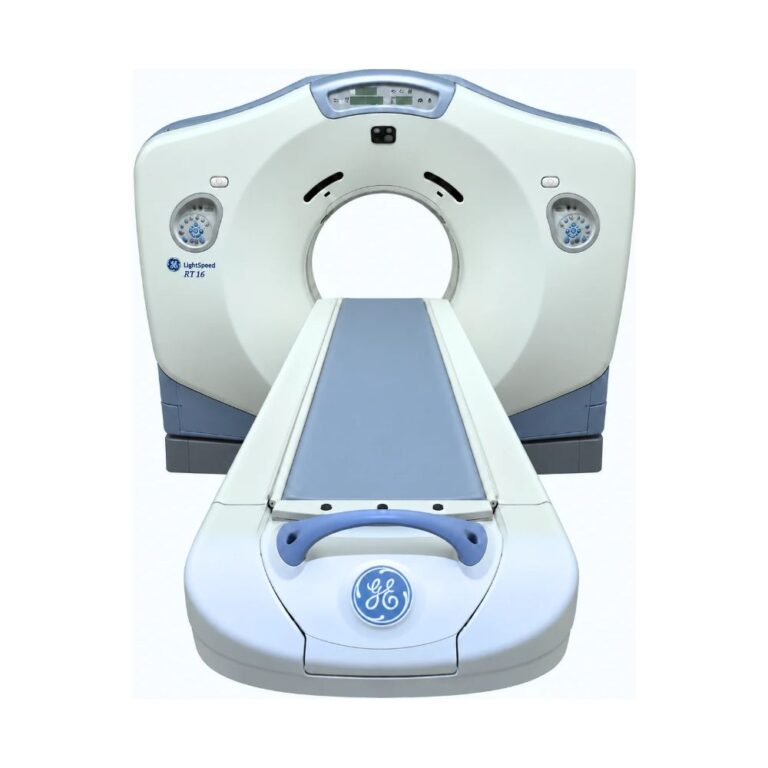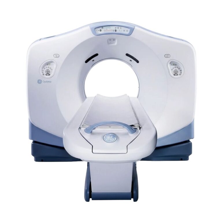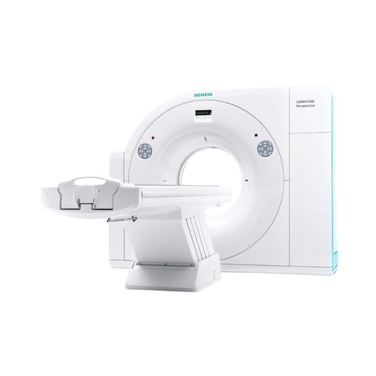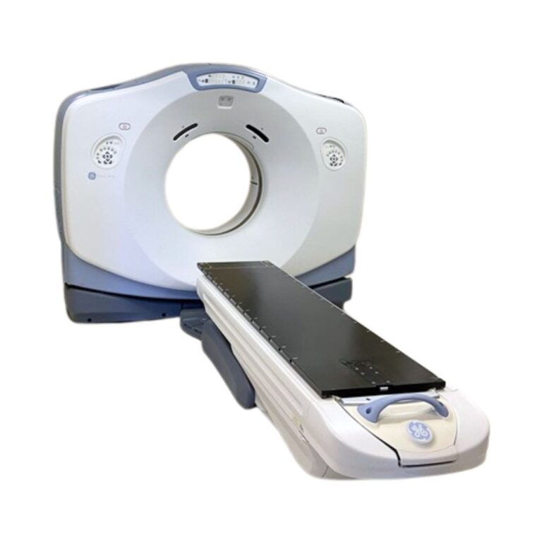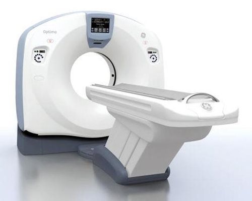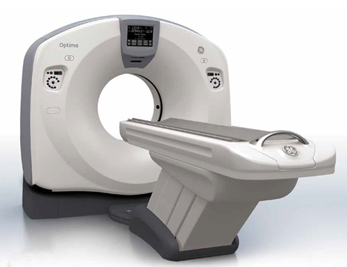
GE Optima CT580 W 16-Slice CT
- Category:
- 16-Slice CT
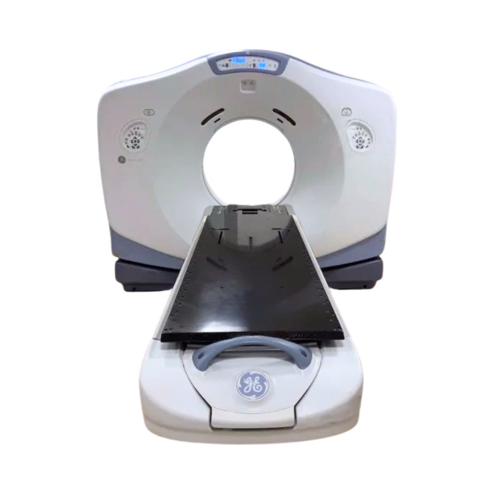
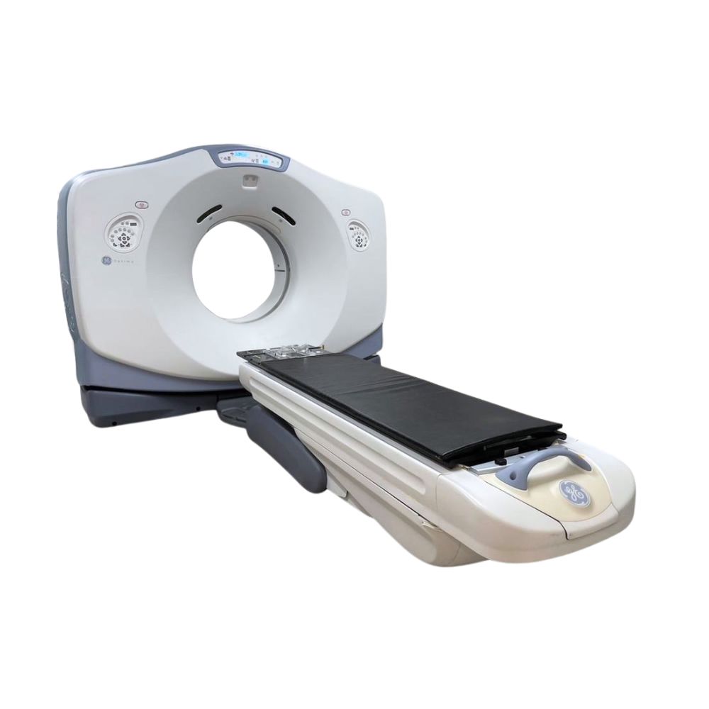
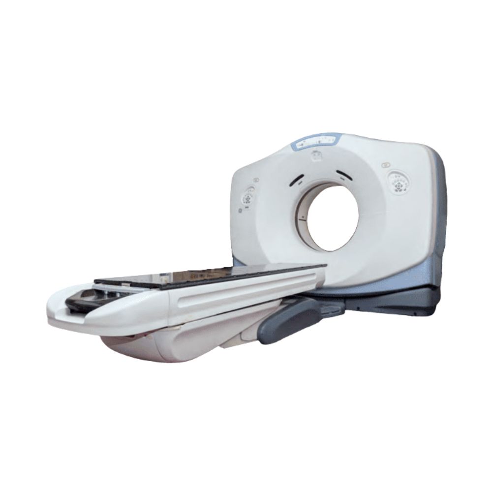
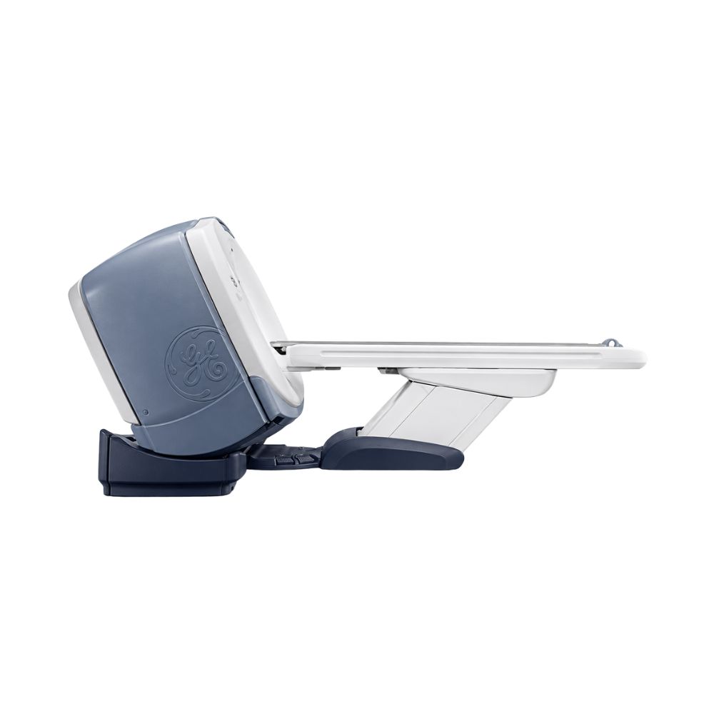
The GE Optima CT580 W 16-slice CT scanner is a wide-bore CT scanner best suited for hospitals, diagnostic imaging centers, and interventional facilities. Its 80 cm bore, 100 kW generator, and optional 650 lb patient table accommodate routine examinations, larger patients, and complex image-guided procedures.
Optima CT580 W Highlights & Benefits
The GE Optima CT580 W 16-slice CT Scanner combines thin-slice diagnostic imaging, wide-bore patient access, and high X-ray power to accommodate routine and complex examinations. Its spacious gantry also provides additional working room for image-guided procedures, allowing facilities to support diagnostic, bariatric, trauma, and interventional imaging within a single CT suite.
Key Features
-
- 80 cm wide-bore gantry: Accommodates larger patients while providing extra room for positioning accessories and interventional instruments.
- 16-slice acquisition: Captures sixteen slices per rotation across up to 20 mm of anatomy.
- microVoxel imaging: Supports acquisition of slices as thin as 0.625 mm for detailed visualization of small anatomical structures.
- 65 cm WideView: Extends anatomical visualization beyond the standard 50 cm scan field, particularly for larger patients.
- 100 kW generator: Provides additional X-ray power for demanding examinations of larger patients.
- 0.5-second rotation: Helps shorten breath holds and reduce patient motion during scanning.
- Up to 650 lb patient capacity: Expands bariatric imaging capabilities with the optional high-capacity table.
- Optional CT fluoroscopy: Provides near-real-time images to guide needle placement during biopsies and other minimally invasive procedures.
Thin-Slice Diagnostic Imaging
HiLight Matrix II Detector for Detailed Imaging
The 24-row HiLight Matrix II detector offers two primary acquisition settings, allowing technologists to balance anatomical detail with scan coverage:
-
- 16 × 1.25 mm: Captures sixteen slices across 20 mm of anatomy per rotation for routine diagnostic examinations.
- 16 × 0.625 mm: Captures thinner slices across 10 mm of anatomy per rotation for more detailed imaging.
These thinner slices work with microVoxel technology to preserve anatomical detail when images are reconstructed from different viewing angles or into three-dimensional images. This improves visualization of small lesions, blood vessels, and bone structures.
Flexible Scan Speeds
VariSpeed allows technologists to adjust gantry rotation times in 0.1-second increments between 0.5 and 1.0 seconds, helping customize scan times and shorten patient breath holds.
Complementing this capability, IQ Enhance (IQE) allows the patient table to move faster during continuous scanning while helping preserve image quality. Together, these technologies support shorter scan times and reduced patient motion, particularly during trauma imaging or examinations of patients who have difficulty remaining still.
X-Ray Power for Demanding Examinations
The 100 kW generator and Performix Pro VCT 100 X-ray tube deliver the power needed to penetrate thicker anatomy and maintain image quality when scanning larger patients. The tube’s 8 MHU heat storage capacity also supports consecutive examinations with fewer interruptions for cooling, helping facilities maintain patient throughput.
Wide-Bore Access for Complex Examinations
Accommodating Larger Patients and Complex Positioning
The 80 cm bore provides a wider opening than conventional 70 cm CT scanners, offering greater flexibility when positioning larger patients or accommodating trauma backboards, monitoring equipment, and specialized accessories. This additional clearance also gives physicians more working space during CT-guided biopsies and other minimally invasive procedures.
WideView for Extended Anatomical Coverage
WideView reconstructs images across a displayed field of view of up to 65 cm, extending visualization beyond the standard 50 cm scan field. This helps capture the outer body contours of larger patients and patients positioned for radiation therapy.
Measurement accuracy may differ outside the standard scan field.
Patient Table Options
The CT580 W supports a 500 lb (227 kg) VT 1700 table and an optional 650 lb (295 kg) high-capacity table. Both configurations accommodate a range of patient sizes, while a low loading height facilitates transfers for patients with limited mobility.
Image Quality & Dose Management
The Optima CT580 W incorporates imaging and dose-management technologies that help maintain diagnostic image quality while tailoring radiation exposure to individual patients and examinations.
Image Quality Technology
-
- Volara 24-bit digital data acquisition: Processes information collected by the detector to reduce image noise and improve visibility in challenging areas, such as the shoulders and hips.
- ASiR (Adaptive Statistical Iterative Reconstruction): Optional software that reduces image noise, helping maintain diagnostic image clarity at lower radiation doses.
Radiation Dose Optimization
-
- Auto mA and Smart mA: Automatically adjust X-ray output according to patient size and the area being scanned, helping reduce unnecessary radiation exposure.
- Z-axis tracking: Automatically adjusts the X-ray beam to concentrate radiation within the intended scanning area.
- Bowtie filtration: Shapes the X-ray beam to reduce unnecessary radiation exposure around the outer areas of the patient’s body.
- Dose reporting: Displays estimated radiation exposure before scanning and records dose information for review and documentation.
Actual dose reduction depends on patient size, examination protocols, and installed software.
CT-Guided Interventional Procedures
The CT580 W’s 80 cm bore provides additional working space for physicians performing biopsies, tumor ablations, and image-guided pain management procedures. Compatible imaging technologies allow physicians to monitor instrument placement using either intermittent or continuously updated CT images.
Real-Time and Intermittent Image Guidance
-
- SmartStep: Captures images on demand, allowing physicians to verify needle placement between adjustments without continuous scanning.
- SmartView Fluoro: Optional CT fluoroscopy provides continuously updated images across three simultaneous views at up to eight frames per second each, helping physicians monitor needle movement during more complex procedures.
- In-room controls: Allow physicians to manage compatible scanning and patient positioning functions directly from the procedure room.
Diagnostic Oncology Imaging & Radiation Therapy Simulation
The GE Optima CT580 W 16-slice CT scanner supports diagnostic oncology imaging for tumor detection, staging, image-guided biopsies, and follow-up examinations.
When equipped with compatible flat tabletops, positioning lasers, immobilization accessories, and treatment-planning software, it can also support CT simulation for radiation therapy. Available radiation therapy table configurations are designed around AAPM TG-66 CT simulation guidelines.
Optional Advantage 4D software works with external respiratory monitoring to organize images by breathing phase. Deviceless 4D uses anatomical movement visible in the CT images instead. Both help radiation oncology teams account for tumor movement during breathing when planning treatment in areas such as the lungs and abdomen.
While the Optima CT580 RT configurations emphasize dedicated radiation oncology workflows, the CT580 W prioritizes multipurpose diagnostic and interventional imaging, with radiation therapy simulation available to accommodate additional clinical needs.
Clinical Environments & Applications
Clinical Environments
-
- Hospitals and medical centers
- Outpatient diagnostic imaging centers
- Emergency and trauma departments
- Interventional radiology suites
- Oncology and cancer treatment centers
- Bariatric imaging facilities
Clinical Specialties
-
- Diagnostic radiology: Head, neck, chest, abdominal, and pelvic imaging.
- Emergency medicine: Trauma assessment and acute diagnostic examinations.
- Interventional radiology: CT-guided biopsies, drainage procedures, and tumor ablations with compatible equipment.
- Pain management: Selected image-guided injections and spinal procedures.
- Oncology: Tumor assessment, staging, follow-up imaging, and compatible radiation therapy simulation.
- Orthopedics: Bone, joint, and spine imaging.
- Neurology: Brain and head imaging.
- Pulmonology: Lung and chest examinations, including low-dose lung cancer screening with compatible software and protocols.
- Urology: Kidney and urinary tract imaging.
- Vascular imaging: CT angiography and vessel assessment with compatible protocols.
- Pediatrics: Age- and size-adjusted diagnostic imaging.
Imaging Workflow & Installed Software
Available imaging applications support the workflow from contrast-enhanced image acquisition through reconstruction, diagnostic review, and image distribution.
- SmartPrep: Monitors the arrival of injected contrast within a selected anatomical area to help determine when to begin scanning.
- Direct MPR (Multiplanar Reconstruction): Automatically creates alternative views of anatomy from existing CT data without requiring additional scans.
- Direct3D: Automatically generates three-dimensional images as part of the scanning workflow, reducing manual image-processing steps.
- Volume Viewer: Allows clinicians to rotate, manipulate, and examine three-dimensional images of anatomical structures.
- Fast reconstruction: Optional processing at up to 16 images per second reduces the time needed to prepare large imaging studies for review.
- DICOM connectivity: Supports image transfer to compatible imaging archives, diagnostic workstations, and treatment-planning systems.
Installed applications vary by system configuration and software release.
GE Optima CT580 Model Comparison
- GE Optima CT580 W 16-Slice CT: Best suited to multidisciplinary imaging departments prioritizing routine diagnostic examinations, bariatric imaging, trauma care, and CT-guided procedures while retaining optional radiation therapy simulation capabilities.
- GE Optima CT580 RT 16-Slice CT: Combines thin-slice diagnostic imaging with a dedicated radiation oncology configuration. Its extended CT number range supports characterization of high-density materials, such as metal implants, for radiation treatment planning.
- GE Optima CT580 RT 8-Slice CT: Offers the RT platform’s radiation oncology capabilities with an acquisition configuration suited to routine diagnostic and simulation protocols that do not require slices thinner than 1 mm.
Radiation therapy accessories, 4D imaging applications, and virtual simulation software vary by installed configuration.
Availability & Project Planning
Pricing and availability for the GE Optima CT580 W vary by system configuration, equipment condition, X-ray tube history, patient table, installed software, interventional accessories, and project scope.
MXR Imaging can help match available systems to your facility’s patient population, clinical applications, imaging volume, and existing space. Eligible equipment may also be available through the Platinum Certified CT and MRI systems program.
From initial room planning, rigging, and installation through ongoing CT maintenance and repair, MXR Imaging provides comprehensive support. Contact our team to discuss available configurations and your facility’s imaging requirements.
GE Optima CT580 W 16-Slice Specifications
CT Acquisition & Detector
-
-
- Slice count: 16
- Detector: HiLight Matrix II, 24 detector rows
- Data acquisition system: Volara 24-bit
- Acquisition configurations: 16 × 1.25 mm and 16 × 0.625 mm
- Maximum acquisition coverage: 20 mm per rotation
- Scan modes: Axial, helical, cine, and compatible gated or fluoroscopic modes
- Gantry rotation: 0.5 to 1.0 seconds in 0.1-second increments; 2, 3, and 4 seconds
-
Gantry & Patient Table
-
-
- Gantry bore: 80 cm (31.5 in)
- Standard scan field of view: Up to 50 cm
- Extended displayed field of view: Up to 65 cm with WideView
- VT 1700 patient table: 500 lb (227 kg)
- High-capacity patient table: 650 lb (295 kg), optional
- Compatible flat tabletops: Configuration-dependent
- X-Ray Generator & Tube
- Generator output: 100 kW
- X-ray tube: Performix Pro VCT 100
- Tube heat capacity: 8 MHU
- Tube voltage: 80, 100, 120, and 140 kVp
- Maximum tube current: 800 mA
-
Image Reconstruction & Performance
-
-
- Reconstruction matrix: 512 × 512
- Minimum acquired slice thickness: 0.625 mm
- Reconstruction speed: Up to 16 images per second, optional
- Reconstruction methods: Conventional filtered back projection and optional ASiR
- CT fluoroscopy: Optional SmartView Fluoro, up to 24 frames per second across three views
- Respiratory-motion imaging: Optional Advantage 4D or Deviceless 4D, depending on software configuration
-
System & Site Requirements
-
-
- Installation: Fixed-site CT suite
- Connectivity: DICOM
- Electrical supply: 380–480 V, three-phase
- Frequency: 50/60 Hz
- Maximum electrical demand: 150 kVA on applicable configurations
- Continuous electrical demand: 30 kVA on applicable configurations
- Recommended dedicated transformer capacity: 225 kVA
- Site preparation: Electrical supply, cooling, radiation shielding, structural support, and equipment clearances based on the selected configuration
-
Specifications and installed capabilities may vary by system and software release.
GE Optima CT580 W OEM Resources
- GE Optima CT580 W Product Brochure: Review its wide-bore imaging capabilities, bariatric accommodations, and CT-guided interventional applications.