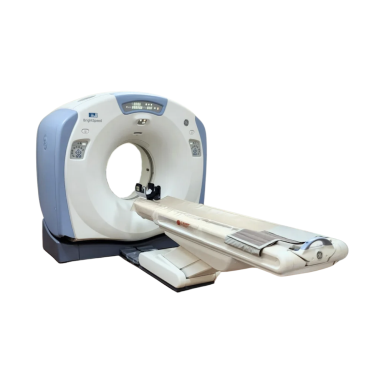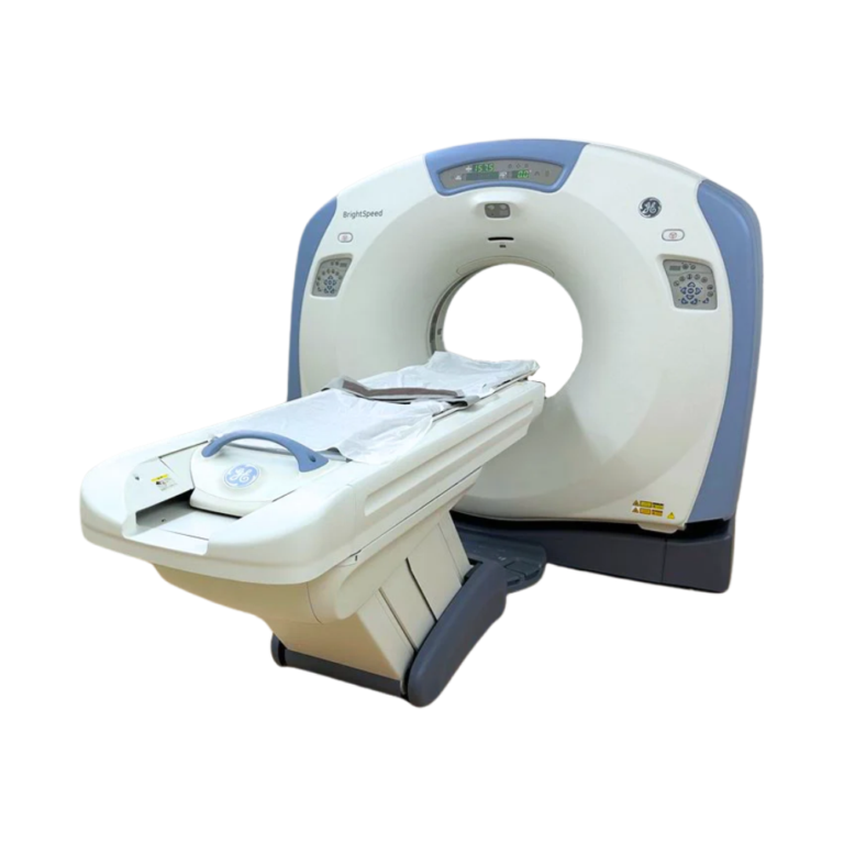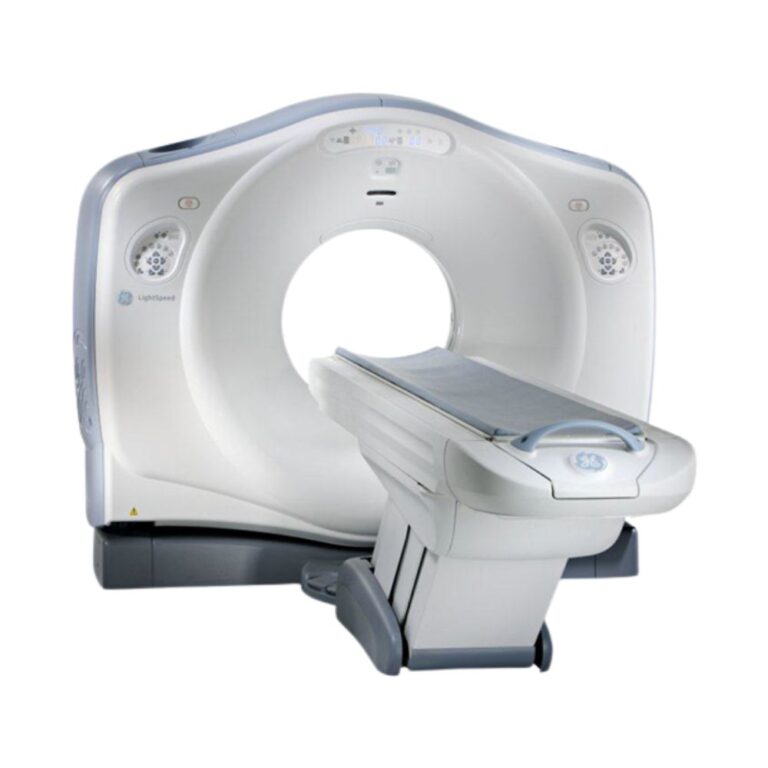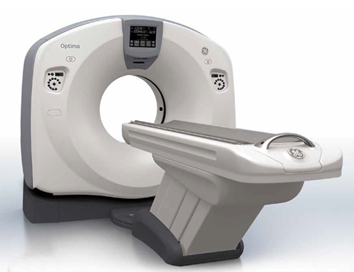
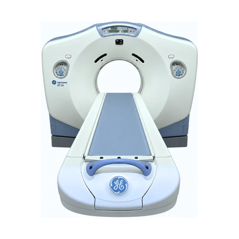
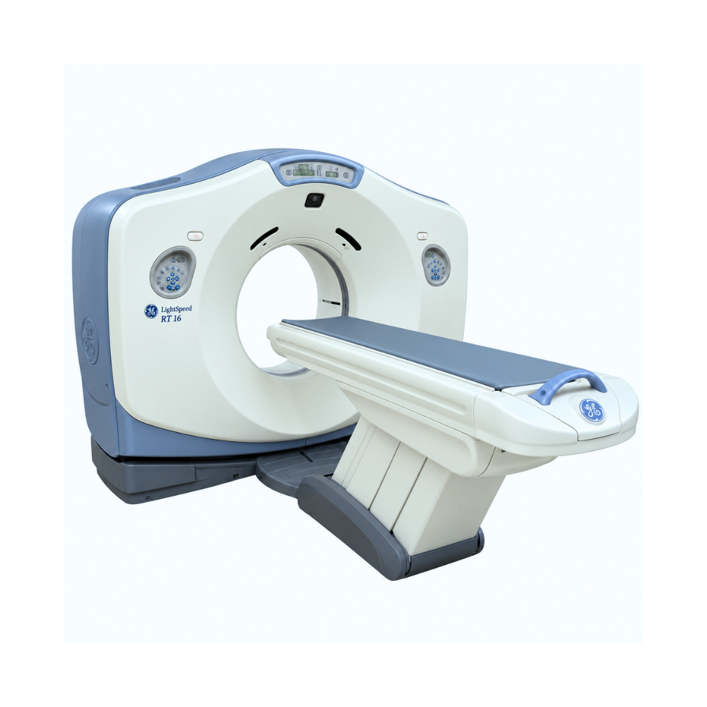
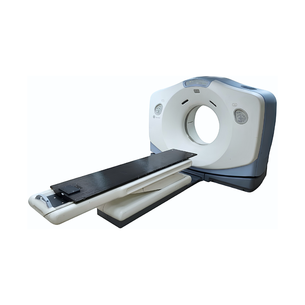
GE LightSpeed RT16 16-Slice CT
- Category:
- CT Scanners
- Brand:
- GE
The GE LightSpeed RT16 is a wide-bore 16-slice CT scanner designed for radiation therapy simulation, oncology, and advanced diagnostic imaging. Best suited for facilities needing flexible treatment positioning and broader patient access, it combines an 80 cm gantry, 65 cm WideView field of view, and 500 lb patient capacity.
LightSpeed RT16 CT Highlights & Benefits
The GE LightSpeed RT16 combines 16-slice CT imaging with the patient access, positioning flexibility, and visualization tools needed for radiation oncology and demanding diagnostic workflows.
The model may also appear in equipment documentation and listings as the GE LightSpeed RT 16.
Key Features & Benefits
-
- 80 cm Wide-Bore Gantry: Provides additional clearance for treatment-position setups, immobilization equipment, larger patients, and interventional access.
- 65 cm WideView Display FOV: Extends displayed anatomy beyond the conventional 50 cm scan field for radiation therapy planning and wide-bore positioning.
- HiLight Matrix II Detector: Supports 16-slice acquisition with available 0.625 mm thin-slice imaging.
- Variable-Speed Acquisition: Compatible configurations support rotation speeds as fast as 0.5 seconds.
- 500 lb Patient Capacity: Supports a broad range of patient sizes with multiple available table configurations.
- GE Radiation Oncology Applications: Compatible AdvantageSim MD and Advantage 4D CT applications support virtual simulation, treatment planning, and respiratory-motion assessment.
- Advanced Reconstruction & Review: Direct3D, Direct MPR, VariViewer, HyperPlane, and CrossBeam support flexible image creation and review on compatible configurations.
Within the GE LightSpeed CT scanner family, the RT16 is differentiated by its 80 cm wide bore, extended WideView display field, higher patient capacity, and radiation therapy simulation workflow.
LightSpeed RT16 CT Imaging Performance
The LightSpeed RT16 uses GE detector, acquisition, and reconstruction technologies to produce volumetric CT datasets for diagnostic imaging and radiation therapy planning.
HiLight Matrix II Detector & microVoxel for Thin-Slice Detail
-
- HiLight Matrix II Detector: Uses 21,888 detector elements for 16-slice CT acquisition. Routine 16 × 1.25 mm acquisition provides up to 20 mm of coverage per rotation, while compatible configurations can acquire 16 × 0.625 mm data for thinner source images.
- microVoxel Imaging: Uses thin acquired data and small reconstructed volumetric elements to preserve detail throughout the scanned volume. This helps coronal, sagittal, oblique, and 3D reconstructions retain useful anatomical information for treatment planning, vascular imaging, and detailed anatomical review.
Variable Rotation & Helical Tilt for Flexible CT Acquisition
-
- Variable Rotation Speed: Compatible LightSpeed RT16 configurations support multiple gantry rotation speeds, reaching 0.5-second rotations when appropriately equipped. Faster acquisition can help reduce scan time and motion during suitable examinations.
- Helical Tilt: Allows helical scanning while the gantry is tilted. This gives technologists more flexibility when the desired imaging plane or patient position does not align with a conventional axial setup.
Helical table travel can reach up to 35 mm per rotation depending on the selected protocol.
HyperPlane & CrossBeam for CT Artifact Reduction
Fast helical acquisition and multislice detector geometry can introduce reconstruction artifacts because CT information is collected across multiple positions and angles.
-
- HyperPlane: Helps manage artifacts associated with higher-pitch helical acquisition while maintaining a consistent reconstructed slice profile.
- CrossBeam: Addresses cone-beam effects associated with multislice detector geometry.
Together, these technologies help maintain more consistent anatomy across reconstructed images for diagnostic interpretation and radiation therapy planning.
LightSpeed RT16 Image Reconstruction & Dose Management
The RT16 includes GE technologies that turn volumetric scan data into clinically useful image sets while helping adapt X-ray output to the patient and examination.
Direct3D, Direct MPR & VariViewer for Flexible Image Review
-
- Direct3D: Uses reconstructed CT images and protocol-defined rendering settings to automatically build a volume-rendered 3D dataset. Incorporating 3D creation into the normal workflow can reduce manual post-processing and make repeatable volumetric views available sooner.
- Direct MPR: Reformats acquired volumetric CT data into coronal, sagittal, oblique, and other viewing planes. Clinicians can evaluate anatomy from more useful orientations without rescanning the patient.
- VariViewer: Allows users to change displayed slice thickness using thin acquired source data. Thin views preserve fine anatomical detail, while thicker views can simplify routine image review.
Available reconstruction applications vary by software release and installed configuration.
Multiple Reconstructions from One CT Acquisition
Prospective Multiple Reconstruction can create multiple image sets from a single acquisition using different image thicknesses, reconstruction intervals, fields of view, or processing settings.
This allows one CT acquisition to support different interpretation, treatment-planning, or post-processing needs without repeating the scan.
SmartmA & 3D Dose Modulation for Patient-Specific Exposure
-
- SmartmA: Adjusts tube current as patient attenuation changes through the scanned anatomy instead of applying one fixed mA throughout the examination.
- 3D Dose Modulation: On compatible configurations, further adapts X-ray output according to differences in patient size and attenuation around the body during each rotation.
Together, these technologies help apply X-ray output more appropriately to the patient and anatomy while working toward the selected image-quality target.
LightSpeed RT16 Wide-Bore Patient Access & Positioning
The RT16 combines an 80 cm gantry opening with extended display coverage and multiple patient-table configurations. These features are especially valuable when patient size, immobilization equipment, or treatment position requires additional space.
80 cm Gantry for Treatment Positioning & Patient Access
The LightSpeed RT16 has an 80 cm (31.5 in) gantry opening, providing substantially more positioning space than a conventional 70 cm diagnostic CT gantry.
The additional clearance can accommodate radiation therapy immobilization devices, larger patients, non-standard treatment positions, and appropriately configured interventional procedures.
WideView for Extended Treatment-Planning Visibility
-
- WideView: Extends the displayed field of view from the conventional 50 cm scan field to as much as 65 cm (25.6 in).
WideView uses reconstruction processing to estimate anatomy extending beyond the measured scan field. This helps radiation oncology teams see more of the patient’s external contour, treatment position, and positioning equipment when those structures extend beyond the conventional field.
The extended region is primarily valuable for visualization and treatment-planning context rather than representing the same directly measured data as anatomy within the standard scan field.
500 lb Patient Table & Available Configurations
The LightSpeed RT16 supports patient loads up to 500 lb (227 kg).
Available configurations include 1700, 2000, and bariatric patient tables, allowing table selection to be matched to radiation oncology, diagnostic imaging, and patient-access requirements.
LightSpeed RT16 Exam Planning & Workflow
The RT16 includes GE workflow technologies for examination setup, contrast timing, patient positioning, reconstruction, and image transfer.
Xtream Workflow for Concurrent Scanning & Processing
GE Xtream allows acquisition, reconstruction, image review, processing, archiving, and networking tasks to operate concurrently rather than forcing one process to finish before another begins.
This can reduce workflow interruptions and help move acquired images toward review and downstream systems more efficiently in busy diagnostic and radiation oncology environments.
SmartPrep for Contrast Timing
GE SmartPrep uses low-dose monitoring images to follow contrast enhancement within a selected region before the diagnostic acquisition.
Scanning can begin when the desired enhancement level is reached rather than relying only on a fixed time delay. This can improve consistency in CT angiography and other contrast-enhanced examinations where timing affects vessel or organ enhancement.
In-Room Controls for Patient Setup
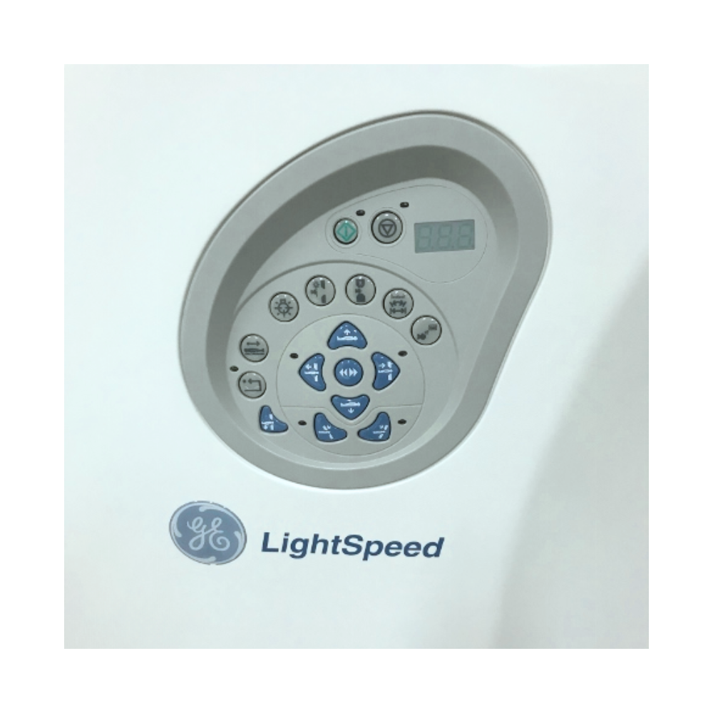
Gantry-mounted positioning controls, alignment lasers, breathing lights, countdown displays, and in-room scan start allow technologists to manage preparation while remaining close to the patient.
These controls are especially useful when precise positioning must be maintained for radiation therapy simulation or other carefully planned examinations.
LightSpeed RT16 CT Simulation & Radiation Therapy Planning
Radiation therapy simulation is the defining clinical focus of the GE LightSpeed RT16. In radiation oncology workflows, the system functions as a wide-bore CT simulator for acquiring planning datasets and supporting the transition from patient positioning to downstream treatment planning.
AdvantageSim MD for Virtual Treatment Simulation
AdvantageSim MD uses the patient’s CT dataset to create a virtual radiation therapy simulation environment.
-
- Multiplanar & 3D Review: Allows clinicians to examine the acquired CT dataset from multiple orientations and in three dimensions.
- Structure Contouring: Supports definition of anatomical structures and treatment targets on the CT dataset.
- Treatment Field & Beam Planning: Provides tools for evaluating treatment fields and beam placement before downstream treatment delivery.
- Digitally Reconstructed Radiographs: Generates projection-style planning images from CT data for treatment localization and comparison with compatible treatment-positioning images.
- DICOM RT Workflow: Supports exchange of compatible treatment-planning information with downstream radiation oncology systems.
AdvantageSim MD availability depends on the installed workstation and software configuration.
Advantage 4D CT for Respiratory Motion
Standard CT captures anatomy at a particular point in time, which can be limiting when a tumor or surrounding structures move during breathing.
-
- Respiratory Phase Imaging: Advantage 4D CT organizes CT information according to different phases of the breathing cycle.
- Motion Assessment: Clinicians can evaluate how tumors and surrounding anatomy move rather than relying on a single static image set.
- Treatment-Planning Support: Motion information can help radiation oncology teams account for respiratory movement when defining treatment targets.
The required software, workstation, and respiratory-motion equipment are configuration-dependent.
CT/MR & CT/PET Fusion for Multimodality Planning
-
- CT/MR Fusion: Aligns the RT16 CT dataset with MRI so treatment-planning teams can combine CT geometry with MRI’s additional soft-tissue information during compatible workflows.
- CT/PET Fusion: Adds PET functional imaging information to the anatomical CT dataset, helping clinicians evaluate metabolically active areas alongside treatment-planning anatomy when compatible software is installed.
Together, these multimodality applications can provide complementary information for target localization and treatment-planning review.
LightSpeed RT16 Advanced Clinical Imaging
The RT16 can extend beyond radiation therapy simulation into diagnostic and image-guided CT applications when equipped with the appropriate acquisition modes and clinical software.
CT Fluoro for Biopsy & Ablation Guidance
-
- CT Fluoro: Provides updated CT imaging during procedural guidance, helping clinicians monitor needle or instrument position during image-guided procedures such as biopsy and tumor ablation.
The RT16’s 80 cm bore also provides additional working and positioning space around the patient during appropriately configured procedures.
CT Angiography, Vessel Analysis & Perfusion Imaging
The RT16 can support several complementary vascular imaging workflows when the applicable acquisition and post-processing options are installed. Each addresses a different clinical need.
-
- CT Angiography: Combines timed intravenous contrast with thin-slice volumetric acquisition to create detailed datasets of contrast-enhanced vessels. These images can be reformatted or rendered in multiple planes for broader evaluation of vascular anatomy.
- Advanced Vessel Analysis: Uses CT angiography data to create 3D vessel views and provide display and measurement tools for selected vessels. This can make it easier to follow vascular anatomy, review vessels from useful orientations, and perform measurements without relying only on individual axial slices.
- CT Perfusion: Analyzes a series of contrast-enhanced images acquired over time rather than relying on one anatomical snapshot. Compatible software can calculate functional information related to tissue blood flow, blood volume, and contrast transit, adding physiological information to structural CT images.
SmartPrep can complement these workflows by monitoring contrast arrival before acquisition so vascular and perfusion imaging can be timed to the desired enhancement.
Clinical Specialties & Applications
The GE LightSpeed RT16 is best suited to facilities that need wide-bore CT access, radiation therapy simulation, advanced treatment-planning workflows, and established 16-slice diagnostic imaging. Its 80 cm gantry, WideView display field, compatible Advantage applications, and CT Fluoro capabilities support a focused range of oncology, diagnostic, interventional, and veterinary environments.
Clinical Environments
-
- Radiation oncology and cancer treatment centers
- Hospital radiology departments with CT simulation or advanced diagnostic imaging needs
- Diagnostic imaging centers requiring wide-bore CT access
- Oncology imaging programs
- Interventional radiology departments using compatible CT-guided workflows
- Veterinary hospitals and specialty referral centers using diagnostic CT
Clinical Specialties
-
- Radiation Oncology: CT simulation, treatment-position planning, target localization, and respiratory-motion assessment with compatible AdvantageSim MD and Advantage 4D CT applications.
- Radiology: Diagnostic head-to-body CT, contrast-enhanced imaging, and multiplanar or 3D reconstruction using the RT16’s 16-slice acquisition platform.
- Oncology: Tumor imaging, treatment-planning datasets, multimodality image fusion, and follow-up assessment.
- Interventional Radiology: CT Fluoro-guided biopsy and tumor ablation on appropriately configured systems, with the 80 cm bore providing additional procedural access around the patient.
- Vascular Imaging: CT angiography, Advanced Vessel Analysis, SmartPrep contrast timing, and compatible CT perfusion workflows.
- Veterinary Medicine: Diagnostic CT in appropriately equipped veterinary hospitals and specialty referral practices.
Common CT Applications
-
- Radiation therapy CT simulation
- Treatment-position and immobilization planning
- Virtual treatment simulation with AdvantageSim MD
- 4D respiratory-motion imaging with Advantage 4D CT
- CT/MR and CT/PET fusion for treatment planning
- Diagnostic head-to-body CT
- Wide-bore imaging for larger patients and complex positioning
- CT angiography and vessel analysis
- CT perfusion on compatible configurations
- CT Fluoro-guided biopsy and tumor ablation
- Multiplanar and 3D image reconstruction
- Veterinary diagnostic CT
LightSpeed RT16 Configurations & Installed Options
Pre-owned GE LightSpeed RT16 systems can vary by software release, patient table, X-ray tube, generator, workstation configuration, and installed clinical applications. MXR Imaging can help match the available configuration to the facility’s radiation oncology, diagnostic imaging, and workflow requirements.
Radiation Oncology & Imaging Software Options
Available configurations may include:
-
- AdvantageSim MD
- Advantage 4D CT
- CT/MR Fusion
- CT/PET Fusion
- microVoxel imaging
- Direct3D
- Direct MPR
- VariViewer
- CT Fluoro
- Advanced Vessel Analysis
- CT perfusion
- Additional visualization and post-processing applications
WideView is a defining RT16 capability and is addressed separately in the patient-access section rather than treated as an optional software package.
Workflow, Dose, & Connectivity Options
Depending on software generation and installed equipment, configurations may also include:
-
- SmartPrep
- SmartmA
- 3D Dose Modulation
- Helical Tilt
- Additional DICOM and workflow capabilities
- Respiratory gating interfaces
- External laser-system connectivity
- Treatment-planning-system connectivity
X-Ray System & Patient Table Options
LightSpeed RT16 systems can be configured with different X-ray power and patient-table setups, including GE 1700, 2000, and bariatric table configurations.
Higher-power configurations can provide up to a 100 kW generator and 800 mA capability. X-ray tube, generator, table, workstation, and software configuration can be matched to the facility’s imaging workload and clinical needs.
GE LightSpeed RT16 vs Other LightSpeed 16-Slice CT Scanners
The LightSpeed RT16 shares GE LightSpeed imaging technology with other 16-slice systems, but each platform is designed around different purchasing priorities.
GE LightSpeed RT16: Best suited to facilities prioritizing radiation therapy CT simulation, an 80 cm wide bore, extended WideView display coverage, treatment-position flexibility, and up to 500 lb patient capacity.
GE LightSpeed 16: A general-purpose 16-slice CT scanner with a 70 cm gantry for routine diagnostic head-to-body imaging, outpatient imaging, hospital radiology, and other established CT workflows.
GE LightSpeed Pro 16 HP: A higher-power diagnostic LightSpeed configuration for facilities that need more X-ray output for demanding diagnostic workloads while retaining the general-purpose 16-slice LightSpeed architecture.
MXR Imaging helps facilities compare LightSpeed configurations based on clinical applications, patient-positioning requirements, imaging workload, X-ray power, software, and available system options to identify the CT scanner that best fits their operational and imaging needs.
LightSpeed RT16 Site Planning Requirements
Site planning for the GE LightSpeed RT16 should account for the selected patient table, 80 cm gantry, operator console, equipment clearances, patient access, and the facility’s radiation oncology or diagnostic imaging workflow.
Additional requirements can include radiation shielding, electrical service, HVAC, treatment-planning equipment, external laser systems, respiratory gating equipment, and other installed accessories. Final room requirements depend on the specific RT16 configuration and installation layout.
MXR Imaging’s turnkey imaging project management services help coordinate equipment selection, room planning, site preparation, delivery, installation, and related project requirements from initial planning through system readiness.
GE LightSpeed RT16 Price & Availability
MXR Imaging offers pre-owned GE LightSpeed RT16 CT scanner and CT simulator configurations for radiation oncology, diagnostic imaging, and related clinical applications.
Pricing depends on equipment condition, X-ray tube history, generator and patient-table configuration, installed software, workstation setup, accessories, installation requirements, and service coverage. MXR Imaging can help match an available RT16 configuration to the facility’s clinical, operational, and budget requirements.
Eligible systems may also be available through MXR Imaging’s Platinum Certified program, providing an additional reconditioning and quality-assurance pathway for pre-owned imaging equipment.
Ongoing CT maintenance and repair services are available to help support equipment performance throughout the scanner’s operating life.
Contact MXR Imaging to discuss current GE LightSpeed RT16 availability, configuration options, pricing, installation planning, and ongoing support.
GE LightSpeed RT16 CT Scanner Specifications
The GE LightSpeed RT16 specifications below summarize its 16-slice acquisition, wide-bore gantry, patient table, X-ray system, imaging technology, radiation oncology capabilities, connectivity, and major configuration differences.
CT Acquisition & Detector
-
- Slice count: 16 slices
- Detector: HiLight Matrix II
- Detector elements: 21,888
- Routine acquisition: 16 × 1.25 mm
- Submillimeter acquisition: 16 × 0.625 mm on applicable configurations
- Maximum detector coverage: 20 mm per rotation
- Acquisition modes: Axial, Cine, Helical, Fluoro, and Gated as configured
- Fastest rotation: Down to 0.5 seconds on compatible configurations
- Maximum helical table travel: Up to 35 mm per rotation
- Scan field of view: Up to 50 cm (19.7 in)
- WideView display field of view: Up to 65 cm (25.6 in)
- Reconstruction matrix: 512 × 512
Gantry & Patient Table
-
- Gantry aperture: 80 cm (31.5 in)
- Gantry tilt: ±30°
- Maximum patient load: 227 kg (500 lb)
- High-accuracy table load: 180 kg (400 lb) with ±0.25 mm positioning accuracy
- Maximum-load positioning accuracy: ±2 mm at 227 kg (500 lb)
- Available patient-table configurations: 1700, 2000, and bariatric
- Positioning: Gantry-mounted controls and laser alignment
X-Ray Generator & Tube
-
- Generator: High-frequency onboard generator
- Standard generator output: Approximately 53.2 kW on applicable configurations
- Higher-power option: Up to 100 kW
- Tube voltage: 80, 100, 120, and 140 kVp
- Standard tube current range: 10 to 440 mA
- Higher-power configuration: Up to 800 mA
- X-ray tube: Performix-family configuration varies by system
- Available higher-power tube: Performix Pro on compatible configurations
- Tube heat capacity: Configuration-dependent
Reconstruction & Imaging Technology
-
- microVoxel: Fine volumetric imaging on compatible configurations
- Direct3D: Automatic protocol-driven 3D volume rendering
- Direct MPR: Multiplanar reconstruction on applicable configurations
- VariViewer: Variable displayed slice-thickness review on applicable configurations
- HyperPlane: Helical artifact management
- CrossBeam: Cone-beam artifact management
- WideView: Up to 65 cm extended display field of view
- Helical Tilt: Helical acquisition with gantry tilt
- Maximum reconstruction speed: Up to 6 images per second
- Multiple reconstruction sets: Supported
Dose, Contrast & Workflow Technology
-
- SmartmA: Automatic tube-current modulation
- 3D Dose Modulation: Patient-responsive exposure adjustment
- SmartPrep: Contrast-enhancement monitoring
- Xtream workflow: Concurrent acquisition, reconstruction, processing, and networking
- In-room controls: Gantry positioning, breathing lights, countdown display, scan start, and alignment tools
Radiation Oncology & Connectivity
-
- DICOM connectivity: Supported
- PACS integration: Supported with compatible systems
- AdvantageSim MD: Configuration-dependent
- Advantage 4D CT: Configuration-dependent
- CT/MR Fusion: Configuration-dependent
- CT/PET Fusion: Configuration-dependent
- Respiratory gating: Supported with compatible equipment
- External laser connectivity: Configuration-dependent
- Treatment-planning-system integration: Configuration-dependent
Electrical & Site Requirements
-
- Nominal electrical input: 380–480 V
- Power: Three-phase
- Frequency: 50/60 Hz
- Site planning: Patient-table and configuration-specific
- GE patient-table configurations: 1700, 2000, and bariatric
- Final room dimensions, shielding, HVAC, electrical service, equipment clearances, and radiation oncology peripherals should be verified for the specific system