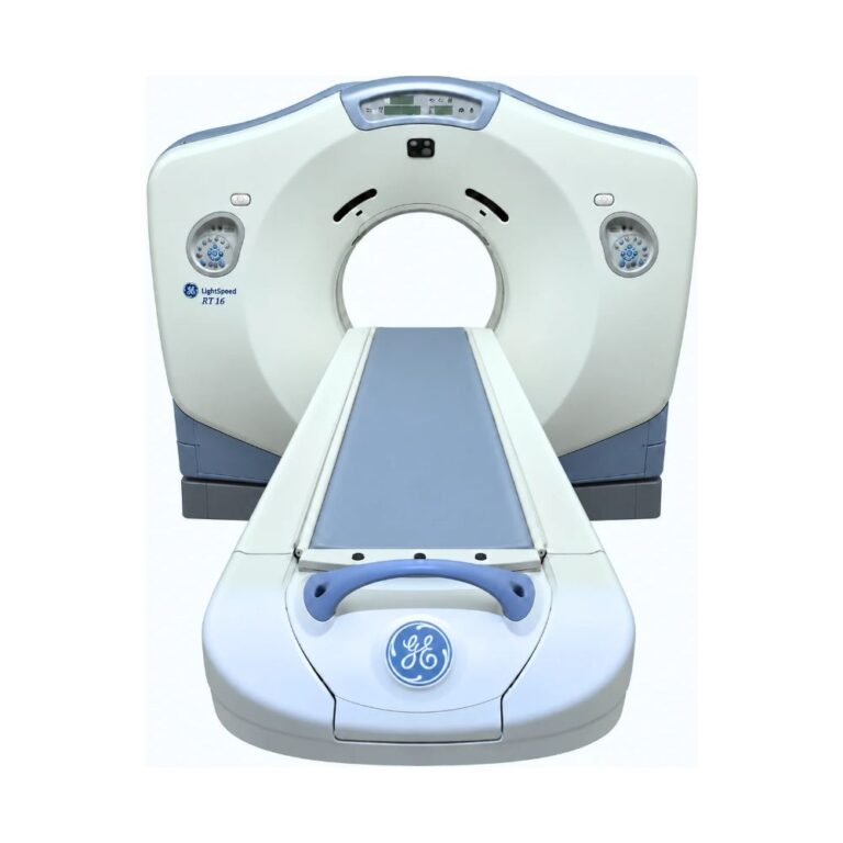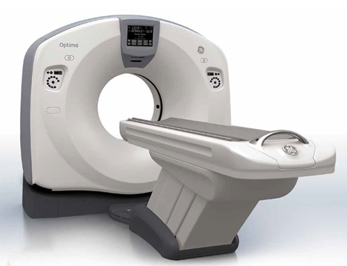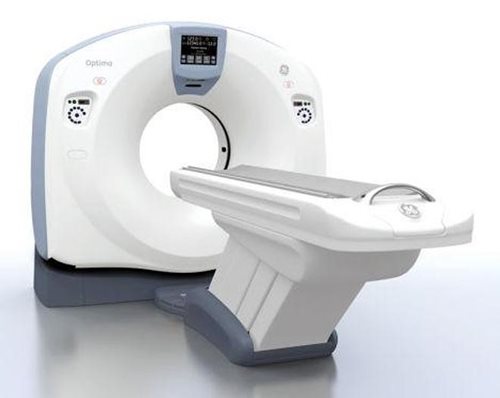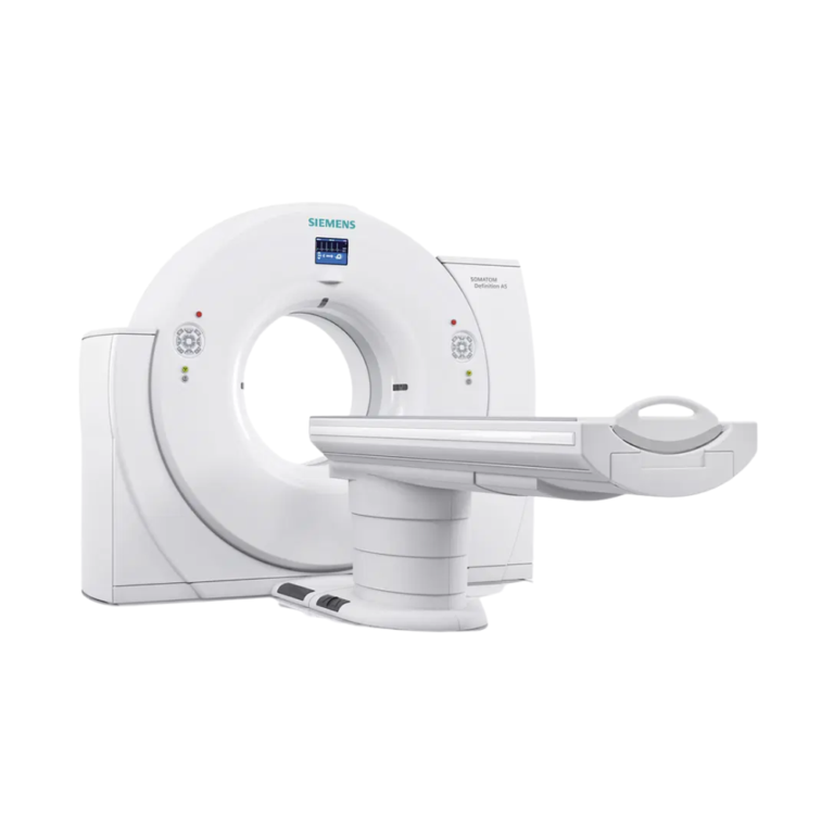
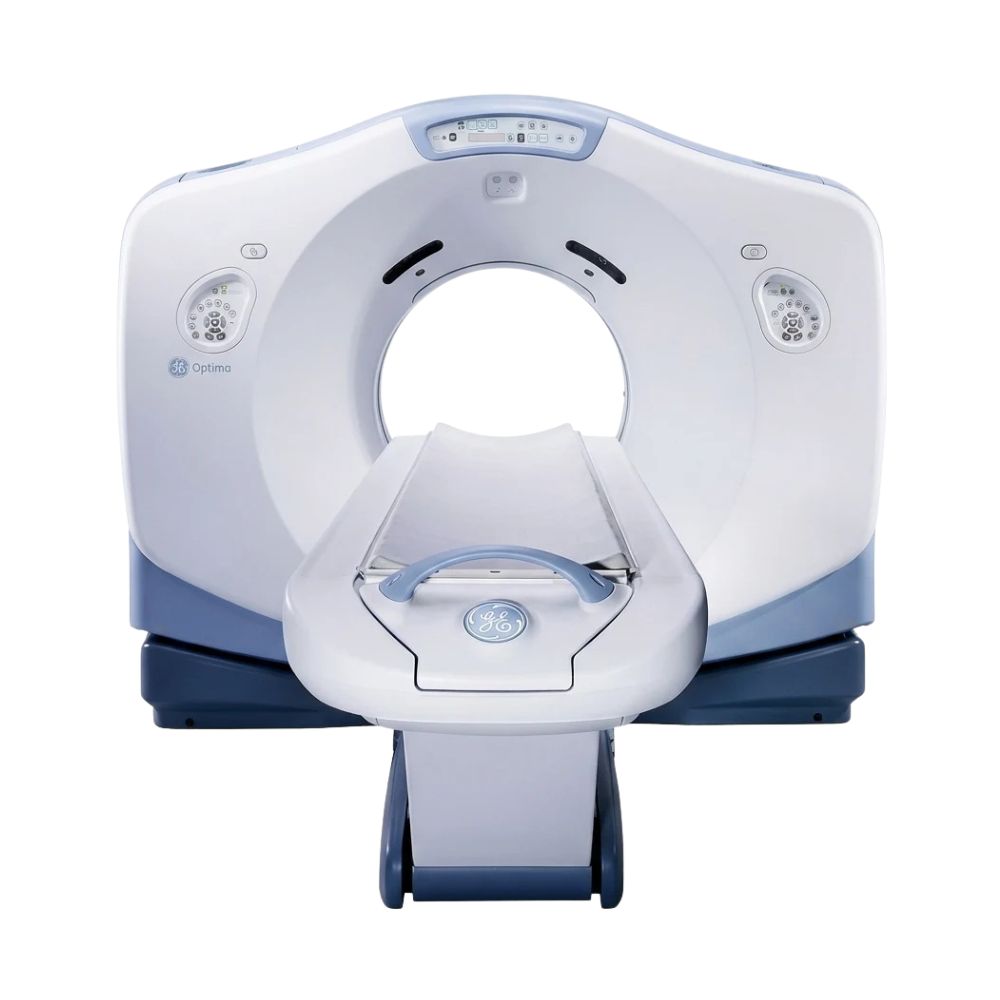
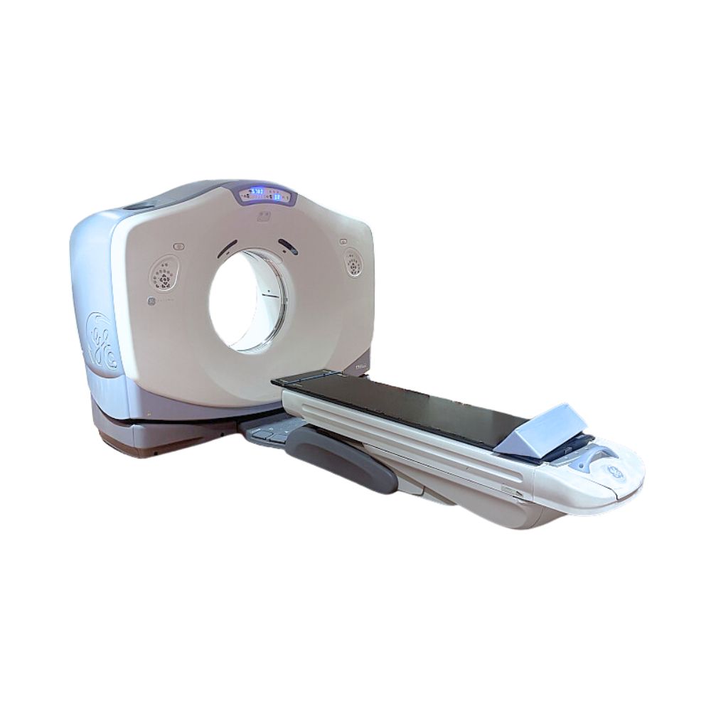
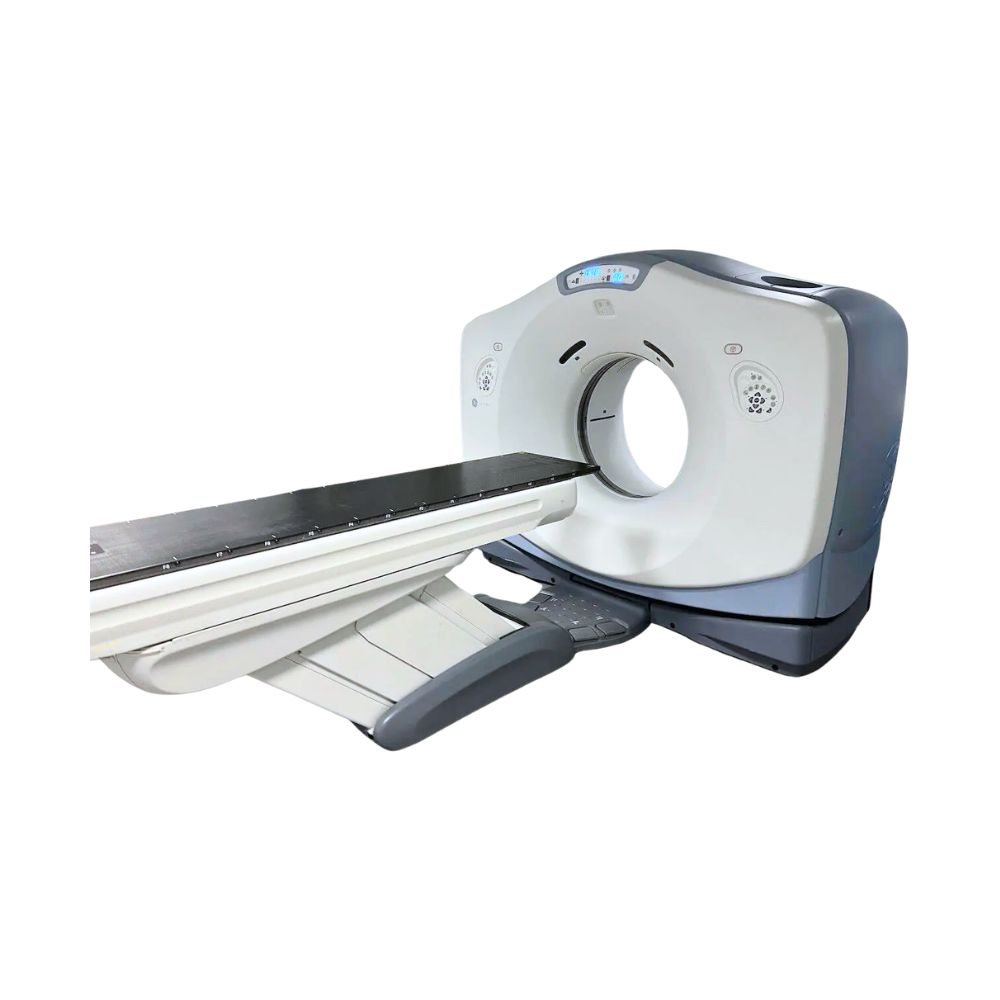
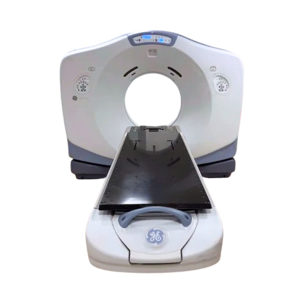
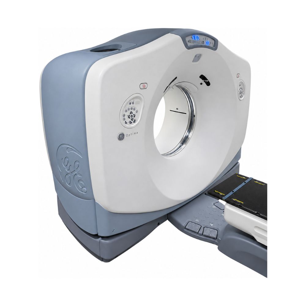
GE Optima CT580 RT 16-Slice CT
- Category:
- CT Scanners
- Brand:
- GE
The GE Optima CT580 RT 16-slice is a wide-bore CT scanner designed for diagnostic imaging and radiation therapy simulation. Best suited to hospitals, imaging centers, and radiation oncology facilities, it combines an 80 cm bore, flexible patient positioning, and available 4D imaging for routine examinations, image-guided procedures, and cancer treatment planning.
Optima CT580 RT 16-Slice Highlights & Benefits
The GE Optima CT580 RT 16 supports routine diagnostic CT, bariatric imaging, and image-guided procedures while functioning as a dedicated radiation therapy CT simulator. Its wide bore and compatible positioning accessories allow patients to be scanned in their planned treatment positions, while available respiratory imaging and virtual simulation software help clinicians locate tumors, assess movement, and plan treatment.
Key Features
-
- 16-slice acquisition: Captures up to 20 mm of anatomy per gantry rotation.
- 80 cm wide bore: Accommodates larger patients, specialized positioning equipment, and interventional access.
- Extended field of view: Optional MaxFOV reconstruction expands anatomical visualization to 80 cm for improved body contour coverage.
- MicroVoxel technology: Provides fine anatomical detail across multiple viewing planes, with optional 0.625 mm slices.
- High-capacity patient table: Optional 650 lb weight capacity expands patient accessibility.
- Radiation therapy positioning: Supports reproducible treatment positioning through compatible flat tabletops, alignment lasers, and immobilization accessories.
- Available 4D CT imaging: Visualizes tumor and organ movement throughout the breathing cycle.
- Advanced image reconstruction: Available ASiR and Smart MAR technologies help reduce image noise and metal-related artifacts.
- Virtual simulation software: Available AdvantageSIM MD supports tumor contouring, treatment center identification, and beam placement planning.
Diagnostic Imaging & Radiation Therapy Simulation
The GE Optima CT580 RT 16-slice combines wide-bore diagnostic CT and radiation therapy simulation in one scanner.
Routine Diagnostic CT & Image-Guided Procedures
The 16-slice imaging platform supports routine head, chest, abdominal, pelvic, spine, and extremity examinations. Its wide bore provides additional access for bariatric imaging and compatible CT-guided procedures.
CT Simulation for Cancer Treatment Planning
For radiation oncology, the scanner images patients in the same position they will occupy during treatment. These images help clinicians locate tumors, identify nearby healthy tissue, and plan where therapeutic radiation will be delivered.
Compatible positioning accessories and alignment lasers help reproduce the planned position during subsequent treatment sessions, while available 4D imaging helps account for tumor movement during breathing. Radiation treatment itself is delivered by a separate treatment system.
16-Slice CT Imaging Performance
Detector Technology for Detailed Imaging
With up to 20 mm of scan coverage per rotation, the 16-slice configuration supports axial and helical imaging for routine diagnostic examinations and treatment planning.
-
- HiLight Matrix II detector: Uses 24 physical detector rows to capture detailed anatomical information for image reconstruction.
- Volara digital data acquisition system (DAS): Processes detector signals while helping reduce electronic image noise.
- MicroVoxel technology: Supports detailed images with similar resolution across viewing planes. Available 0.625 mm slices help visualize small anatomical structures and tumor boundaries.
Scan Speed & Power for Demanding Examinations
Standard gantry rotation times start at 0.8 seconds, with optional settings as fast as 0.5 seconds to shorten acquisition times and help reduce motion-related image distortion. To support repeated or demanding scans, the Performix Pro VCT 100 X-ray tube provides an 8 MHU (million heat units) heat storage capacity.
Facilities that frequently image larger patients or perform demanding thin-section examinations may also benefit from the optional 100 kW generator, which provides additional X-ray power.
Wide-Bore Patient Access & Positioning
80 cm Bore & Extended Imaging Coverage
The 80 cm opening provides additional room for larger patients, clinician access during image-guided procedures, and specialized radiation therapy positions, including arms-up positioning and setups using breast boards or vacuum cushions.
-
- Bore diameter (80 cm): Provides physical clearance for patients and positioning equipment.
- Native scan field of view (50 cm): Defines the primary area from which CT data is acquired.
- Displayed field of view (up to 65 cm): Extends anatomical visualization beyond the native scan field of view.
- MaxFOV reconstruction (optional): Extends the displayed field of view to 80 cm by estimating anatomy beyond standard scan coverage, helping visualize the patient’s external body contour for bariatric imaging and radiation therapy planning.
Patient Table Capacity & Treatment Positioning
The GE Optima CT580 RT 16 offers two patient-table configurations designed to support patient positioning and maintain stability under load.
-
- GT1700 table: Supports patients weighing up to 500 lb (227 kg).
- High-capacity table: Optional configuration supporting up to 650 lb (295 kg).
For radiation therapy simulation, available tables designed to meet TG-66 CT simulation guidelines, flat tabletops, and indexed immobilization accessories help maintain consistent positioning between scanning and treatment. Integrated alignment lasers identify scan planes, while compatible external RT lasers help establish reference points for subsequent treatment sessions.
Image Clarity & Radiation Dose Management
The scanner offers reconstruction and exposure management technologies that support both diagnostic imaging and radiation therapy simulation.
Reconstruction Technologies for Clearer Images
-
- Smart Metal Artifact Reduction (Smart MAR): Optional software that helps reduce streaks from dental restorations, hip prostheses, and other dense implants, improving visibility of nearby anatomy.
- Extended CT-number range: Expands the range of tissue and material densities represented in CT images, helping clinicians account for dense implants during radiation therapy planning.
- Adaptive Statistical Iterative Reconstruction (ASiR): Optional software that reduces image noise and may support lower-dose scanning protocols.
Patient-Specific Exposure Control
-
- 3D automatic tube current modulation: Adjusts X-ray output according to patient size, shape, and tissue density during scanning.
- Pediatric color-coded protocols: Help teams select appropriate pediatric imaging techniques.
- Dose Check: Alerts operators when planned scan exposure exceeds configured thresholds.
- DICOM dose reporting and optional DoseWatch: Support recording, monitoring, and analysis of radiation exposure.
4D CT Imaging for Respiratory Motion
Optional 4D CT simulation captures anatomy over time, showing how tumors and surrounding organs move during breathing. This is particularly useful for radiation therapy planning in the chest and upper abdomen.
Respiratory Motion Imaging Technologies
-
- Cine 4D: Collects repeated CT images during breathing to capture anatomical movement.
- Advantage 4D: Organizes images by breathing phase for review and treatment planning.
- Respiratory gating: Uses compatible external monitoring equipment, including Varian RPM, to control scan timing or sort images by breathing phase afterward.
- Auto 4D: Automates image sorting and transfer, making respiratory-correlated datasets available within 90 seconds on supported configurations.
- Smart Deviceless 4D: Derives breathing information directly from CT images without an external belt or marker, simplifying setup and allowing natural breathing.
Virtual Simulation & Radiation Therapy Planning
AdvantageSIM MD Treatment Planning Tools
Available AdvantageSIM MD software connects CT imaging with virtual radiation therapy simulation. Its tools support:
-
- Patient marking and identification of the planned treatment center (isocenter)
- Contouring of tumors and surrounding organs
- Definition of treatment beam geometry
- Review of 4D respiratory motion datasets
- Digitally reconstructed radiographs (DRRs), which create X-ray-like projection images from CT data for treatment setup and verification
- Automated organ segmentation on compatible software releases
Multimodality Imaging & Treatment System Connectivity
Available software and connectivity tools help clinicians review and transfer imaging information throughout the oncology workflow.
-
- Integrated Registration: Aligns CT, MRI, PET, and SPECT images, helping clinicians identify tumor boundaries and surrounding anatomy using information from multiple imaging modalities.
- MD Connect: Provides access to imaging data from multiple modalities.
- DICOM connectivity: Supports image exchange with compatible picture archiving and communication systems (PACS), virtual simulation applications, and radiation therapy planning systems. RT data exchange depends on installed software and workstations.
Clinical Environments & Applications
Clinical Environments
-
- Hospitals and health systems
- Outpatient diagnostic imaging centers
- Hospital radiation oncology departments
- Dedicated cancer centers
- Freestanding radiation therapy facilities
- Academic medical centers
Diagnostic & Interventional Specialties
-
- Diagnostic radiology: Routine head, chest, abdominal, pelvic, and extremity CT
- Neurology: Brain and spine imaging
- Orthopedics: Bone, joint, and musculoskeletal imaging
- Pulmonology: Lung and chest imaging
- Urology: Kidney and urinary tract imaging
- Interventional radiology: CT-guided biopsies and drainage procedures with compatible options
The wide bore and optional high-capacity table also support bariatric imaging across applicable specialties.
Radiation Oncology Applications
The RT configuration supports CT simulation for 3D conformal radiation therapy (3D-CRT), intensity-modulated radiation therapy (IMRT), and other compatible external-beam treatment workflows.
-
- Thoracic oncology: Lung and other chest tumors
- Abdominal oncology: Liver, pancreatic, and other abdominal tumors
- Breast oncology: Breast and chest wall treatment planning
- Pelvic oncology: Prostate, gynecologic, and colorectal cancers
- Neuro-oncology: Brain and skull base tumors
- Head and neck oncology: Treatment planning with specialized immobilization
Optima CT580 Model Comparison & Available Options
CT580 RT 8-Slice, RT 16-Slice, & W 16-Slice
The GE Optima CT580 platform offers three configurations with distinct acquisition capabilities and clinical applications.
-
-
- GE Optima CT580 RT 8-Slice CT: The original RT configuration supports routine radiation therapy simulation with a minimum slice thickness of 1.25 mm. It is best suited to facilities with less demanding acquisition requirements that still need wide-bore access and repeatable treatment positioning. A 16-slice upgrade was originally available.
- GE Optima CT580 RT 16-Slice CT: Combines radiation therapy simulation with greater thin-section imaging flexibility. Available 0.625 mm acquisition improves visualization of small anatomical structures, making it particularly useful for detailed tumor contouring and diagnostic examinations.
- GE Optima CT580 W 16-Slice CT: Designed primarily for diagnostic radiology, bariatric imaging, and image-guided procedures. It offers submillimeter imaging, faster standard rotation settings, and wide-bore patient access without the dedicated radiation therapy simulation configuration.
-
Although all three share an 80 cm bore, their acquisition performance, installed software, and treatment-planning capabilities differ.
Installed Hardware & Software Options
Available systems may differ in generator power, rotation speed, slice thickness, patient-table capacity, positioning accessories, and installed software. Radiation therapy configurations may also vary in respiratory monitoring equipment, external positioning lasers, and treatment-planning integration.
Additional diagnostic applications may include:
-
- SmartPrep: Uses a series of monitoring scans to track injected contrast as it reaches a selected blood vessel or anatomical region. This helps time the diagnostic scan for the desired level of contrast enhancement.
- Direct MPR (Multiplanar Reconstruction): Automatically generates coronal, sagittal, and oblique image views from acquired CT data, reducing manual processing and allowing anatomy to be examined from multiple perspectives.
- Volume Viewer: Creates interactive 3D representations from CT images, helping clinicians visualize anatomical relationships, assess blood vessels, and review complex structures.
- Real-time CT fluoroscopy: Provides rapidly updated CT images during image-guided procedures, allowing clinicians to monitor needle placement and progression during biopsies, drainage procedures, and other compatible interventions.
Optima CT580 RT 16-Slice Availability & Project Planning
The GE Optima CT580 RT 16-slice CT scanner is available as a pre-owned system, subject to availability. Pricing varies by equipment age, tube condition, hardware, software, accessories, reconditioning, and project scope.
MXR Imaging can help match available systems to dedicated diagnostic imaging, radiation therapy simulation, or shared-use workflows. Eligible systems may also be available through the Platinum Certified CT and MRI Systems program.
Turnkey imaging project management supports room planning, rigging, installation, and project coordination, with ongoing support available through MXR’s CT maintenance and repair services. For radiation oncology installations, commissioning and clinical acceptance testing should be coordinated with the facility’s medical physics team.
GE Optima CT580 RT 16-Slice Specifications
Specifications describe the 16-slice RT configuration. Installed options and site requirements vary by unit.
CT Acquisition & Detector
-
- Acquisition: 16-slice axial and helical CT
- Detector: HiLight Matrix II
- Physical detector rows: 24 (16 central 0.625 mm rows and eight outer 1.25 mm rows)
- Maximum acquisition coverage: 20 mm
- Minimum slice thickness: 1.25 mm standard; 0.625 mm optional
- Standard rotation settings: 0.8, 0.9, 1.0, 2.0, 3.0, and 4.0 seconds
- Optional rotation settings: 0.5, 0.6, and 0.7 seconds
- Helical pitch: 0.5625:1, 0.9375:1, 1.375:1, and 1.75:1
Gantry & Patient Table
-
- Bore diameter: 80 cm (31.5 in)
- Gantry tilt: ±30°
- Native scan field of view: 50 cm
- Displayed field of view: Up to 65 cm; up to 80 cm with applicable MaxFOV option
- GT1700 table capacity: 500 lb (227 kg)
- High-capacity table option: 650 lb (295 kg)
- Positioning: Integrated lasers and compatible indexed RT accessories
X-Ray Generator & Tube
-
- X-ray tube: Performix Pro VCT 100
- Tube construction: Metal-ceramic
- Tube heat storage: 8 MHU
- Generator power: Approximately 53 kW standard; 100 kW optional
- Tube voltage: 80, 100, 120, or 140 kV
- Tube current: 10–440 mA; up to 800 mA with high-power option
Image Reconstruction & Performance
-
- Reconstructed slice thicknesses: 0.625 mm (optional), 1.25, 2.5, 3.75, 5, 7.5, and 10 mm
- Iterative reconstruction: ASiR, optional
- Metal artifact reduction: Smart MAR, optional
- Extended CT-number range: RT configuration
- Respiratory imaging: Advantage 4D and Smart Deviceless 4D, optional
- Virtual simulation: AdvantageSIM MD, optional
- Connectivity: DICOM
System & Site Requirements
-
- Gantry width × depth: 244.8 × 101.3 cm (96.4 × 39.9 in)
- Gantry weight with covers: Approximately 1,816 kg (4,003 lb)
- Gantry heat dissipation: Up to 8.96 kW
- Electrical supply: 380–480 V three-phase, 50/60 Hz, configuration dependent
- Documented power demand: 150 kVA maximum; 30 kVA average
- Minimum overhead gantry service clearance: 90 in (2.286 m)
- Site planning: Radiation shielding, structural anchoring, HVAC, electrical distribution, equipment access, and compatible RT laser and network provisions
GE Optima CT580 RT Resources
- GE Optima CT580 RT Product Brochure (PDF): Clinical capabilities, imaging technologies, and specifications.
- GE Discovery & Optima RT Pre-Installation Manual (PDF): Electrical, structural, and environmental installation requirements.
- GE Optima CT580 RT/W Site Planning Drawings (PDF): Equipment dimensions, room layouts, and clearances for the GT1700 table.