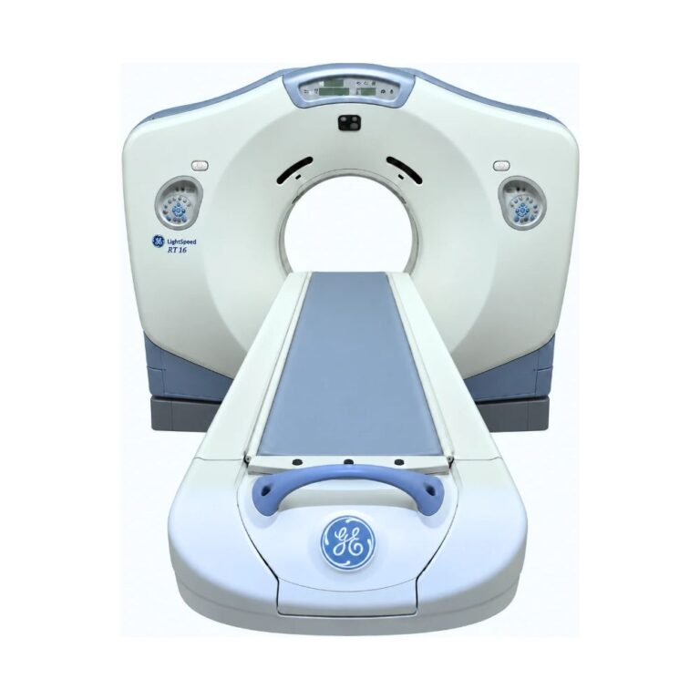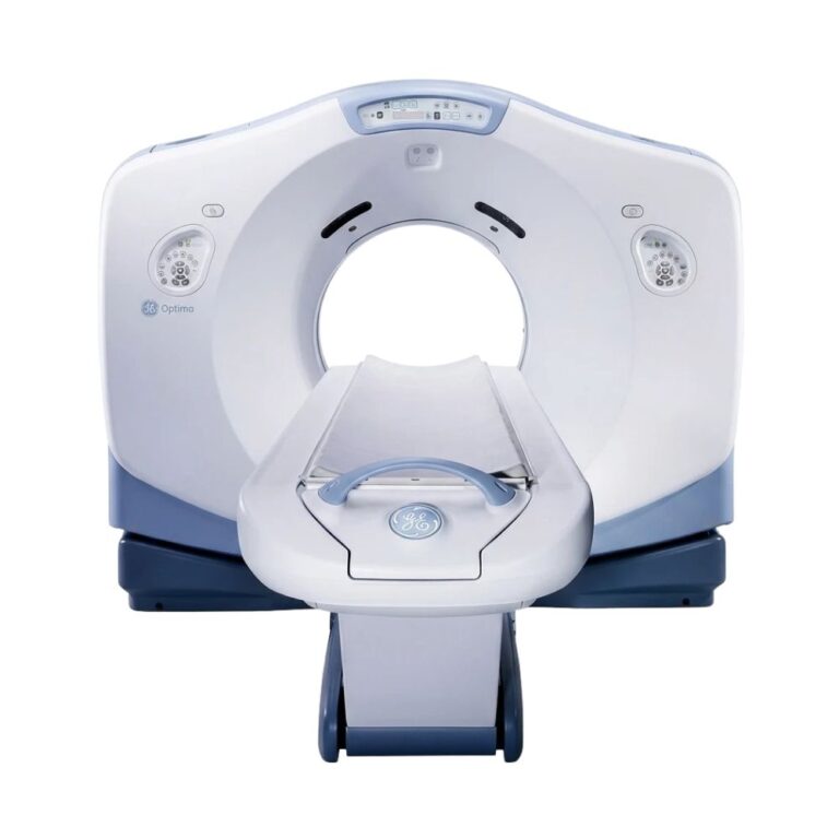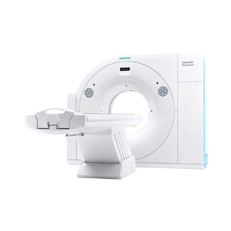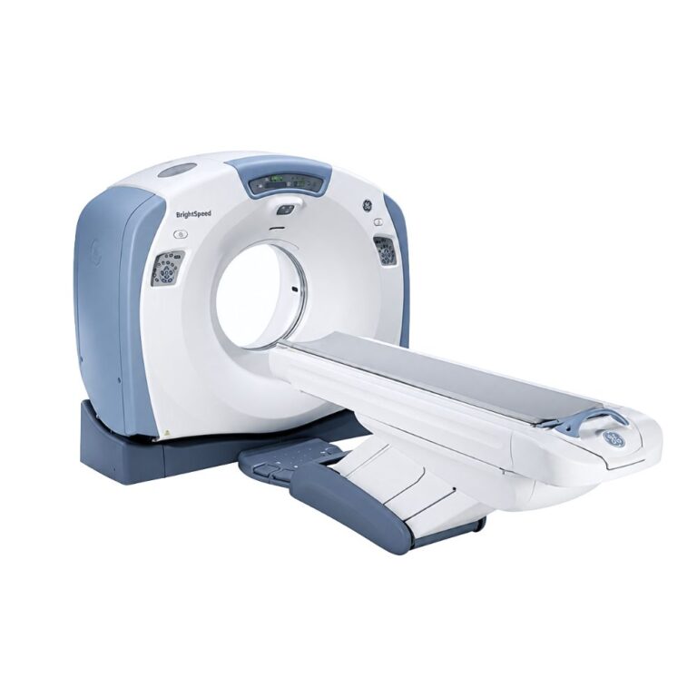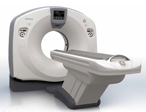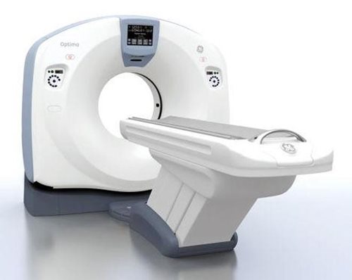
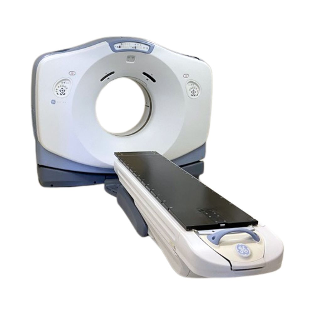
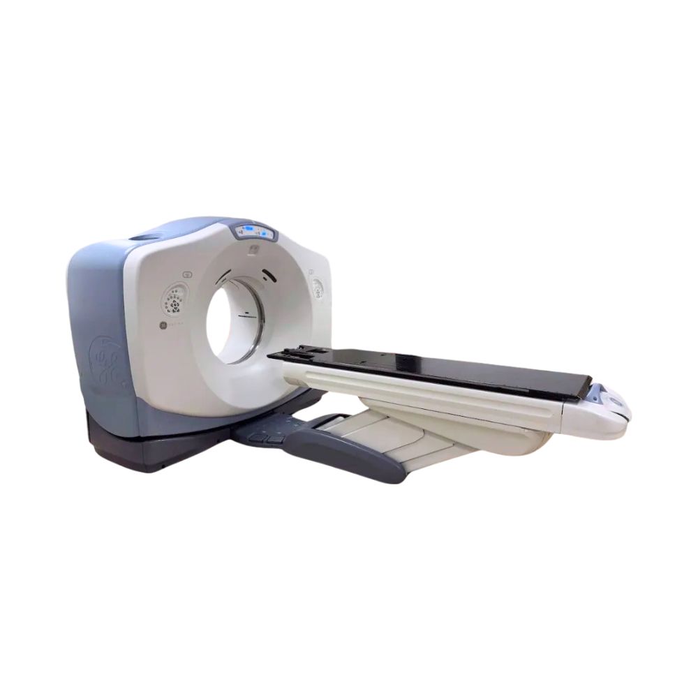
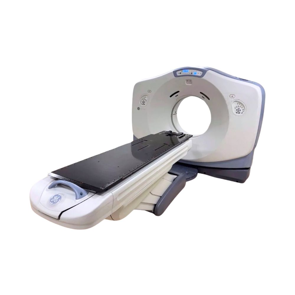
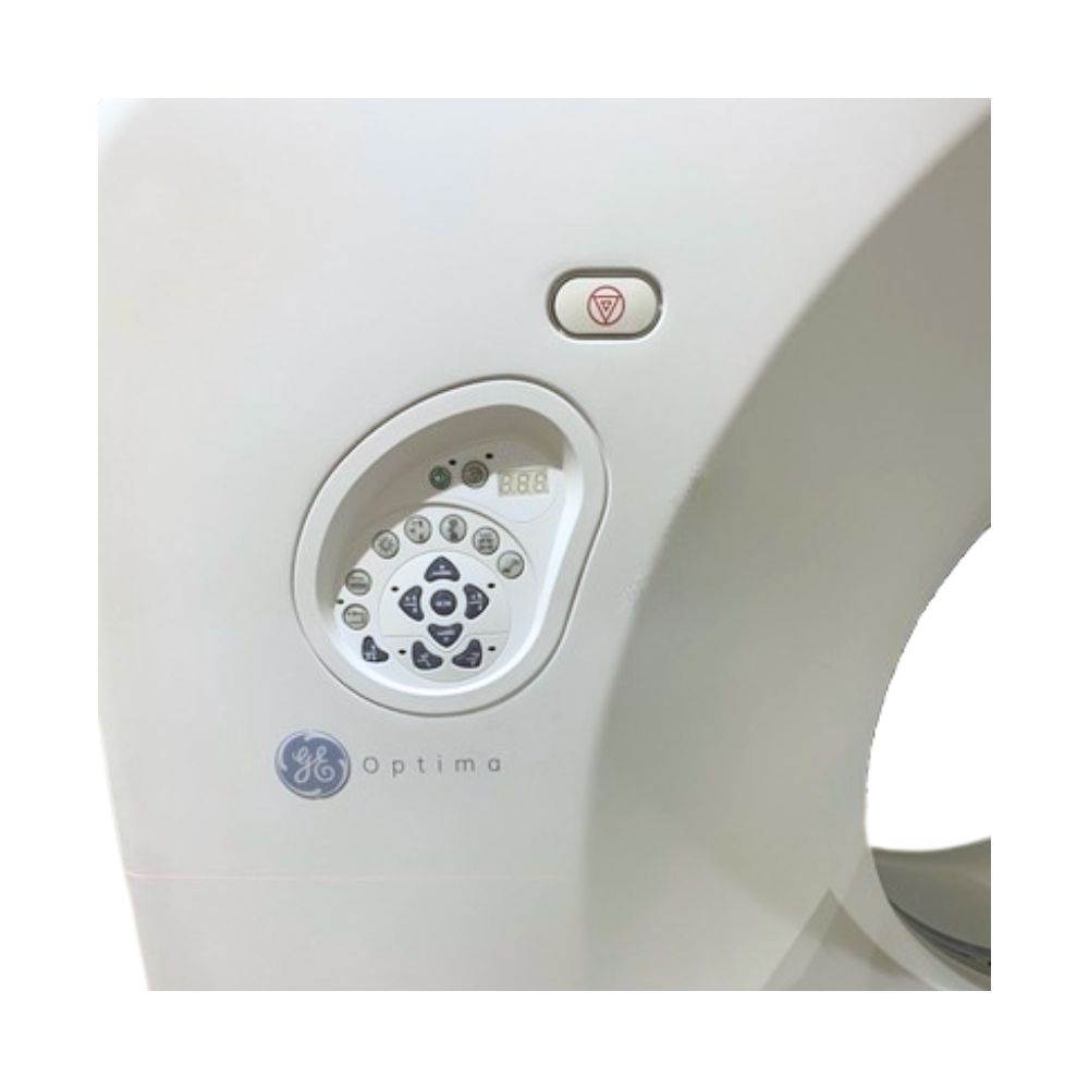
GE Optima CT580 RT 8-Slice CT
- Category:
- CT Scanners
- Brand:
- Siemens
The GE Optima CT580 RT is an 8-slice, wide-bore CT scanner for diagnostic imaging and radiation therapy simulation. Best suited for hospitals, cancer centers, and imaging facilities, it combines an 80 cm patient opening with flexible scan coverage and optional oncology-specific treatment planning technologies.
Optima CT580 RT 8-Slice Highlights & Benefits
The GE Optima CT580 RT 8-slice CT scanner combines everyday head and body CT imaging with the ability to scan patients in the position required for radiation therapy. Its wide bore accommodates larger patients, positioning accessories, and immobilization devices, while optional simulation software supports cancer treatment planning.
Key Features
-
- Eight-slice acquisition: Select between thinner slices for anatomical detail and wider coverage for routine examinations.
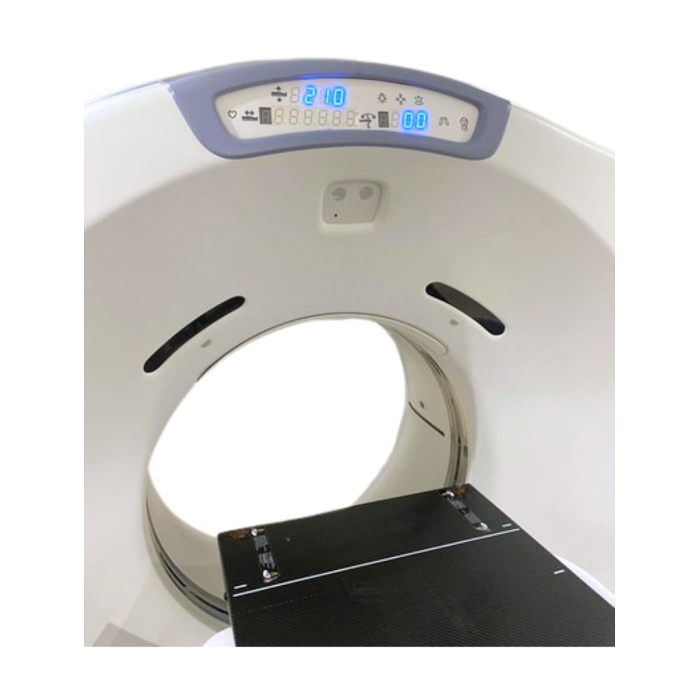
- 80 cm wide bore: Provides additional clearance for larger patients and specialized positioning.
- Up to 650 lb patient capacity: Available with the high-capacity table for accommodating a wider patient population.
- Extended field of view: Helps visualize anatomy that extends outside the conventional scanning area.
- Radiation therapy positioning: Compatible flat tabletops and external positioning lasers support reproducible patient setup.
- Optional 4D CT: Tracks anatomical movement during breathing to support radiation treatment planning.
Diagnostic Imaging and Radiation Therapy Simulation
Routine Diagnostic CT
The CT580 RT supports head and whole-body imaging with or without contrast. Its eight-slice acquisition is suitable for routine neurological, chest, abdominal, pelvic, and musculoskeletal examinations.
CT Simulation for Cancer Treatment
The Optima CT580 RT captures three-dimensional images of patients in their intended treatment position, allowing oncology teams to accurately identify tumors, delineate surrounding organs, and plan targeted radiation delivery while minimizing exposure to healthy tissue.
8-Slice CT Imaging Performance
Selectable Scan Coverage
The GE Optima CT580 RT 8-slice uses a 24-row detector with two primary acquisition settings, allowing facilities to balance anatomical detail with scan coverage:
-
- 8 × 1.25 mm: Captures thinner slices across 10 mm of anatomy per rotation for more detailed imaging.
- 8 × 2.5 mm: Covers 20 mm of anatomy per rotation when broader coverage is preferred.
Both settings support axial imaging (scanning in individual sections) and helical imaging (continuous scanning as the patient table moves). This flexibility allows technologists to adjust scan coverage and anatomical detail based on the examination or radiation therapy planning requirements.
Scan Speed and Available X-Ray Power
Rotation speed determines how quickly the scanner collects images, while X-ray power helps maintain image quality when imaging thicker anatomy or larger patients.
Available generator and tube upgrades provide additional power for demanding examinations. Rotation speeds and maximum tube output depend on the installed configuration.
Flexible Patient Access with an 80 cm Bore
Wide-Bore Patient Positioning
The GE Optima CT580 RT 8-slice CT scanner’s 80 cm gantry opening provides additional clearance for patients who may be difficult to accommodate in a conventional CT scanner. This is particularly useful for bariatric imaging, patients with limited mobility, and radiation therapy simulation requiring arms-up positioning or immobilization accessories.
Extended Anatomical Visualization
The scanner’s conventional 50 cm scan field of view can be supplemented by extended reconstruction capabilities. A 65 cm displayed field of view helps visualize anatomy extending beyond the standard scan area.
Compatible MaxFOV reconstruction can extend the displayed area to 80 cm on appropriately equipped systems. This is useful for capturing external body contours during radiation therapy planning. Extended images are reconstructed beyond the conventional measurement area and should not be interpreted as equivalent to the standard scan field.
Patient Table and Treatment Positioning
Available table configurations include a 500 lb (227 kg) VT 1700 table and a 650 lb (295 kg) high-capacity table.
Compatible flat tabletop inserts, immobilization accessories, and external laser systems help reproduce patient positioning between CT simulation and radiation treatment. Available radiation therapy table configurations are designed to meet AAPM TG-66 CT simulation guidelines.
Optimizing Image Quality at Lower Doses
Available dose management and reconstruction technologies help tailor imaging exposure to patient size and examination requirements:
- Auto mA and Smart mA: Adjust X-ray output according to patient anatomy and attenuation, helping avoid unnecessary exposure.
- ASiR (Adaptive Statistical Iterative Reconstruction): Reduces image noise through advanced reconstruction, potentially allowing lower radiation exposure while maintaining diagnostic image quality.
- Smart MAR (Metal Artifact Reduction): Reduces streaking caused by metal implants, helping improve visibility of nearby anatomical structures.
- Dose Check: Provides notifications or alerts when planned exposure exceeds configured dose thresholds.
- Pediatric color-coded protocols: Help technologists select imaging settings suited to smaller patients.
Actual dose reduction and image quality depend on patient size, clinical protocols, and installed software.
Virtual Simulation for Precise Radiation Therapy Planning
AdvantageSim MD Virtual Simulation Software
AdvantageSim MD is optional software specifically designed for planning radiation therapy for cancer. It uses CT images to create a virtual representation of the patient, allowing oncology teams to visualize treatment areas before radiation is delivered.
Depending on the software release, available functions include:
-
- Contouring and segmentation: Identify and outline tumors and nearby organs to help distinguish treatment targets from healthy tissue.
- Virtual beam placement: Visualize proposed radiation beam directions relative to the patient’s anatomy.
- Digitally reconstructed radiographs (DRRs): Generate X-ray-like images from CT data to help verify patient positioning during treatment.
Multimodality imaging: Incorporate compatible MRI and PET/CT images to provide additional information about tumor location and surrounding anatomy.
4D CT Simulation for Respiratory Motion
Tumors and surrounding organs in the chest and upper abdomen can shift as patients breathe. Optional Advantage 4D respiratory gating captures images at different stages of the breathing cycle, allowing the treatment team to evaluate this motion when planning radiation delivery.
Compatible Smart Deviceless 4D software can derive respiratory motion information without attaching external breathing-monitoring devices to the patient.
Connecting CT Simulation to Treatment Planning
DICOM connectivity allows images to be transferred to compatible imaging archives, workstations, and radiation therapy planning systems.
Optional GE Integrated Registration software aligns CT images with compatible MRI, PET, or SPECT datasets, helping oncology teams compare anatomical and functional information in the same spatial reference.
Clinical Environments & Applications
Clinical Environments
-
- Hospitals and medical centers
- Radiation oncology departments
- Cancer treatment centers
- Outpatient diagnostic imaging centers
- Multidisciplinary imaging facilities
Clinical Specialties & Applications
-
- Radiation oncology: CT simulation and external beam radiation therapy planning.
- Diagnostic radiology: Routine head, chest, abdominal, and pelvic imaging.
- Neurology: Routine brain and head examinations.
- Orthopedics: Bone, joint, and spine imaging.
- Pulmonology: Chest and lung imaging.
- Urology: Renal and urinary tract imaging.
- Emergency medicine: Routine trauma and acute diagnostic examinations.
- Interventional radiology: Selected CT-guided procedures, such as biopsy, with compatible equipment.
Installed Hardware & Software Options
Pre-owned GE Optima CT580 RT 8-slice CT scanner systems may differ in tube capacity, generator power, patient table, reconstruction software, oncology accessories, and installed clinical applications.
Additional configuration-dependent capabilities include:
- SmartPrep: Measures contrast enhancement in a selected anatomical region during preliminary monitoring scans to help time diagnostic imaging with contrast arrival.
- Multiplanar reconstruction (MPR): Reconstructs images in different anatomical planes without repeating the scan.
- 3D visualization: Creates volumetric views of anatomy for reviewing spatial relationships.
- CT-guided intervention: Supports compatible image-guided procedural workflows.
- Advanced vascular and perfusion applications: Provide specialized image analysis where supported by installed software and acquisition capabilities.
GE Optima CT580 Model Comparison
- GE Optima CT580 RT 8-Slice CT: Offers up to 20 mm of anatomical coverage per rotation using eight 2.5 mm slices, or 10 mm using eight 1.25 mm slices. Best suited to routine diagnostic imaging and radiation therapy simulation protocols that do not require thinner acquisition.
- GE Optima CT580 RT 16-Slice CT: Captures sixteen 1.25 mm slices across the same 20 mm coverage, providing finer anatomical detail without reducing coverage. Compatible submillimeter acquisition options provide additional flexibility for examinations requiring thinner slices.
- GE Optima CT580 W 16-Slice CT: A wide-bore configuration positioned primarily for diagnostic radiology, larger-patient imaging, and selected image-guided procedures. Facilities requiring dedicated CT simulation should evaluate the installed radiation therapy accessories and software.
Availability & Project Planning
Pricing varies by system age, tube condition, patient table, installed software, accessories, reconditioning, and project scope.
MXR Imaging can help evaluate available configurations against your facility’s clinical needs, patient volume, and existing space. Eligible systems may also be available through the Platinum Certified CT and MRI systems program.
Turnkey imaging project management is available for facilities requiring room planning, rigging, installation, and project coordination.
GE Optima CT580 RT 8-Slice Specifications
Specifications describe the eight-slice RT configuration and shared platform hardware. Installed options vary by unit.
CT Acquisition & Detector
-
-
- Acquisition: 8-slice axial and helical CT
- Detector: HiLight Matrix II platform
- Physical detector rows: 24
- Multislice configurations: 8 × 1.25 mm and 8 × 2.5 mm
- Maximum acquisition coverage: 20 mm per rotation
- Minimum slice thickness: 1.25 mm
- Standard rotation settings: 0.8, 0.9, 1.0, 2.0, 3.0, and 4.0 seconds
- Optional rotation settings: 0.5, 0.6, and 0.7 seconds
- Helical pitch: 0.625:1, 0.875:1, 1.35:1, and 1.675:1
-
Gantry & Patient Table
-
-
- Bore diameter: 80 cm (31.5 in)
- Gantry tilt: ±30°
- Scan field of view: Up to 50 cm
- Displayed field of view: Up to 65 cm
- GT1700 table capacity: 500 lb (227 kg)
- High-capacity table option: 650 lb (295 kg)
- Positioning: Integrated lasers and compatible RT positioning accessories
-
X-Ray Generator & Tube
-
-
- Standard generator: 53.2 kW
- Optional high-power generator: 100 kW
- Tube voltage: 80, 100, 120, or 140 kV
- Tube current: Up to 440 mA or 800 mA with applicable high-power configuration
- X-ray tube: Performix Pro VCT 100 on applicable configurations
- Tube heat storage: Up to 8 MHU with applicable tube
-
Reconstruction & Clinical Software
-
-
- Reconstructed slice thicknesses: 1.25, 2.5, 3.75, 5, 7.5, and 10 mm
- Iterative reconstruction: ASiR, optional
- Respiratory imaging: Advantage 4D, optional
- Virtual simulation: AdvantageSim MD, optional
- Connectivity: DICOM
-
System & Site Requirements
-
-
- Gantry width × depth: Approximately 244.8 × 101.3 cm (96.4 × 39.9 in)
- Gantry weight with covers: Approximately 1,816 kg (4,003 lb)
- Electrical supply: 380–480 V three-phase, 50/60 Hz, configuration dependent
- Documented maximum power demand: Up to 150 kVA, configuration dependent
- Site planning: Radiation shielding, structural anchoring, HVAC, equipment access, and compatible RT laser and network provisions
-
GE Optima CT580 RT OEM Resources
- GE Optima CT580 RT Product Brochure: Learn about the scanner’s imaging capabilities, key technologies, and radiation therapy applications.
- GE Optima CT580 RT Pre-Installation Manual: Review the electrical, environmental, and structural requirements for system installation.
- GE Optima CT580 RT Site Planning Drawings: View equipment dimensions, recommended room layouts, and required installation clearances.