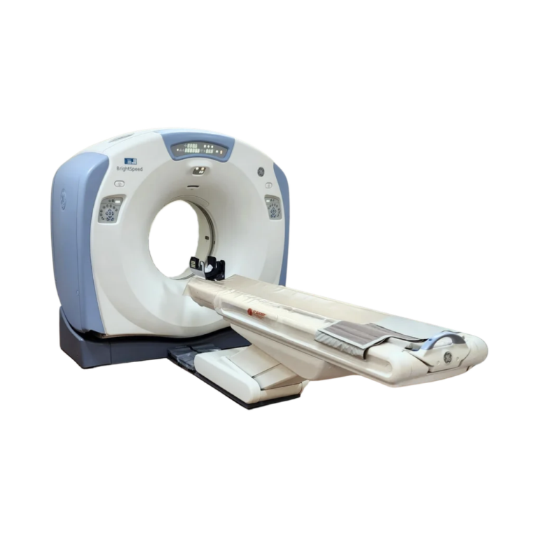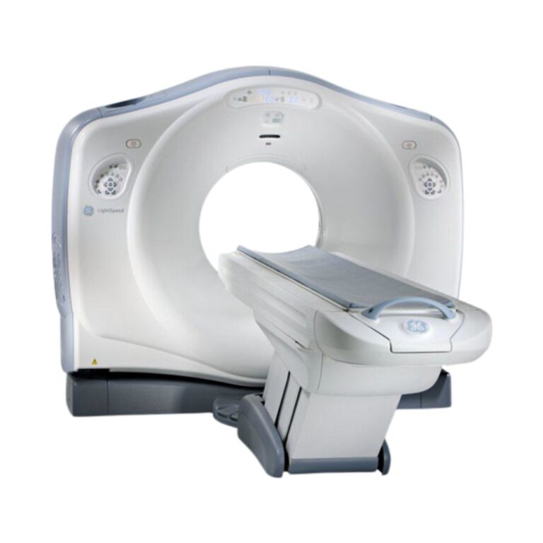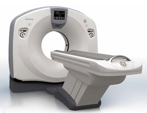
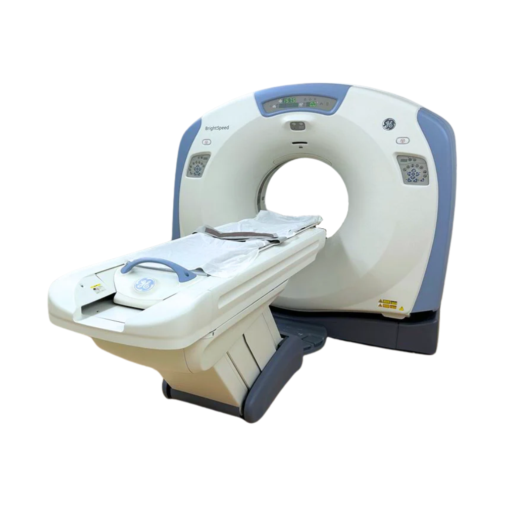
GE BrightSpeed Elite Select 16-Slice CT
- Category:
- CT Scanners
- Brand:
- GE
The GE BrightSpeed Elite Select is a 16-slice CT scanner well suited for hospitals, outpatient imaging centers, and specialty practices. Its 3.5 MHU Solarix X-ray tube manages heat during repeated scans, while thin-slice imaging and a compact design support detailed diagnostics and efficient everyday workflows.
BrightSpeed Elite Select CT Highlights & Benefits
The GE BrightSpeed Elite Select 16 combines detailed multislice imaging, patient-specific scanning controls, and compact system design. Its X-ray generator and 3.5 MHU tube support routine diagnostic CT with configurable clinical software.
Key BrightSpeed Elite Select Capabilities
-
- 16-slice CT acquisition: Captures 16 slices per rotation, with sections as thin as 0.625 mm for detailed anatomical imaging.
- 3.5 MHU Solarix 350 X-ray tube: Uses heat-management and cooling technology to support repeated examinations with fewer potential cooling delays.
- X-ray generator: Supports routine head-to-body CT, with output and maximum tube current dependent on the installed configuration.
- 0.8-second gantry rotation: Supports efficient scanning for chest, abdominal, pelvic, and other examinations.
- 70 cm (27.6 in) gantry opening: Accommodates patient positioning for routine diagnostic imaging.
- Automatic exposure control: Available Auto mA technology adjusts X-ray output according to patient anatomy.
- Advanced software options: Compatible vascular, lung, reconstruction, and image-review tools extend clinical capabilities when installed.
BrightSpeed Elite Select CT Acquisition & Image Detail
HiLight Detector for Detailed 16-Slice Imaging
The BrightSpeed Elite Select uses GE’s HiLight solid-state detector to collect X-ray measurements during scanning.
Its 24 physical detector rows support simultaneous acquisition of 16 slices. The 16 × 0.625 mm mode captures thin sections of anatomy, helping radiologists evaluate fine structures in the lungs, bones, blood vessels, and other tissues.
A 16 × 1.25 mm acquisition mode provides greater anatomical coverage per rotation when thicker sections are appropriate.
These settings allow technologists to balance anatomical detail and scanning coverage according to the examination.
0.8-Second Rotation for Efficient Routine CT
The BrightSpeed Elite Select completes a full gantry rotation in as little as 0.8 seconds.
During helical scanning, the patient table moves continuously through the rotating X-ray beam, allowing the scanner to image larger anatomical regions without stopping between rotations.
This supports routine chest, abdominal, pelvic, and spine imaging, including contrast-enhanced studies when appropriately configured.
The selectable acquisition settings help facilities manage examination times while maintaining the image detail needed for clinical interpretation.
Available IQ Enhance technology helps reduce image distortions during faster helical scanning, supporting greater anatomical coverage while preserving detail in thin-slice images.
Multiple Image Views from a Single CT Scan
The BrightSpeed Elite Select reconstructs acquired scan data into cross-sectional images for clinical interpretation.
With reconstruction speeds up to six images per second, the system can process acquired images efficiently for clinical review.
Multiplanar reconstruction allows clinicians to examine the same anatomy from multiple directions, including front-to-back and side views, without repeating the original scan.
Compatible software can also produce three-dimensional images for evaluating complex anatomy.
These capabilities provide greater flexibility during image review and help clinicians assess different structures using the same examination data.
BrightSpeed Elite Select Image Quality & Dose Management
Volara Digital Processing for Image Clarity
GE’s Volara digital data acquisition technology converts X-ray measurements collected by the detector into information used to create CT images.
Its digital processing helps reduce electronic noise, which can appear as unwanted graininess or interference in an image. This helps preserve anatomical detail for clinical review.
Selectable reconstruction settings allow images to be tailored to different tissues, including lung, bone, and soft-tissue anatomy.
Automatic Exposure & Patient-Specific Dose Control
Available Auto mA technology automatically adjusts X-ray tube current, which controls the amount of radiation produced during scanning, according to patient anatomy and the image quality required.
For example, thicker areas of the body may require more X-ray output than thinner areas. Auto mA helps make these adjustments rather than relying on one fixed exposure level throughout the examination.
This supports patient-specific radiation-dose management while maintaining the image detail needed for diagnosis.
Pediatric and other examination-specific protocols provide additional flexibility when available on the installed system.
Optional ASiR Reconstruction for Dose Reduction
Some BrightSpeed Elite Select configurations may include Adaptive Statistical Iterative Reconstruction (ASiR), GE’s image-processing technology designed to reduce image noise.
ASiR helps reduce the grainy appearance of CT images, potentially allowing certain examinations to use less radiation while preserving the detail needed for diagnosis.
The benefits depend on the patient, examination, and selected scanning settings. ASiR availability must be confirmed for the individual system.
BrightSpeed Elite Select Patient Access & CT Workflow
70 cm Gantry for Patient Access & Positioning
The BrightSpeed Elite Select has a 70 cm (27.6 in) gantry opening for routine head-to-body diagnostic imaging.
Its maximum scan field of view of 50 cm (19.7 in) defines the largest anatomical area captured within an image.
The gantry also tilts up to 30° in either direction, allowing technologists to adjust the scanning angle for head imaging and other positioning needs.
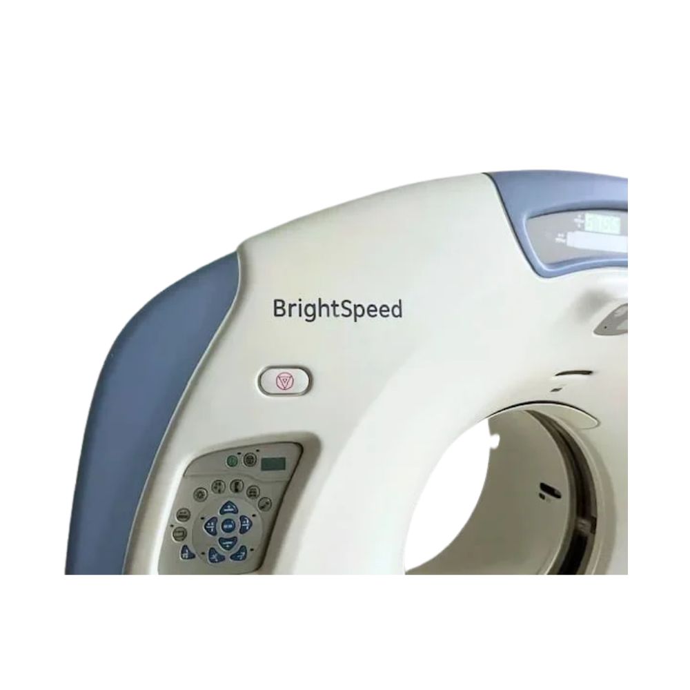
Patient alignment lights, gantry controls, and positioning features help technologists prepare examinations and maintain consistent patient setup.
Patient Table Capacity & Scanning Range
The motorized patient table supports positioning for head, chest, abdominal, pelvic, spine, and extremity examinations.
Compatible configurations accommodate patient loads up to 450 lb (205 kg), providing flexibility for different patient sizes.
Actual patient capacity, scanning range, table movement, and accessories depend on the installed table configuration.
SmartPrep for Accurate Contrast Scan Timing
Programmable examination protocols help technologists select appropriate scanning settings for different body regions and clinical applications.
Available SmartPrep technology helps time CT examinations that use an injected contrast agent, a substance that makes blood vessels and certain tissues easier to see.
SmartPrep monitors when the contrast reaches a selected area of the body so the scan can begin at the appropriate moment. This helps capture the intended anatomy when contrast visibility is most useful, rather than relying solely on a preset delay.
Compatible console software also supports image reconstruction, examination review, and transfer to connected imaging systems.
Clinical Environments & Applications
The GE BrightSpeed Elite Select 16 supports routine diagnostic CT across hospital, outpatient, and specialty imaging environments. Its thin-slice acquisition, 70 cm gantry, and available clinical software make it well suited to facilities seeking established 16-slice imaging capabilities.
Clinical Environments
-
- Hospitals and diagnostic imaging departments
- Outpatient imaging centers
- Emergency departments
- Multispecialty medical practices
- Specialty diagnostic and surgical facilities
- Oncology and cancer care centers
- Veterinary hospitals and specialty referral centers
Clinical Specialties
-
- Radiology: Routine head, chest, abdominal, pelvic, and spine examinations.
- Neurology & Neurosurgery: Brain, skull, and spine evaluation.
- Orthopedics: Fractures, joints, bone anatomy, and preoperative assessment.
- Oncology: Tumor evaluation, disease staging, and treatment follow-up.
- Pulmonology: Lung nodules, pulmonary disease, and thoracic imaging.
- Urology: Kidney stones, renal abnormalities, and urinary tract imaging.
- Gastroenterology: Abdominal and pelvic assessment.
- Emergency Medicine: Evaluation of acute medical conditions and selected traumatic injuries.
- Vascular Medicine: CT angiography and blood vessel evaluation when the required software and contrast protocols are available.
- Veterinary Medicine: Diagnostic CT for companion animals, including neurological, orthopedic, thoracic, and abdominal examinations.
Common CT Applications
-
- Routine noncontrast and contrast-enhanced CT
- Head, brain, and spine imaging
- Chest, abdomen, and pelvis examinations
- Musculoskeletal and extremity CT
- Lung nodule evaluation
- Renal stone assessment
- Oncology staging and follow-up
- Selected trauma examinations
- CT angiography when appropriately equipped
- Veterinary head, spine, thoracic, abdominal, and orthopedic imaging
BrightSpeed Elite Select Advanced Imaging Options
The BrightSpeed Elite Select supports compatible clinical software that can extend its capabilities beyond routine diagnostic examinations.
These applications depend on the installed hardware, workstation, software version, and active licenses.
CT Angiography & Vascular Imaging
CT angiography uses injected contrast material to make blood vessels more visible during scanning.
Compatible systems may include GE’s Advanced Vessel Analysis software, which helps clinicians view blood vessels from different angles and measure features such as vessel diameter.
These tools can support selected vascular examinations when the required software and scanning protocols are installed.
Lung, Bone & 3D Image Review
Compatible imaging software provides additional tools for evaluating lung, bone, and other anatomical structures.
Advanced Lung Analysis supports the assessment and follow-up of lung nodules. AutoBone helps remove bone structures from certain reconstructed images, improving visibility of nearby anatomy. Volume Viewer creates three-dimensional images that clinicians can rotate and examine from different angles.
These applications make complex anatomy easier to review and can support clinical interpretation or surgical planning when installed.
Specialized Clinical Software
Certain configurations may include additional applications such as CT perfusion, which analyzes how injected contrast moves through tissue over time to help assess blood flow.
The available software package determines which specialized examinations and image-review tools the system can support.
MXR Imaging helps facilities select configurations aligned with their clinical applications and imaging requirements.
BrightSpeed Elite Select Configurations & Installed Options
Pre-owned GE BrightSpeed Elite Select scanners may vary by production year, X-ray tube condition, workstation, patient table, software version, and installed clinical applications.
Solarix X-Ray Tube & Hardware Configurations
The BrightSpeed Elite Select uses GE’s Solarix 350 X-ray tube, designed to manage the heat generated during CT scanning.
Its 3.5 million heat unit (MHU) capacity measures how much heat the tube can store. An advanced cooling design helps transfer that heat away from the tube between examinations, reducing potential delays before the next scan.
The Solarix 350 also features a graphite-backed target to help withstand high operating temperatures and a bearing design intended to support reliable operation.
Together, these features support efficient routine diagnostic CT workflows. For pre-owned systems, X-ray tube age, usage history, maintenance, and overall condition remain important considerations.
Clinical Software & Workstation Options
Depending on the system configuration, available options may include:
-
- Auto mA exposure control
- SmartPrep contrast timing
- Multiplanar and 3D reconstruction
- ASiR reconstruction
- Advanced Vessel Analysis
- Advanced Lung Analysis
- AutoBone
- Volume Viewer
- CT perfusion and other specialized applications
These technologies represent possible installed options rather than a standard software package included with every pre-owned scanner.
DICOM Connectivity & Image Management
The BrightSpeed Elite Select supports Digital Imaging and Communications in Medicine (DICOM), the standard used to exchange medical images and related information between compatible systems.
DICOM connectivity supports integration with picture archiving and communication systems (PACS), where images are stored and accessed for clinical review.
Workstation software, image-storage capacity, network services, and other connectivity features vary by installed configuration.
MXR Imaging works with facilities to identify a scanner that fits their clinical applications, examination volume, and existing imaging infrastructure.
BrightSpeed Elite Select vs. Other BrightSpeed Models
The GE BrightSpeed Elite Select provides 16-slice imaging with a 3.5 MHU X-ray tube and configurable software. Its primary distinction from the BrightSpeed Elite is the lower-output hardware, while the Edge Select and Excel Select offer fewer imaging slices.
- GE BrightSpeed Elite 16-Slice CT: Higher-output 16-slice scanner with a 6.3 MHU tube, faster 0.5-second rotation, and broader available advanced imaging options.
- GE BrightSpeed Edge Select 8-Slice CT: Eight-slice configuration for routine diagnostic CT, with different scanning coverage and acquisition capabilities.
- GE BrightSpeed Excel Select 4-Slice CT: Four-slice configuration for facilities whose examination requirements are well matched to lower-slice-count CT imaging.
The Elite Select is well suited to facilities seeking detailed 16-slice imaging without the higher-output hardware of the BrightSpeed Elite.
BrightSpeed Elite Select Installation & Site Requirements
The BrightSpeed Elite Select’s compact gantry design makes it a practical option for facilities replacing an existing CT scanner or planning an imaging suite with limited space.
Installation requirements depend on the selected gantry, patient table, electrical configuration, and supporting equipment.
Site planning includes radiation shielding, structural support, electrical service, cooling, ventilation, patient access, and adequate service clearances.
MXR Imaging offers turnkey imaging project management to help coordinate equipment selection, site preparation, installation, and related project requirements.
BrightSpeed Elite Select 16 CT Price & Availability
MXR Imaging offers pre-owned GE BrightSpeed Elite Select 16-slice CT scanners for healthcare facilities seeking established diagnostic imaging capabilities and flexible clinical software options.
Pricing depends on system condition, X-ray tube history, software licenses, patient table, workstation, accessories, installation requirements, and service coverage.
Eligible systems may also be available through the Platinum Certified CT program, providing an additional reconditioning and quality-assurance pathway.
Ongoing CT maintenance and repair services are available to support equipment performance throughout its operating life.
Request a quote for the GE BrightSpeed Elite Select 16 to compare available pre-owned configurations, clinical software options, pricing, and installation support for your facility.
GE BrightSpeed Elite Select 16 CT Specifications
Specifications may vary by hardware revision, X-ray tube, patient table, software release, and installed options.
CT Acquisition & Detector
-
- OEM model: GE BrightSpeed Elite Select
- Slice count: 16
- Detector: GE HiLight solid-state detector
- Detector configuration: 24 physical detector rows, with 16 simultaneous data acquisition channels
- Thin-slice acquisition: 16 × 0.625 mm
- Additional acquisition mode: 16 × 1.25 mm
- Scanning modes: Helical, axial, and scout
- Minimum gantry rotation: 0.8 seconds
- Acquisition coverage: 10 mm or 20 mm per rotation, depending on detector mode
Gantry & Patient Table
-
- Gantry opening: 70 cm (27.6 in)
- Maximum scan field of view: 50 cm (19.7 in)
- Gantry tilt: ±30°
- Patient capacity: Up to 205 kg (450 lb) on compatible configurations
- Patient positioning: Motorized table
- Table movement and accessories: Configuration-dependent
- Horizontal table travel: Dependent on installed table
X-Ray Generator & Tube
-
- X-ray tube: GE Solarix 350
- Tube heat storage: 3.5 MHU
- Generator output: Up to 42 kW, configuration-dependent
- Tube voltage: 80, 100, 120, and 140 kVp
- Tube current: Up to 260 mA or 350 mA, depending on configuration
- Tube cooling: High-efficiency heat-transfer and cooling design
Image Reconstruction & Performance
-
- Image matrix: 512 × 512
- Minimum acquired slice thickness: 0.625 mm
- Reconstruction speed: Up to 6 images per second
- Multiplanar reconstruction: Supported
- Digital data acquisition: GE Volara
- Automatic exposure control: Available by configuration
- SmartPrep contrast monitoring: Available on equipped systems
- 3D reconstruction: Available with compatible software
- ASiR reconstruction: Configuration-dependent
- IQ Enhance: Available on compatible configurations
- Specialized clinical software: Dependent on installed applications
System & Site Requirements
-
- Image connectivity: DICOM
- PACS integration: Supported with compatible network configuration
- Electrical service: Three-phase, configuration-dependent
- Radiation shielding: Site-specific
- Cooling and ventilation: Based on installed equipment and room requirements
- Structural support: Required for gantry, patient table, and supporting components
- Room dimensions and service clearances: Based on selected equipment and site-planning requirements