Konica Minolta DDR
- Category:
- X-Ray & U-Arm Systems
- Brand:
- Konica Minolta
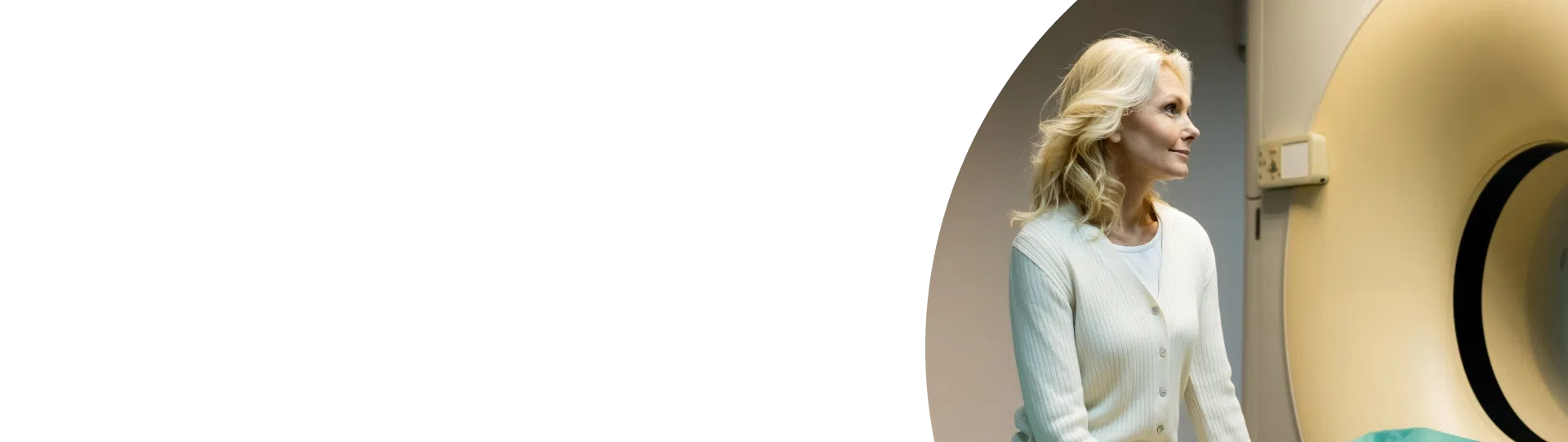
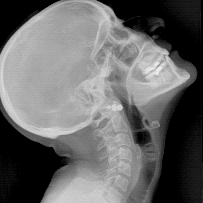
See anatomy in motion with Dynamic Digital Radiography. Konica Minolta DDR captures high-speed X-ray image sequences that allow clinicians to evaluate anatomical structures during movement. By combining anatomical visualization with functional assessment, DDR provides insight into biomechanics, joint mechanics, and physiological motion that may not be visible on static radiographs. DDR integrates into familiar digital radiography workflows and supports musculoskeletal and pulmonary imaging applications.
What Is Dynamic Digital Radiography (DDR)?
Traditional digital radiography captures a single moment in time. Konica Minolta DDR expands on conventional X-ray imaging by acquiring a sequence of images that can be viewed individually or played as a cine loop, allowing clinicians to evaluate anatomical structures during motion.
This additional functional perspective can help clinicians assess movement patterns, joint mechanics, and physiological motion that may not be fully appreciated on static imaging alone.
Key Benefits of Dynamic Digital Radiography
-
- Captures anatomy in motion using X-ray technology
- Produces cine-loop image sequences for dynamic review
- Supports image analysis and motion assessment
- Acquired in natural upright or weight-bearing positions when appropriate
- Enhances evaluation of biomechanics and musculoskeletal function
DDR is not fluoroscopy. It is X-ray that moves. While fluoroscopy and cineradiography have long been used to examine joints and anatomical structures at rest and in motion, DDR provides dynamic X-ray imaging within a familiar digital radiography environment, enabling clinicians to visualize movement while maintaining standard radiographic workflows.
Watch Dynamic X-Ray Imaging in Motion
The video below showcases Dynamic Digital Radiography examinations across several anatomical regions, including the shoulder, elbow, wrist, cervical spine, and lower extremities.
As each motion sequence plays, you can see how DDR captures movement throughout a range of motion rather than a single moment in time. This ability to visualize anatomical relationships during movement has generated significant interest among orthopedic, sports medicine, chiropractic, musculoskeletal, rehabilitation, and radiology professionals.
By combining traditional X-ray imaging with motion visualization, DDR provides additional functional information that may support clinical evaluation, treatment planning, rehabilitation monitoring, and follow-up assessment.
How DDR Provides Functional Imaging Beyond Static X-Rays
Many imaging modalities provide excellent anatomical detail but limited insight into how structures function during movement. DDR helps bridge that gap by combining anatomical visualization with motion assessment.
Clinical Benefits of Motion-Based X-Ray Imaging
-
- Visualize anatomy throughout a range of motion
- Review movement retrospectively after image acquisition
- Evaluate dynamic interactions between bone, muscle, and joint structures
- Analyze biomechanics and musculoskeletal function
- Support communication between imaging providers, physicians, and patients
Because motion is captured as a sequence of images, clinicians can observe relationships between anatomical structures that may be difficult to assess using static radiographs alone.
Clinical Applications
Dynamic Digital Radiography (DDR) can support a wide range of clinical imaging environments where evaluating anatomy during motion may provide additional diagnostic insight. By capturing a sequence of X-ray images during movement, DDR helps clinicians visualize functional anatomy alongside traditional static radiographs.
Common Clinical Applications
-
- Orthopedic imaging and joint biomechanics
- Sports medicine and musculoskeletal injury evaluation
- Chiropractic imaging and spinal motion assessment
- Rehabilitation and treatment follow-up
- Pulmonary imaging and respiratory motion evaluation
- General radiology and diagnostic imaging
- Preoperative planning and postoperative assessment
- Common Anatomical Areas Evaluated
- Shoulder motion and scapulohumeral rhythm
- Cervical spine movement
- Lumbar spine movement
- Knee motion and patellar tracking
- Wrist and hand motion
- Elbow movement
- Ankle motion under load
- Thoracic and respiratory motion
Common Practice Environments
-
- Hospitals
- Imaging centers
- Orthopedic practices
- Sports medicine clinics
- Chiropractic clinics
- Outpatient radiology centers
- Rehabilitation facilities
DDR’s ability to visualize anatomy throughout movement makes it a valuable complement to conventional radiography across a variety of musculoskeletal and pulmonary imaging applications.
Integrating DDR Into Existing Radiology Workflows
One of DDR’s advantages is its ability to fit within established radiology workflows.
DDR-capable systems continue to perform standard static radiographic examinations while providing the option to acquire dynamic image sequences when clinically appropriate.
Workflow Advantages of DDR Technology
-
- Familiar radiographic acquisition process
- No separate imaging room required
- Retrospective review of captured motion
- Supports collaboration between radiologists and referring physicians
- Enables dynamic and static imaging from the same platform
Unlike fluoroscopic procedures, DDR examinations do not require physician presence during image acquisition, making implementation practical for many imaging departments.
AI-Powered Motion Analysis & Quantitative Imaging
DDR provides a foundation for advanced image analysis and future AI-assisted applications.
The motion data captured during DDR studies can be analyzed to quantify anatomical movement patterns, supporting objective evaluation and future automation opportunities. Ongoing research continues to explore machine learning applications for motion assessment and measurement.
Emerging Applications for AI and Motion Tracking
-
- Automated motion measurements
- Quantitative biomechanics analysis
- Shoulder kinematics assessment
- Scapular motion tracking
- Advanced clinical decision support
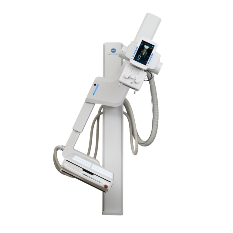
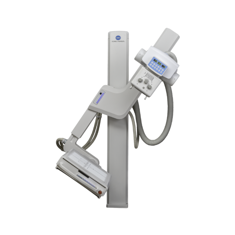
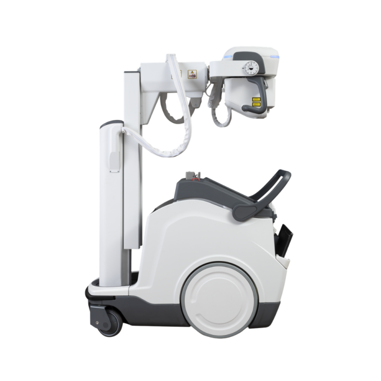
DDR-Compatible X-Ray Systems and Configurations
Dynamic Digital Radiography requires DDR-capable Konica Minolta imaging technology.
X-Ray System Types That Support DDR
-
- Fixed digital radiography rooms
- Overhead tube crane systems
- Floor-mounted radiography systems
- Upright imaging configurations
- Mobile and portable imaging environments, depending on system configuration
MXR can help determine DDR compatibility for new imaging installations and existing Konica Minolta DR environments.
Specifications
DDR Imaging Technology Features
-
- Dynamic Digital Radiography (DDR)
- High-speed sequential image acquisition
- Cine-loop image review capability
- Static and dynamic imaging from the same platform
- Motion-based X-ray visualization
- Functional imaging assessment capabilities
Image Acquisition and Frame Rate Capabilities
-
- Acquires a sequence of individual digital radiographs
- Available frame rates include 6 fps and 15 fps depending on exam protocol
- Supports retrospective review of motion sequences
Clinical Imaging Areas Supported by DDR
-
- Upper extremities
- Lower extremities
- Spine
- Thorax
- Musculoskeletal imaging
- Pulmonary imaging
Workflow and Motion Analysis Features
-
- Static and dynamic imaging on DDR-capable systems
- Upright and weight-bearing imaging capabilities where applicable
- Quantitative motion analysis support
- AI-ready imaging platform