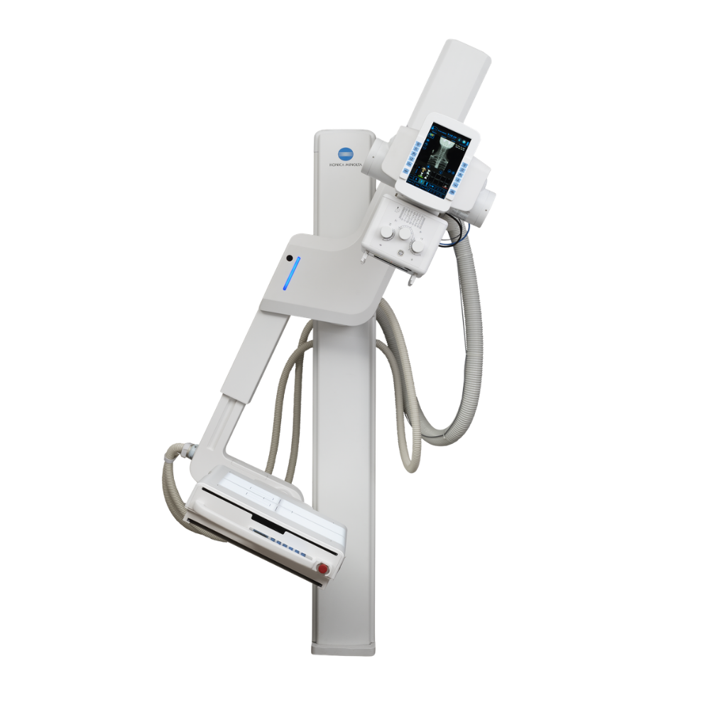
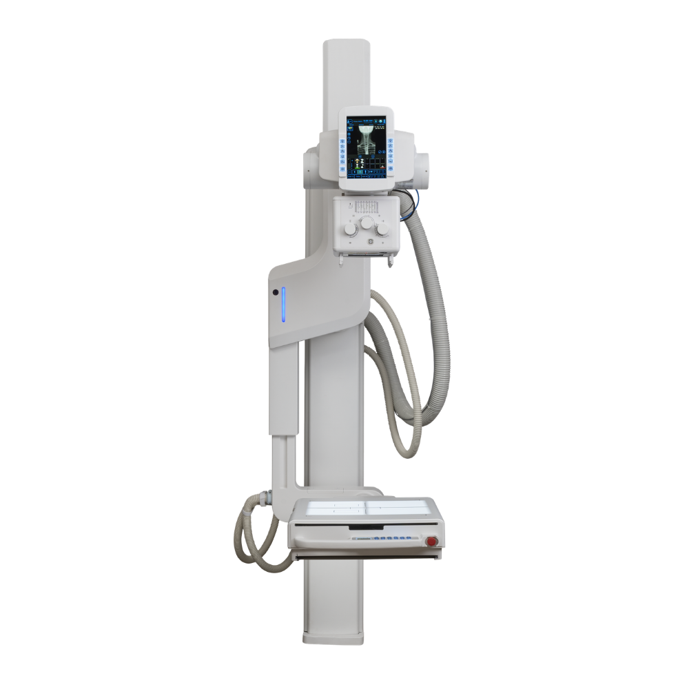
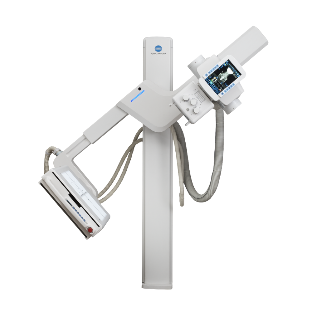
Konica Minolta KDR Advanced U-Arm System
- Category:
- X-Ray & U-Arm Systems
- Brand:
- Konica Minolta
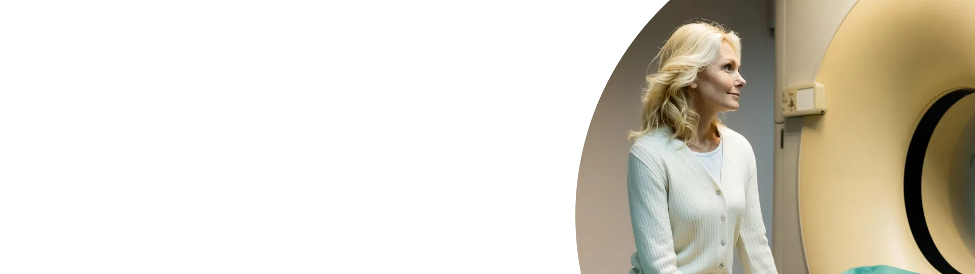
The Konica Minolta KDR Advanced U-Arm System is a compact, high-performance digital radiography system designed to deliver fast clinical answers without sacrificing space. Perfect for facilities with limited square footage, this advanced floor-to-wall mounted X-ray system combines state-of-the-art imaging technology with motorized positioning to optimize patient throughput and enhance clinical workflow.
Video Tour & In-Depth System Overview
Watch this video to see the Konica Minolta KDR Advanced U-Arm System in action, highlighting key clinical benefits and featuring a live demonstration of a technologist operating the system.
Space-Saving Design & Easy Installation
Low Ceiling Compatibility & Floor Mounting
The floor-mounted architecture is engineered to fit into tight spaces. It easily accommodates ceiling heights as low as 8 feet, allowing clinical practices to maximize existing office square footage without requiring expensive room renovations or structural ceiling modifications.
Compact Streamlined Detector Design
The streamlined detector frame is built to optimize access around the patient. By allowing better head-end and foot-end access, the design simplifies patient positioning and makes the system comfortable to navigate for both technologists and patients.
Superior Image Quality & Advanced Imaging Technology
High-Definition Cesium Iodide Detector
Equipped with a premium 17″ x 17″ Cesium Iodide (CsI) detector, the system maximizes clinical efficiency and provides exceptional visualization of both bone and soft tissue from a single study exposure. This high-efficiency design helps clinicians reach accurate diagnoses sooner. Additionally, a durable, protective panel enclosure helps to increase system longevity and stability, protecting your technology investment over years of clinical operation.
Direct X-ray & Panel Alignment
To prevent geometric distortion and ensure perfect image capture, the system maintains constant alignment. The X-ray tube and DR panel remain perfectly aligned regardless of the swivel arm tilt or panel angle.
Dynamic Digital Radiography Compatibility & Motion Imaging
The system is DDR-ready, allowing facilities to upgrade to Dynamic Digital Radiography (DDR) to view anatomical motion in real-time, expanding diagnostic capabilities. This novel, low-dose X-ray imaging technique enables medical professionals to observe physiological movement like never before.
The DDR option can acquire up to 15 sequential radiographs per second and play them back as a cine loop, allowing clinicians to observe the physiological cycle and individual radiographic images up to 17″ x 17″ in size. This rapid acquisition delivers up to 20 seconds of physiological movement in less than a minute. The exam is performed quickly by standard radiology staff without requiring a physician to be present. Additionally, because the radiation dose is significantly lower than an average fluoroscopy exam, patients receive safer, highly targeted care. The system remains incredibly versatile, allowing these motion images to be captured standing, seated, or on a table.
Optimized Patient Care & Rapid Workflow
Automated Swivel Technology
The system automatically moves to predetermined positions based on selected exam information. This automated positioning minimizes manual adjustments, saving valuable time during busy clinical days.
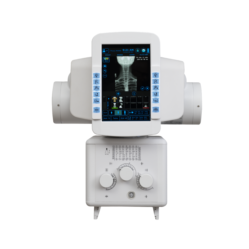
Rapid Three-Second Image Review
Captured images are displayed for on-screen review in under three seconds. This instantaneous feedback allows technologists to confirm positioning immediately, resulting in faster patient turnover and reduced exam times.
Tube-Mounted User Interface
With an integrated touchscreen control on the tube head, technologists can confirm patient data and review images at the point of care. This minimizes the need to repeatedly walk back and forth to the control booth.
Effortless Patient Transitions
Clinicians can transition smoothly from PA to lateral positions without moving the patient. Combined with the standard 440 lb. capacity mobile patient table, this feature ensures safe, dignified, and comfortable imaging for patients with limited mobility.
Real-World Clinical Applications
Specialized Orthopedics & Chiropractic Care
For orthopedic clinics and chiropractic offices, the system offers optional automatic stitching features. This capability allows for highly accurate, long-bone and full-spine imaging required for joint assessment and spinal alignment analysis.
High-Throughput Diagnostic Centers & Hospitals
In busy diagnostic centers and hospital emergency settings, speed is essential. The rapid three-second preview and automated positioning protocols allow staff to process high volumes of patients efficiently while maintaining reliable image quality.
Technical Specifications
- Main Column: Floor-to-wall-mounted main column supporting vertical movement with a dual-speed, motorized swivel arm.
- Swivel Arm Range: 135-degree range of motion with convenient detents at 0, 30, 90, and 120 degrees.
- Vertical Travel: 39 inches of motorized vertical arm movement to accommodate patients of all heights.
- Patient Table: 440 lb. capacity mobile patient table included as a standard feature.
- Collimation: Advanced linear laser collimator with a light field indicator and a three-knob manual control.
- Imaging Software: ULTRA Acquisition Software for advanced image processing and maximum clarity.