KA Imaging Reveal Mobi Pro Mobile X-Ray
- Category:
- X-Ray Systems
- Brand:
- KA Imaging
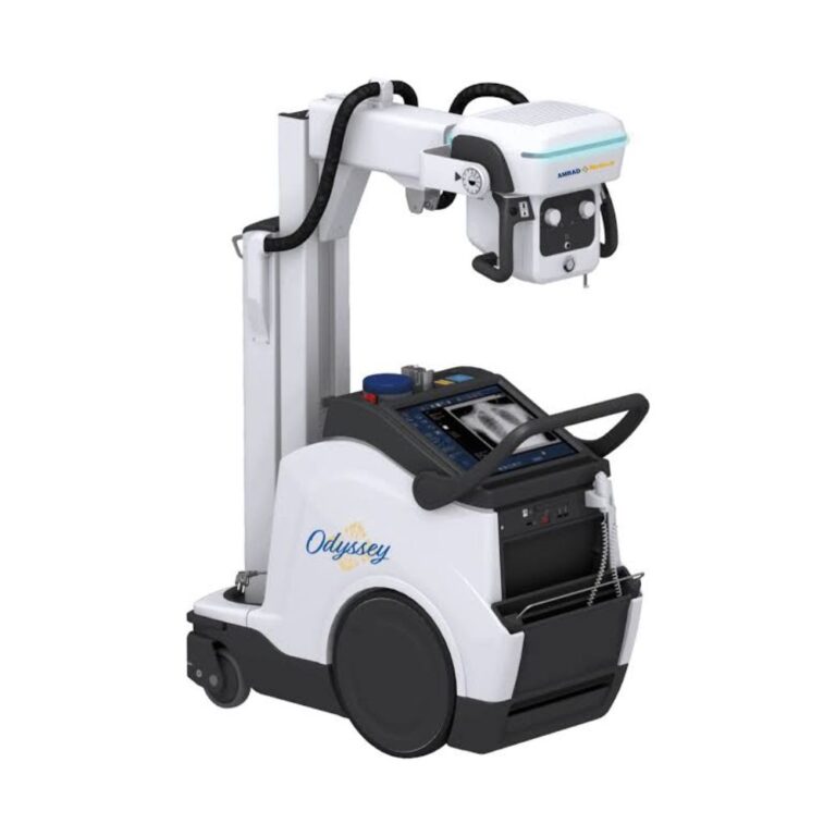
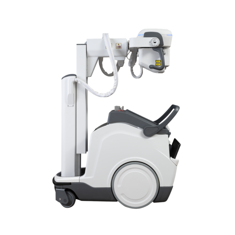
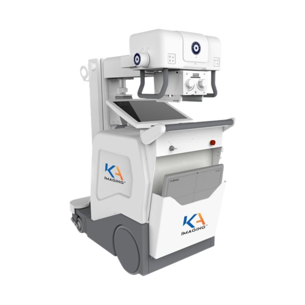
The KA Imaging Reveal Mobi Pro is a motor-assisted mobile X-ray system best suited for hospital bedside imaging, including intensive care and emergency department use. It combines a 40 kW generator with the Reveal 35C detector to produce standard, bone, and soft-tissue images from a single X-ray exposure.
Reveal Mobi Pro Mobile X-Ray Highlights & Benefits
The KA Imaging Reveal Mobi Pro combines a complete mobile X-ray system with the Reveal 35C digital detector and KA Imaging’s dual-energy technology. From one X-ray exposure, the system produces a standard radiograph plus separate bone and soft-tissue images without requiring additional exposures for the supplemental views.
Reveal Mobi Pro Key Benefits
-
- Standard, bone, and soft-tissue images from one X-ray exposure
- Reveal 35C 14 x 17 in. (35 x 43 cm) digital X-ray detector
- 40 kW X-ray generator
- Motor-assisted drive for moving between imaging locations
- Collapsible column for a lower transport profile
- Integrated 19 in. color touchscreen workstation
- Programmable anatomical exam settings
- Telescopic arm and flexible tube-head positioning
- Anti-collision bumpers
- Battery-powered mobile operation
The system brings additional dual-energy image information directly to the bedside while maintaining a workflow similar to conventional mobile X-ray imaging.
KA Imaging Reveal Mobi Video Preview
See the Reveal Mobi Pro’s mobility, bedside positioning, integrated workstation, and imaging workflow.
Reveal 35C Dual-Energy Imaging: 3 Images from 1 Exposure
SpectralDR is KA Imaging’s dual-energy X-ray technology. Integrated into the Reveal 35C detector, it separates X-ray information during a single exposure to create a standard radiograph plus supplemental bone and soft-tissue images.
Standard, Bone & Soft-Tissue Images from 1 Exposure
All three images are acquired simultaneously from one standard X-ray exposure. No second exposure or patient repositioning is required to produce the additional bone and soft-tissue views.
This gives clinicians additional image information while preserving a familiar mobile radiography acquisition process.
Bone & Soft-Tissue Differentiation at the Bedside
Separating bone and soft-tissue information can improve visualization when overlapping anatomy makes a finding more difficult to see on a standard radiograph.
This capability is particularly relevant to bedside and chest imaging, where ribs and other dense structures can overlap the lungs, lines, tubes, and other areas of interest.
Reveal 35C Detector & Connectivity
The Reveal 35C is a 14 x 17 in. (35 x 43 cm) digital X-ray detector with a 140 µm pixel pitch and cesium iodide scintillator.
The detector supports wired Gigabit Ethernet and IEEE 802.11 ac/n Wi-Fi connectivity. Hot-swappable batteries allow a depleted battery to be exchanged rather than waiting for the detector to complete a full recharge.
For additional detector details, view the KA Imaging Reveal 35C X-Ray Detector.
Reveal Mobi Pro Clinical Environments & Bedside Applications
The KA Imaging Reveal Mobi Pro supports hospital mobile X-ray imaging when radiography needs to be brought directly to the patient. Its motor-assisted drive, bedside positioning range, integrated detector, and single-exposure dual-energy imaging are particularly useful when patient transport to a fixed X-ray room is undesirable or impractical.
Intensive Care, Emergency Department & Bedside Imaging
The system is particularly well suited for:
-
- Intensive care units
- Emergency departments
- Critical care areas
- Other hospital bedside imaging environments
The mobile design allows imaging to take place where the patient is being treated while keeping acquisition, image review, and detector access together on the cart.
SpectralDR Clinical Applications
The supplemental bone and soft-tissue images are particularly relevant to chest imaging applications where overlapping anatomy can limit visualization on a standard radiograph.
Documented applications include:
-
- Lung nodule visualization
- Pneumonia imaging
- Pneumothorax imaging
- Line and tube tip visualization
- Retained surgical object visualization
- Calcification visualization
Coronary calcium visualization is also being studied as an emerging application for Reveal 35C dual-energy imaging.
Reveal Mobi Pro Mobility & Bedside Positioning
Collapsible Column for Transport Visibility
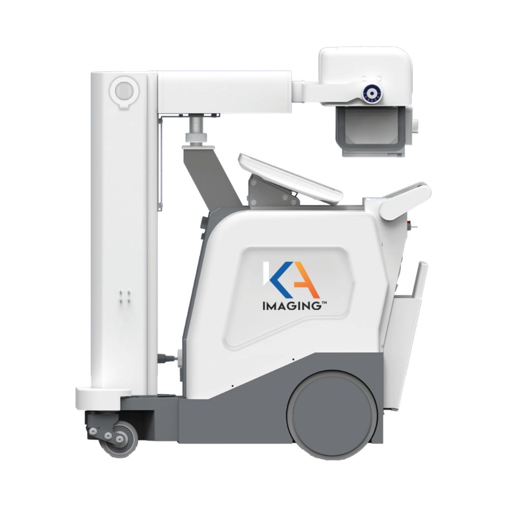
The KA Imaging Reveal Mobi Pro uses a collapsible column to lower its transport profile and improve the operator’s forward view while moving through corridors and patient-care areas.
In transport position, the system measures approximately 22.7 x 46.2 x 53.3 in. (57.6 x 117.3 x 135.4 cm). The transport handle is positioned 35 in. (89 cm) from the floor.
Motor-Assisted Drive & Anti-Collision Bumpers
Motor-assisted movement helps operators transport the 959 lb. (435 kg) system between imaging locations.
Travel speeds include:
-
- Forward in transport position: 0 to 3 mph
- Forward in working position: 0 to 1.6 mph
- Reverse: 0 to 1.6 mph
Anti-collision bumpers help protect the system while maneuvering around beds, equipment, doorways, and other obstacles.
Telescopic Arm & Tube-Head Positioning
The telescopic arm provides 14.2 in. (36 cm) of extension to help position the X-ray tube around beds and patients.
Positioning ranges include:
-
- Front range: 28 to 42 in. (71 to 107 cm)
- Lateral range: 20 to 34.5 in. (51.7 to 87.7 cm)
- Tube focal spot height from the floor: 27.2 to 80.5 in. (69 to 204.5 cm)
- Maximum system height: 86 in. (217.6 cm)
The tube head also provides multi-directional rotation for adapting the system to different bedside positions.
Reveal Mobi Pro Imaging Controls & Operator Workflow
40 kW Generator & Exposure Controls
The KA Imaging Reveal Mobi Pro uses a high-frequency 40 kW X-ray generator.
Operators can use:
-
- 2-point technique, selecting kV and mAs
- 3-point technique, selecting kV, mA, and exposure time
- Programmable anatomical exam presets
The 2-point and 3-point controls give technologists direct control over the exposure factors needed for different examinations, while programmable presets support repeatable technique selection.
Integrated 19-Inch Touchscreen Workstation
A 19 in. color LCD touchscreen provides the primary system interface for image acquisition and review.
The integrated computer includes:
-
- Windows 10 Pro 64-bit
- 3.0 GHz Intel Core i5 processor
- 8 GB RAM
- 500 GB SSD
Keeping the workstation on the mobile system gives the operator access to acquisition controls and images at the bedside.
Battery Operation & Charging
The KA Imaging Reveal Mobi Pro is battery powered for mobile use throughout the facility.
The system is rated for an average of approximately 500 exposures between charges, depending on exposure conditions. Full system recharge time is approximately five hours.
The Reveal 35C detector uses its own hot-swappable battery system and can also operate while connected to AC power.
KA Imaging Reveal Mobi Pro Specifications
X-Ray Generator & Exposure Controls
-
- Generator: High-frequency monoblock, a compact integrated generator design
- Maximum generator power: 40 kW
- 2-point technique: kV and mAs
- 3-point technique: kV, mA, and exposure time
- Programmable anatomical exam settings
Operator Interface & Computer
-
- Display: 19 in. color LCD touchscreen
- Operating system: Windows 10 Pro 64-bit
- Processor: 3.0 GHz Intel Core i5
- Memory: 8 GB RAM
- Storage: 500 GB SSD
System Dimensions & Weight
-
- Transport dimensions: 22.7 x 46.2 x 53.3 in. (57.6 x 117.3 x 135.4 cm)
- Transport handle height: 35 in. (89 cm)
- Maximum height: 86 in. (217.6 cm)
- System weight with batteries: 959 lb. (435 kg)
Telescopic Arm & Tube Positioning
-
- Telescopic extension: 14.2 in. (36 cm)
- Front positioning range: 28 to 42 in. (71 to 107 cm)
- Lateral positioning range: 20 to 34.5 in. (51.7 to 87.7 cm)
- Tube focal spot height from floor: 27.2 to 80.5 in. (69 to 204.5 cm)
- A-swivel: ±180°
- B-swivel: ±320° from transport position
- C-swivel: 127°, including 90° forward and 37° backward
Motor-Assisted Drive & Battery
-
- Forward speed in transport position: 0 to 3 mph
- Forward speed in working position: 0 to 1.6 mph
- Reverse speed: 0 to 1.6 mph
- Anti-collision bumpers
- System power: Battery
- Exposures per charge: Approximately 500, depending on exposure conditions
- System recharge time: Approximately 5 hours
Reveal 35C Detector
-
- Detector size: 14 x 17 in. / 35 x 43 cm
- Detector weight: 8 lb. (3.6 kg), including battery
- Pixel pitch: 140 µm
- Sensor: Amorphous silicon
- Scintillator: Cesium iodide (CsI)
- Image digitization: 16-bit analog-to-digital conversion
- Wired connectivity: Gigabit Ethernet, 1000BASE-T
- Wireless connectivity: IEEE 802.11 ac/n Wi-Fi
- Power: Hot-swappable battery or tethered AC
- AC input: 100-240 VAC, 50-60 Hz
- Charging time: Approximately 2.5 hours from empty to full
- Battery operation: Up to 5 hours, depending on imaging technique
Regulatory Status
-
- Reveal Mobi Pro: FDA 510(k) cleared
- Reveal 35C: FDA 510(k) cleared
For additional information and specifications, access the PDFs provided below.