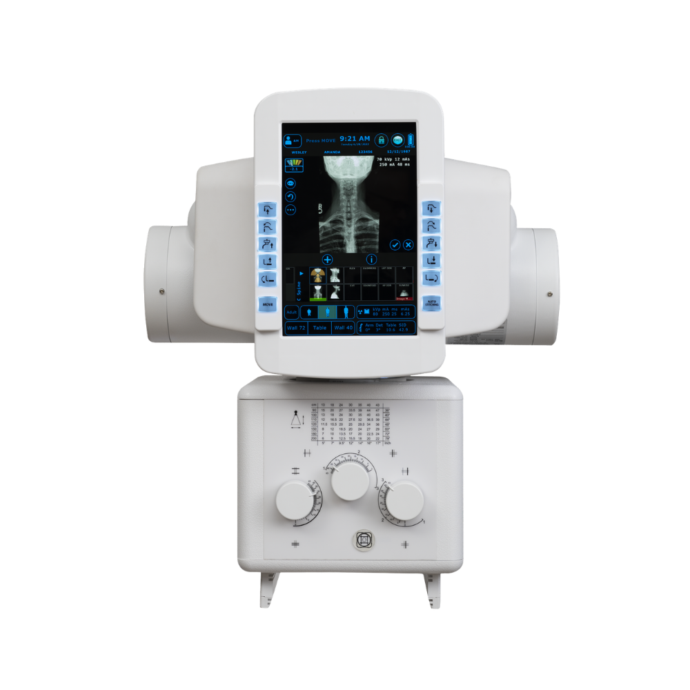
Konica Minolta KDR Advanced U-Arm System
- Category:
- X-Ray Systems
- Brand:
- Konica Minolta
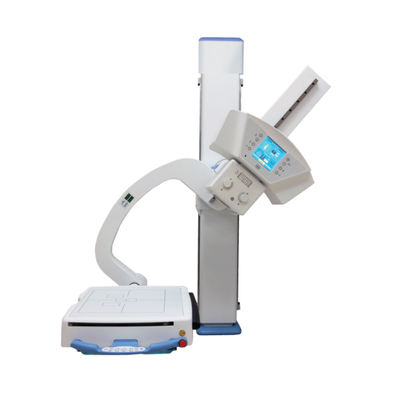
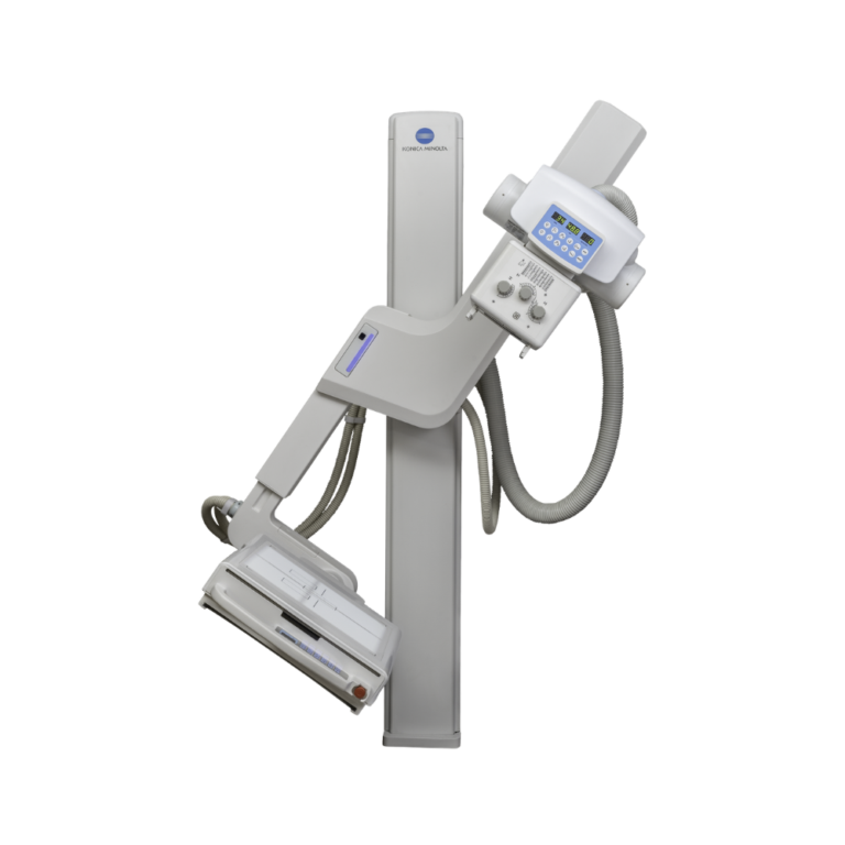
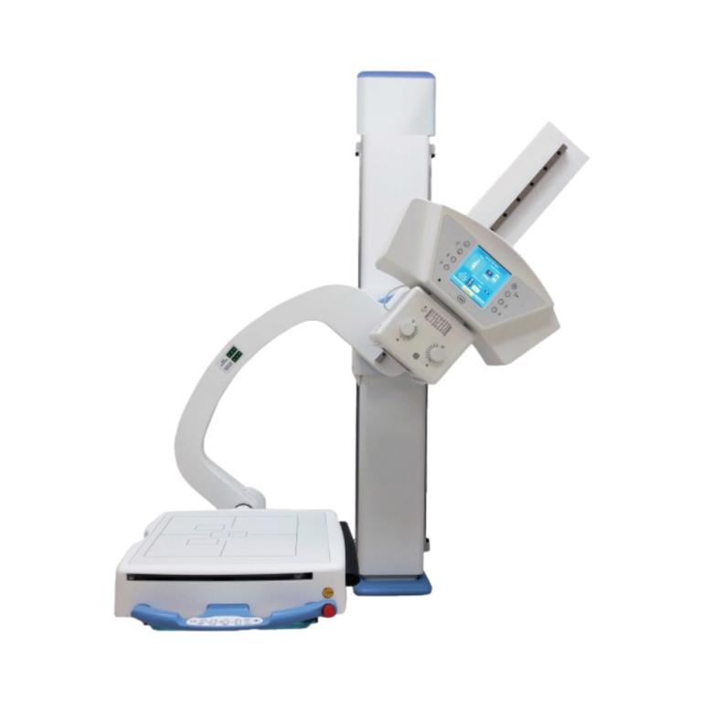
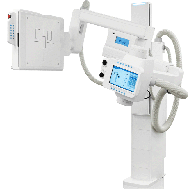
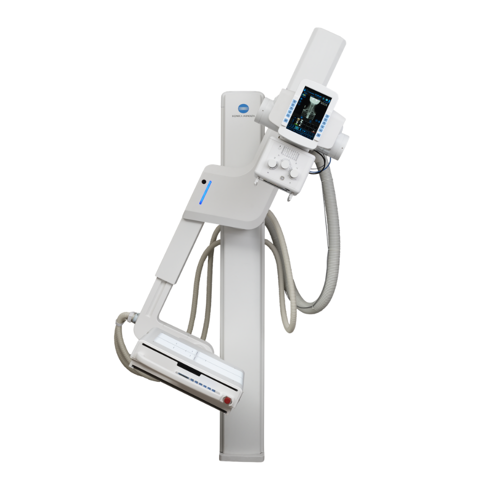
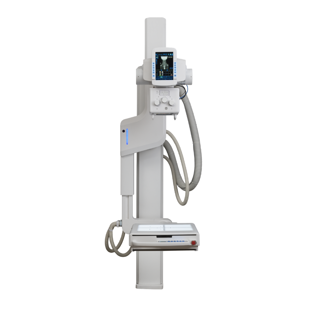
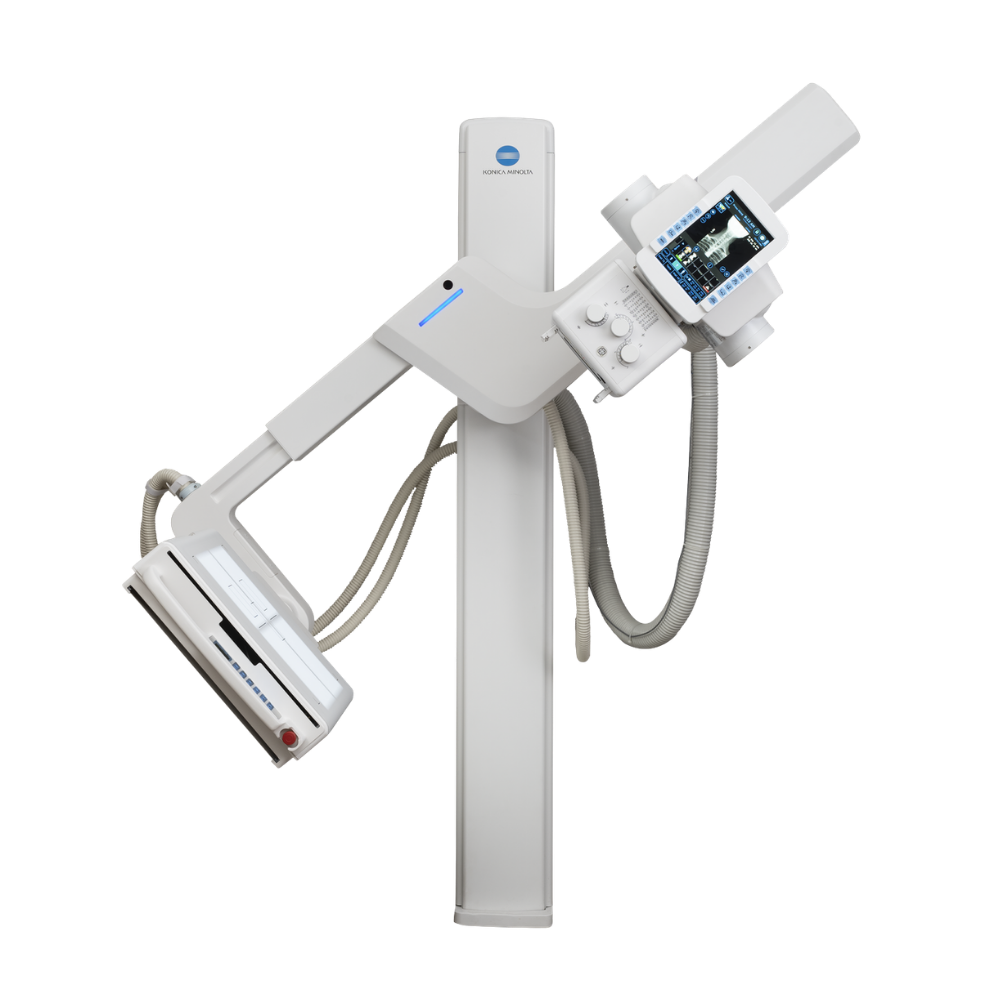
The Konica Minolta KDR Advanced U-Arm System is a compact, high-performance digital radiography system designed for practices, imaging centers, and hospital radiology departments. Its floor-to-wall-mounted design combines motorized positioning, a 17 × 17-inch CsI detector, and optional DDR to support versatile radiography in space-conscious X-ray rooms.
KDR Advanced U-Arm Highlights & Benefits
The KDR Advanced is a motorized U-arm X-ray system designed to keep the X-ray tube and detector aligned as the system moves through different imaging positions.
Key Features & Benefits
-
- Floor-to-wall-mounted design compatible with rooms with 8-foot ceilings
- Motorized 135° swivel-arm range for flexible patient positioning
- 17 × 17 in. (43 × 43 cm) Cesium Iodide detector with AeroDR technology
- Automated positioning based on selected exam information
- Image display for on-screen review in under three seconds
- PA-to-lateral positioning without moving the patient
- Table, standing, seated, and wheelchair imaging capabilities
- Optional Dynamic Digital Radiography (DDR) and automatic stitching
Video Tour & In-Depth System Overview
Watch this video to see the Konica Minolta KDR Advanced U-Arm System in action, highlighting key clinical benefits and featuring a live demonstration of a technologist operating the system.
Space-Saving Design & Easy Installation
Low Ceiling Compatibility & Floor Mounting
The floor-mounted architecture is engineered to fit into tight spaces. It easily accommodates ceiling heights as low as 8 feet, allowing clinical practices to maximize existing office square footage without requiring expensive room renovations or structural ceiling modifications while maintaining the positioning flexibility of a U-arm X-ray system.
Compact Streamlined Detector Design
The streamlined detector frame is built to optimize access around the patient. Improved head-end and foot-end access simplifies patient positioning and allows patients to be positioned in front of or behind the detector as needed for the exam.
17 × 17-Inch CsI Detector & Advanced Imaging Technology
High-Definition Cesium Iodide Detector
Equipped with a premium 17″ x 17″ Cesium Iodide (CsI) detector featuring AeroDR technology, the KDR Advanced provides detailed bone and soft-tissue visualization from a single study exposure. A durable protective panel enclosure also helps support detector stability and system longevity. Additionally, a durable, protective panel enclosure helps to increase system longevity and stability, protecting your technology investment over years of clinical operation.
Constant X-Ray Tube & Detector Alignment
The system maintains alignment between the X-ray tube and DR panel throughout changes in panel angle and swivel-arm position. This constant alignment supports consistent image geometry across a range of radiographic views.
Optional Dynamic Digital Radiography
The KDR Advanced U-Arm is available with Dynamic Digital Radiography (DDR) to view anatomical motion in real-time, which acquires sequential X-ray images that can be reviewed individually or played as a cine loop to visualize anatomy in motion.
The DDR option can acquire up to 15 sequential radiographs per second and play them back as a cine loop, allowing clinicians to observe the physiological cycle and individual radiographic images up to 17″ x 17″ in size. This rapid acquisition delivers up to 20 seconds of physiological movement in less than a minute. The exam is performed quickly by standard radiology staff without requiring a physician to be present. Additionally, because the radiation dose is significantly lower than an average fluoroscopy exam, patients receive safer, highly targeted care. The system remains incredibly versatile, allowing these motion images to be captured standing, seated, or on a table.
Automated Positioning & Exam Workflow
Automated Swivel Positioning
The system automatically moves to predetermined positions based on selected exam information. This automated positioning minimizes manual adjustments, saving valuable time during busy clinical days.
Rapid Three-Second Image Review
Captured images are displayed for on-screen review in under three seconds. This instantaneous feedback allows technologists to confirm positioning immediately, resulting in faster patient turnover and reduced exam times.
Tube, Detector & Remote Controls
A tube-mounted user interface allows technologists to confirm patient information without repeatedly returning to the main workstation. Controls at the tube and detector, along with remote operation, provide additional flexibility when positioning the system around the patient.
Effortless Patient Transitions
Clinicians can transition smoothly from PA to lateral positions without moving the patient. Combined with the standard 440 lb. capacity mobile patient table, this feature ensures safe, dignified, and comfortable imaging for patients with limited mobility.
PA, Lateral, Table & Wheelchair Positioning
The KDR Advanced can move from PA to lateral positions without requiring the patient to reposition. Independent positioning controls also support other common radiographic views, including table and wheelchair exams.
Clinical Environments & Applications
Specialized Orthopedic, Chiropractic & Musculoskeletal Imaging
For orthopedic clinics, chiropractic practices, and other musculoskeletal imaging environments, optional automatic stitching can capture and combine multiple images for long-bone, spine, and full-body radiography. The system also supports standing and weight-bearing positioning when appropriate for the examination.
General Radiography in Practices, Imaging Centers & Hospitals
The KDR Advanced supports routine digital radiography across practices and organizations of different sizes. Automated positioning, flexible U-arm movement, and rapid image review support chest, skeletal, extremity, and other general radiographic examinations in outpatient and hospital environments.
Seated, Wheelchair & Limited-Mobility Imaging
The system’s positioning range supports patients who need to remain seated or in a wheelchair as well as patients positioned on the mobile table. Moving from PA to lateral positioning without moving the patient can also simplify exams when patient mobility is limited.
System Equipment & Options
The KDR Advanced U-Arm can be configured around the imaging requirements of the facility.
-
- 440 lb. (200 kg) capacity mobile patient table included
- Advanced three-knob collimator with linear laser
- Optional collimator camera
- Optional automatic stitching for long-bone, spine, and full-body imaging
- Optional Dynamic Digital Radiography (DDR)
Konica Minolta KDR Advanced U-Arm Specifications
System Design & Positioning
-
- System Type: Floor-to-wall-mounted U-arm digital X-ray system
- Ceiling Compatibility: Accommodates rooms with ceilings as low as 8 ft. (2.44 m)
- Swivel Arm Range: 135° range of motion
- Positioning Detents: 0°, 30°, 90°, and 120°
- Vertical Arm Travel: 39 in. (99 cm) of motorized vertical movement
- Positioning: Automated movement to predetermined exam positions and source-to-image-receptor distance
- Tube & Detector Alignment: Maintained throughout changes in detector angle and swivel-arm position
Detector & Image Acquisition
-
- Detector Size: 17 × 17 in. (43 × 43 cm)
- Detector Type: Cesium Iodide (CsI) digital detector
- Imaging Technology: Konica Minolta AeroDR
- High-Definition Imaging: 100 μm at 72% DQE at 0 cycle/mm
- Image Review: Captured image displayed for on-screen review in under three seconds
- Image Processing: Konica Minolta ULTRA acquisition software
Controls & Patient Support
-
- System Controls: Tube-mounted interface, detector-side controls, and remote control
- Patient Positioning: Standing, seated, table, and wheelchair imaging
- PA-to-Lateral Positioning: Supports transitions without repositioning the patient
- Patient Table: Mobile patient table included
- Table Capacity: 440 lb. (200 kg)
- Collimation: Three-knob collimator with linear laser
- Collimator Camera: Optional
Optional Imaging Capabilities
-
- Dynamic Digital Radiography: Optional DDR for sequential motion X-ray acquisition
- DDR Acquisition: Up to 15 sequential radiographs per second
- DDR Positioning: Standing, seated, and table imaging
- Automatic Stitching: Optional long-length image acquisition for long-bone, spine, and full-body imaging