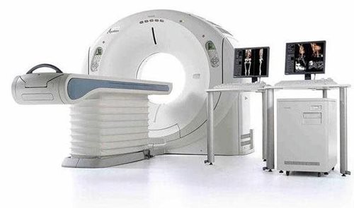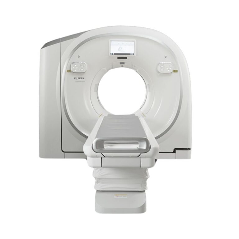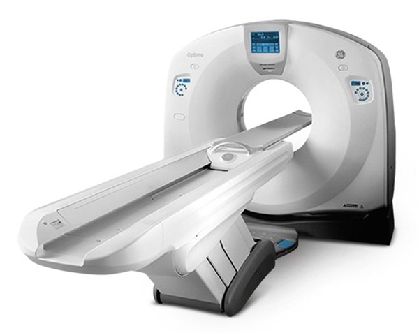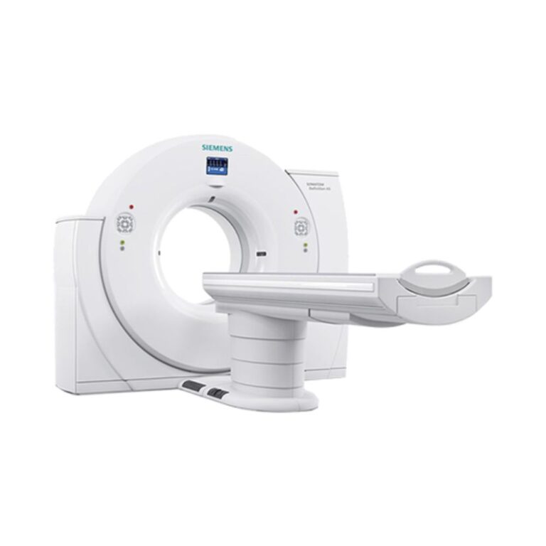
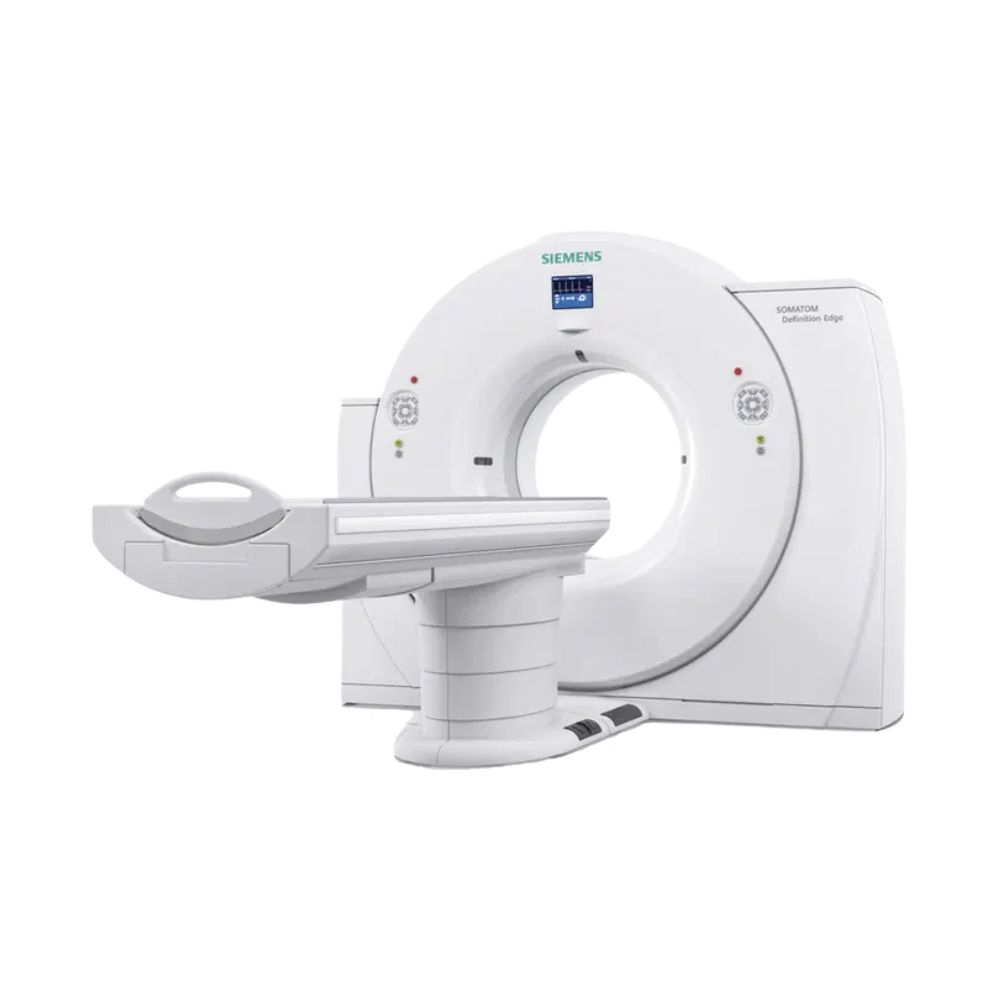
Siemens SOMATOM Definition Edge 128-Slice CT
- Category:
- CT Scanners
- Brand:
- Siemens
The Siemens SOMATOM Definition Edge is a 128-slice CT scanner for routine and advanced diagnostic imaging across diverse clinical settings. Its Stellar detector, fast acquisition, and high-resolution imaging support cardiac, vascular, emergency, and oncology examinations, with available technologies for dose reduction and specialized applications.
SOMATOM Definition Edge Highlights & Benefits
The Siemens SOMATOM Definition Edge combines 128-slice CT acquisition, Stellar detector technology, and fast scanning in a single-source system. With a 78 cm gantry opening and advanced clinical options, it supports routine diagnostic imaging and specialized examinations requiring speed, anatomical detail, and flexible patient positioning.
Key Benefits
-
- 128-slice CT acquisition: Uses 64 physical detector rows and advanced sampling technology to produce 128 slices per rotation.
- Stellar detector: Reduces electronic noise to support detailed imaging at lower radiation doses.
- Fast scanning: Rotation times as low as 0.28 seconds and scan speeds up to 23 cm/s (9.1 in/s).
- Fine anatomical detail: Approximately 0.30 mm spatial resolution helps visualize smaller structures.
- Cardiac CT imaging: Temporal resolution as low as 142 milliseconds helps limit motion-related image blur.
- 78 cm (30.7 in) gantry opening: Provides room for a broad range of patient sizes and examination positions.
- High-capacity patient table: Optional configurations accommodate patients up to 676 lb (307 kg).
- Advanced imaging options: Available TwinBeam Dual Energy, metal artifact reduction, perfusion imaging, and CT-guided procedures.
128-Slice CT Acquisition & Scan Speed
Stellar Detector & 128-Slice Imaging
The SOMATOM Definition Edge uses a Siemens Stellar detector with 64 physical detector rows and advanced sampling technology to acquire 128 slices per rotation. The detector covers 38.4 mm (1.5 in) of anatomy per rotation.
This 128-slice acquisition is achieved through additional sampling of the detector data rather than 128 separate physical detector rows. The system can generate up to 384 reconstructed slices, depending on the reconstruction method.
TrueSignal technology processes signals within the detector electronics to reduce electronic noise, which can appear as unwanted graininess in CT images.
Combined with approximately 0.30 mm spatial resolution, this supports detailed imaging of small blood vessels, bones, and complex anatomical structures.
0.28-Second Rotation & Fast Scanning
Rotation times as low as 0.28 seconds and scan speeds up to 23 cm/s (9.1 in/s) allow the Definition Edge to cover large anatomical areas quickly.
Fast acquisition is especially beneficial for emergency CT, chest and abdominal imaging, and CT angiography, where minimizing scan time can help reduce the effects of patient movement.
High-pitch scanning, which moves the patient table farther through the scanner during each gantry rotation, supports rapid coverage during compatible examination protocols.
Cardiac Imaging & Motion Reduction
The Definition Edge provides in-plane temporal resolution as low as 142 milliseconds, helping reduce image blur caused by the movement of the heart.
With compatible software, electrocardiogram (ECG)-synchronized scanning coordinates image acquisition with the patient’s heartbeat.
This supports cardiac CT examinations, including coronary CT angiography for evaluating the blood vessels supplying the heart.
Image quality and available cardiac protocols depend on patient characteristics, examination settings, and installed software.
Stellar Detector Image Quality & Radiation Dose
CARE Dose Management & Low-Dose Imaging
Siemens CARE dose-management technologies adjust scanning settings according to the patient and examination, helping maintain diagnostic image quality while managing radiation exposure.
CARE Dose4D automatically adjusts X-ray output as the scanner moves through different parts of the body. CARE kV helps select the appropriate X-ray energy based on patient anatomy and examination requirements.
These technologies support patient-specific, low-dose CT protocols for routine examinations, pediatric imaging, oncology follow-up, and other applications where radiation management is important.
SAFIRE & ADMIRE Image Quality
SAFIRE (Sinogram Affirmed Iterative Reconstruction) and ADMIRE (Advanced Modeled Iterative Reconstruction) are Siemens image-processing technologies that help reduce graininess in CT images while preserving anatomical detail.
They can help maintain diagnostic image quality when lower-dose scanning protocols are used.
Availability depends on the scanner’s software generation and installed licenses.
Tin Filter Technology & Metal Artifact Reduction
Available Tin Filter technology removes lower-energy X-rays that contribute less useful diagnostic information, helping reduce radiation exposure during selected examinations, including lung and sinus CT.
Optional iMAR (iterative Metal Artifact Reduction) reduces streaking and distortion in CT images caused by metal implants, including orthopedic hardware and dental restorations.
These capabilities can improve image clarity when scanning patients with implants or performing examinations where reducing radiation exposure is especially important.
78 cm Gantry, Patient Capacity & Workflow
78 cm Gantry Opening
The Definition Edge features a 78 cm (30.7 in) gantry opening, providing additional room for patient positioning during routine and specialized CT examinations.
The opening supports a broad range of patient sizes and examinations requiring additional positioning space. Actual patient fit also depends on body dimensions, table configuration, and positioning accessories.
Patient Table Capacity & Positioning
Available high-capacity patient tables accommodate patients weighing up to 676 lb (307 kg), supporting imaging for larger patients within the installed table’s limits.
This capacity, combined with the 78 cm gantry opening, makes the Definition Edge a useful option when bariatric CT imaging and flexible patient positioning are important.
Table weight limits, travel range, and positioning accessories vary by configuration.
FAST CARE Scanning & Automation
Siemens FAST CARE provides automated scanning and dose-management tools to simplify CT examination setup and support consistent imaging protocols.
Available features assist with patient-specific scan settings, automatic exposure adjustments, and guided examination planning.
FAST Cardio Wizard provides step-by-step assistance for cardiac CT preparation on systems equipped with the appropriate software.
These technologies help reduce repetitive setup tasks and support efficient examination workflows.
Clinical Environments & Applications
The SOMATOM Definition Edge supports routine and advanced CT imaging across a wide range of clinical settings. Its acquisition speed, detailed imaging, and available clinical software accommodate different patient populations and diagnostic requirements.
Clinical Environments
-
- Hospitals and health systems
- Diagnostic and outpatient imaging centers
- Emergency departments and trauma centers
- Cardiac and cardiovascular imaging facilities
- Oncology and cancer treatment centers
- Veterinary hospitals and specialty imaging centers
- Specialty clinics and facilities providing diagnostic CT
Clinical Specialties
-
- Radiology: General diagnostic and contrast-enhanced CT examinations.
- Cardiology: Coronary artery assessment and structural heart imaging.
- Vascular Medicine: Blood vessel evaluation and procedural planning.
- Neurology & Neurosurgery: Brain, spinal, and neurovascular imaging.
- Oncology: Cancer detection, staging, and treatment monitoring.
- Orthopedics: Bone, joint, fracture, and postoperative assessment.
- Pulmonology: Lung disease and thoracic imaging.
- Gastroenterology & Urology: Abdominal, gastrointestinal, pelvic, and urinary tract imaging.
- Emergency Medicine: Rapid CT for trauma and acute conditions.
- Pediatrics: Clinically appropriate CT examinations using patient-specific dose protocols.
- Veterinary Medicine: Orthopedic, neurological, oncology, thoracic, abdominal, and vascular CT imaging for animal patients.
Common CT Applications
-
- Head, chest, abdomen, and pelvis CT
- Coronary and vascular CT angiography
- Pulmonary embolism evaluation
- Emergency and trauma imaging
- Stroke and neurovascular assessment
- Oncology staging and follow-up examinations
- Kidney stone and urinary tract imaging
- Musculoskeletal and postoperative CT
- Pediatric CT with appropriate dose-management protocols
- Bariatric CT within installed patient table and positioning limits
- Veterinary diagnostic CT using appropriate animal positioning and examination protocols
Specialized cardiac, Dual Energy, perfusion, and interventional examinations require the corresponding installed software and hardware.
Advanced Cardiac, Dual Energy & Interventional Imaging
Available hardware and software packages expand the Definition Edge beyond routine CT, supporting specialized cardiovascular examinations, material analysis, blood flow assessment, and image-guided procedures.
Advanced Cardiac & Vascular Imaging
Available cardiac software supports coronary CT angiography, coronary plaque assessment, and imaging used to plan structural heart procedures.
Low-voltage scanning can improve the visibility of iodine-based contrast during selected vascular examinations, including planning for transcatheter aortic valve replacement (TAVR).
Appropriately equipped systems can also support triple-rule-out CT, a combined examination evaluating the coronary arteries, pulmonary arteries, and aorta.
TwinBeam Dual Energy Imaging
Siemens TwinBeam Dual Energy uses two different X-ray energy ranges during a scan to provide additional information about tissue and material composition.
Unlike conventional CT, which primarily shows differences in X-ray absorption, Dual Energy imaging can help distinguish materials that appear similar in standard CT images.
With compatible software, applications include gout assessment, material differentiation, and specialized vascular imaging.
Virtual noncontrast reconstruction can also generate images resembling a scan performed without contrast material, potentially reducing the need for a separate scan during selected examinations.
TwinBeam requires compatible hardware and installed software licenses.
Blood Flow & Dynamic CT Imaging
Dynamic CT captures a series of images over time rather than a single snapshot. This helps evaluate blood flow and the movement of injected contrast material through tissues.
Available Adaptive 4D Spiral technology and compatible imaging applications support specialized examinations such as stroke-related blood flow assessment and selected vascular or oncology studies.
These applications provide functional information beyond the anatomical detail captured during routine CT.
CT-Guided Procedures
The optional Adaptive 3D Intervention Suite provides imaging and planning tools for procedures performed under CT guidance.
Applications include biopsies, fluid drainage, and other minimally invasive procedures where accurate instrument positioning is important.
Image Processing & System Connectivity
The SOMATOM Definition Edge supports image reconstruction, clinical review, and connectivity with existing diagnostic imaging systems.
Image Reconstruction & Advanced Review
Multiplanar reconstruction allows acquired CT data to be reviewed from different angles, including cross-sectional, front, and side views.
Depending on the installed workstation and software, additional capabilities include three-dimensional visualization, blood vessel analysis, cardiac assessment, and specialized image processing.
PACS Connectivity & Clinical Integration
DICOM is the standard used to store and exchange medical images. It supports image transfer from the scanner to compatible picture archiving and communication systems (PACS) for storage and diagnostic review.
Available connectivity options also support radiology information systems (RIS), electronic examination worklists, and existing clinical workflows.
MXR can help align workstation, software, and connectivity options with the facility’s imaging infrastructure.
Available Configurations & Installed Options
Pre-owned Siemens SOMATOM Definition Edge CT scanners may differ by model year, installed hardware, X-ray tube history, software generation, and clinical applications.
MXR helps facilities select equipment based on imaging requirements, patient needs, site compatibility, and budget. Available configurations can include different performance and software options within the Definition Edge platform.
Scanner Hardware & Patient Table
Important configuration details include:
-
- Model year and installed hardware revisions
- STRATON MX X-ray tube condition, usage, and replacement history
- 80 kW generator or optional 100 kW configuration
- Patient table load capacity and travel range
- Included patient positioning accessories
Reconstruction, Dose & Imaging Software
Installed software may include:
-
- SAFIRE or ADMIRE image reconstruction
- CARE dose-management applications
- Tin Filter technology
- iMAR metal artifact reduction
- Software versions and licensed imaging features
- Advanced Clinical Applications
- Available software and hardware packages may include:
- Cardiac CT and cardiovascular imaging applications
- TwinBeam Dual Energy
- Adaptive 4D Spiral and perfusion imaging
- Adaptive 3D Intervention Suite
- Vascular and oncology post-processing applications
Workstations, Accessories, & Service History
Pre-owned configurations can also differ in acquisition console hardware, post-processing workstations, software licenses, network components, and maintenance history.
MXR Imaging can help identify a system with the appropriate clinical applications, accessories, installation requirements, and service support. For veterinary imaging, this includes evaluating animal positioning accessories and compatibility with the facility’s anesthesia and examination workflows.
Facilities comparing Siemens 128-slice CT platforms can also explore the Siemens SOMATOM Definition AS 128, which offers a different range of detector, hardware, and software configurations.
CT Installation & Site Requirements
The Definition Edge requires a CT examination room with suitable electrical service, cooling, radiation shielding, and sufficient space for the gantry, patient table, and operator workstation.
Room layout, utility connections, and installation requirements depend on the selected equipment configuration and facility conditions.
MXR Imaging’s turnkey imaging project management services can help coordinate site preparation, equipment delivery, installation, and project requirements.
Final installation planning should follow the technical documentation for the selected scanner.
CT Scanner Price & Availability
Pre-owned Siemens SOMATOM Definition Edge pricing depends on equipment age, system configuration, X-ray tube condition, installed clinical software, and included accessories.
MXR Imaging helps healthcare facilities evaluate available 128-slice CT scanner options based on clinical capabilities, patient needs, operational requirements, and budget.
Select equipment may qualify for MXR’s Platinum Certified imaging equipment options, depending on the specific system and available service packages.
MXR Imagin also provides CT and PET/CT maintenance and repair services to support ongoing equipment performance.
Contact MXR Imaging to discuss available system configurations, pricing, installation, and service options.
SOMATOM Definition Edge Specifications
Specifications represent platform capabilities and available options. Individual pre-owned configurations may differ.
CT Acquisition & Detector
-
- Scanner type: Single-source CT
- Slice acquisition: 128 slices per rotation
- Physical detector rows: 64
- Detector element width: 0.6 mm
- Detector coverage: 38.4 mm (1.5 in) per rotation
- Reconstructed slices: Up to 384
- Detector: Siemens Stellar detector
- Detector signal technology: TrueSignal
- Rotation time: As low as 0.28 seconds
- Maximum scan speed: 23 cm/s (9.1 in/s)
- Maximum pitch factor: Up to 1.7
- In-plane temporal resolution: As low as 142 milliseconds
- Spatial resolution: Approximately 0.30 mm
Gantry & Patient Table
-
- Gantry opening: 78 cm (30.7 in)
- Maximum patient table capacity: Up to 676 lb (307 kg), optional
- Patient table travel: Configuration-dependent
- Patient positioning accessories: Vary by installed system
X-Ray Generator & Tube
-
- X-ray tube: Siemens STRATON MX
- Generator power: 80 kW; optional 100 kW
- Tube voltage settings: 70, 80, 100, 120, and 140 kV
- Maximum tube current at 70 kV: 500 mA
- Maximum tube current at 80 kV: 650 mA
- Tin Filter technology: Available option
Image Reconstruction & Performance
-
- Reconstruction technologies: Conventional reconstruction; SAFIRE or ADMIRE depending on software configuration
- Dose management: CARE Dose4D and CARE kV
- Metal artifact reduction: iMAR, optional
- Dual Energy: TwinBeam, optional
- Dynamic imaging: Adaptive 4D Spiral, configuration-dependent
- Interventional imaging: Adaptive 3D Intervention Suite, optional
- Cardiac imaging: ECG-synchronized acquisition and licensed cardiac applications
- Post-processing: Multiplanar reconstruction and optional advanced applications
System & Site Requirements
-
- Installation type: Fixed CT scanner
- Electrical service: Configuration-dependent
- Cooling requirements: Based on the installed system
- Radiation shielding: Site-specific requirements
- Network connectivity: DICOM/PACS and optional RIS integration
- Installation clearance: Determined by equipment configuration and room layout