JPI DeteCT Vet
- Category:
- Veterinary Imaging
- Brand:
- JPI
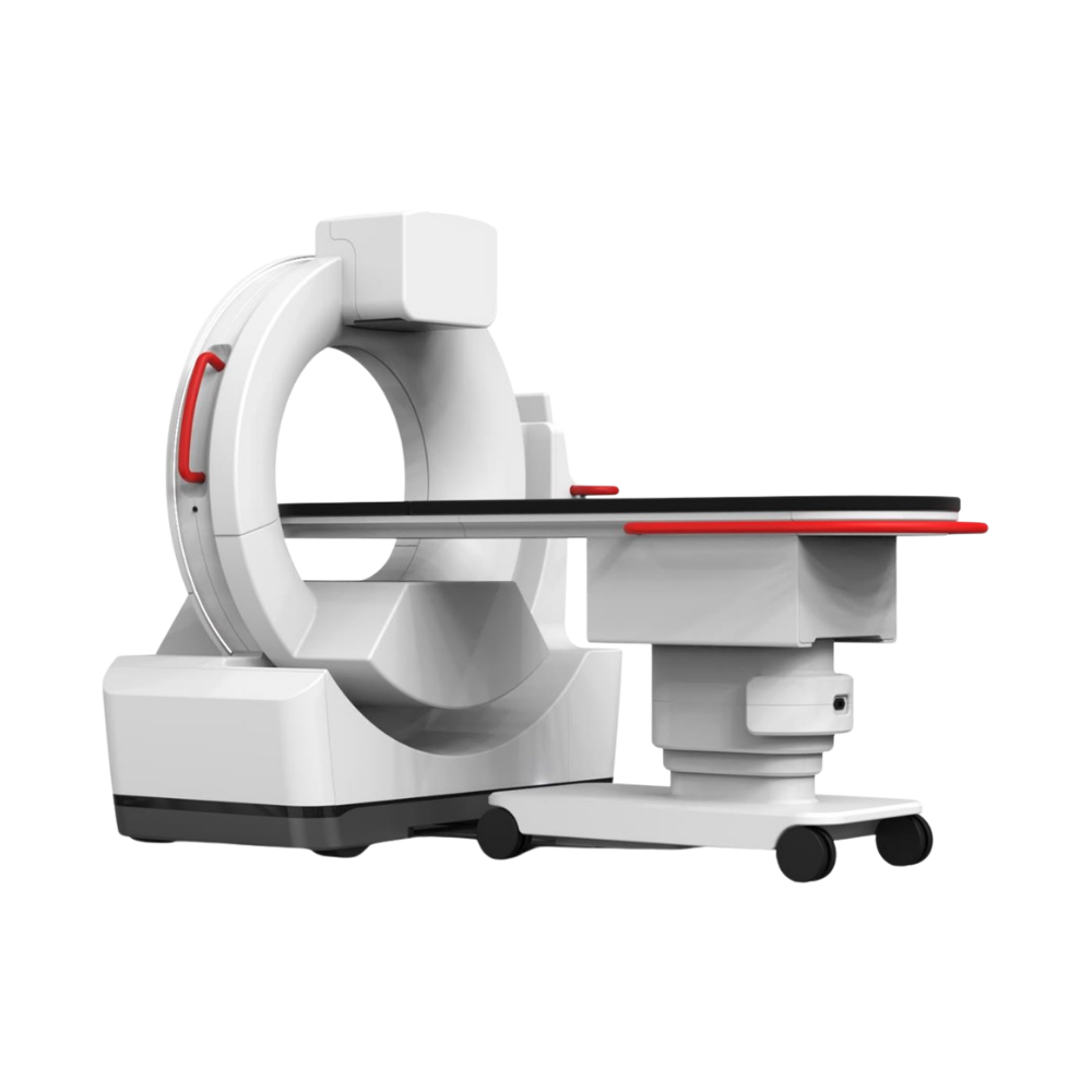
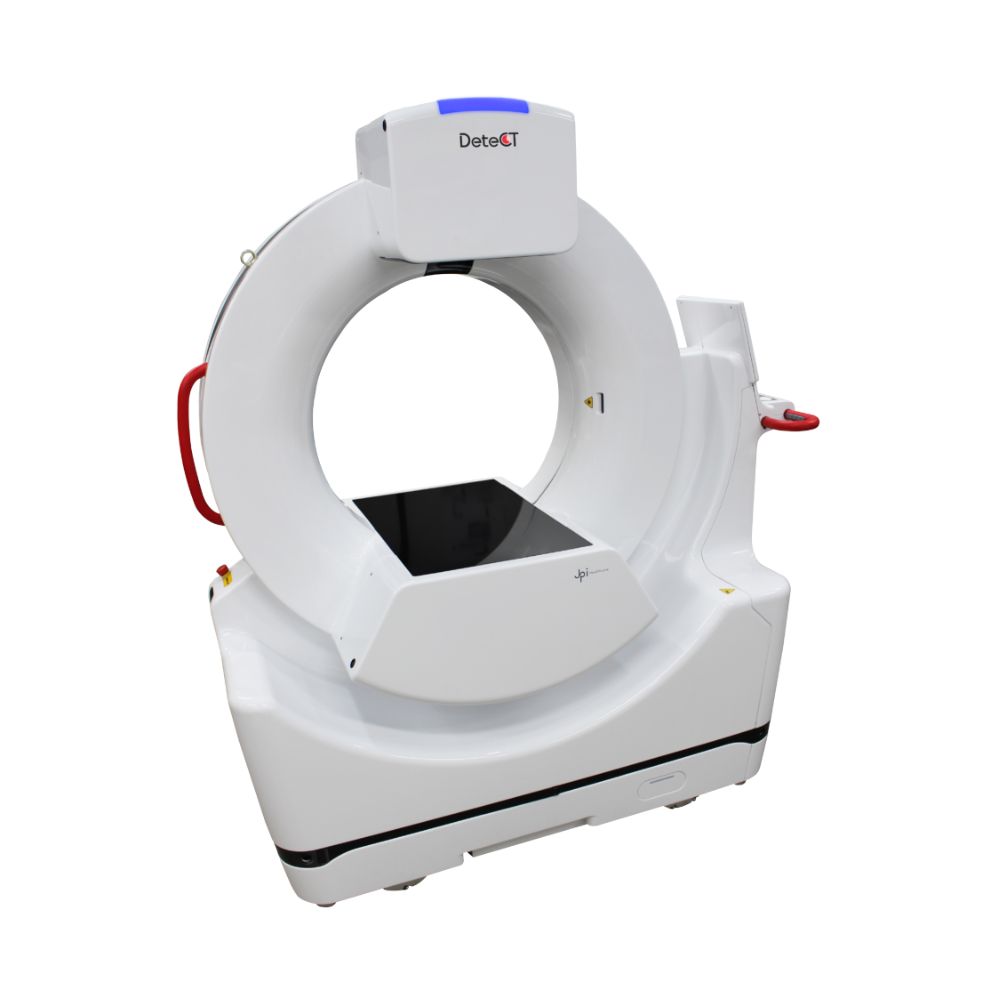
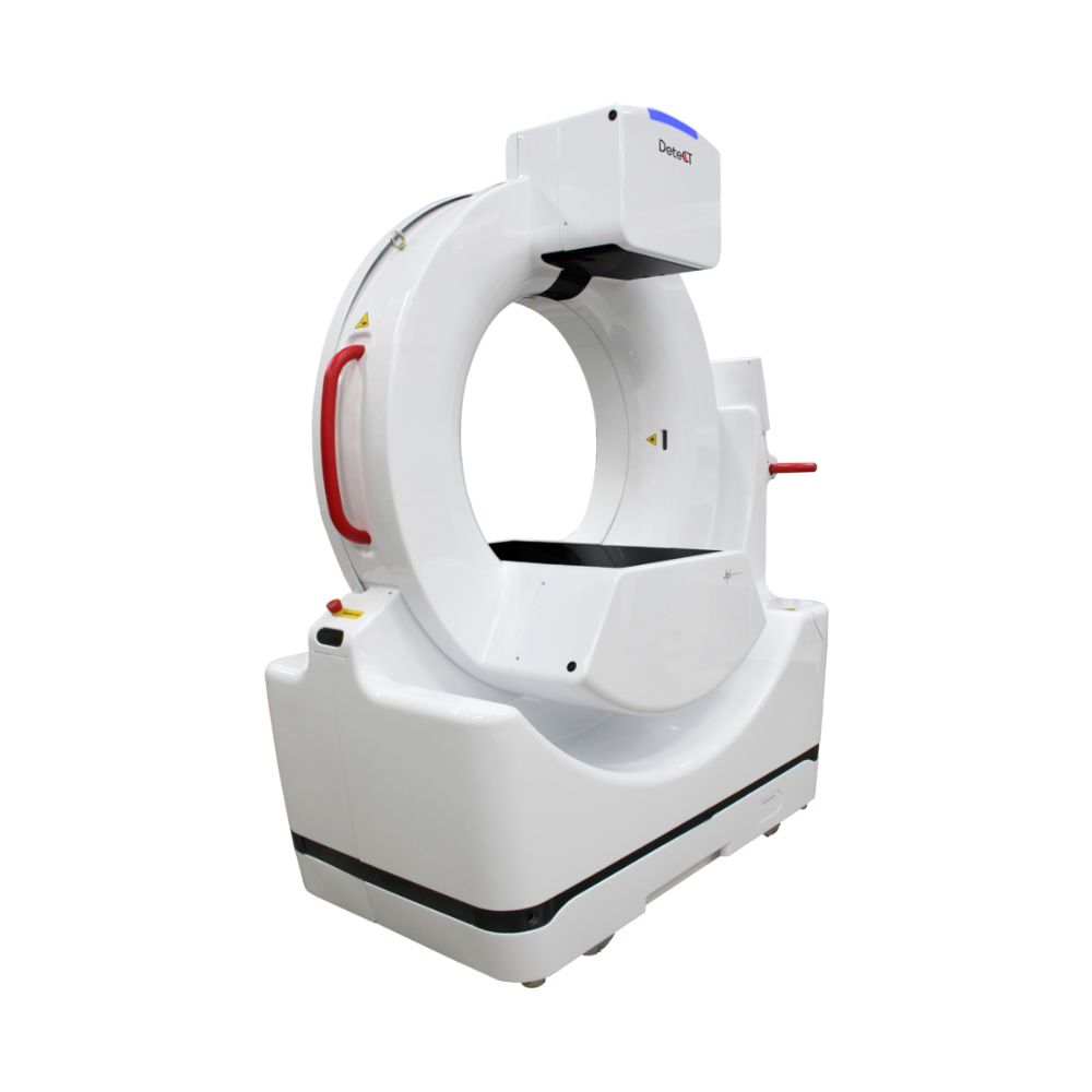
The JPI DeteCT Vet is a high-definition, mobile hybrid 3-in-1 CT system designed specifically for modern veterinary practices. It integrates Cone Beam Computed Tomography (CBCT), 17 x 17 inch live video X-ray (fluoroscopy), and digital radiography into a single mobile platform that fits comfortably into a standard 8 x 12 foot room. Powered by ExamVue Duo acquisition software, it delivers highly accurate 3D images, rapid scan times, and automated workflows to minimize patient downtime and maximize diagnostic accuracy without the infrastructure costs of conventional CT scanners.
Overview & Key Advantages of the JPI DeteCT Vet
Three Powerful Diagnostic Modalities in One Footprint
Equip your clinic with high-definition Cone Beam Computed Tomography (CBCT), real-time fluoroscopy, and digital radiography. This hybrid architecture eliminates the need to purchase, maintain, and house multiple distinct imaging systems, saving valuable clinical space and minimizing capital expenditures.
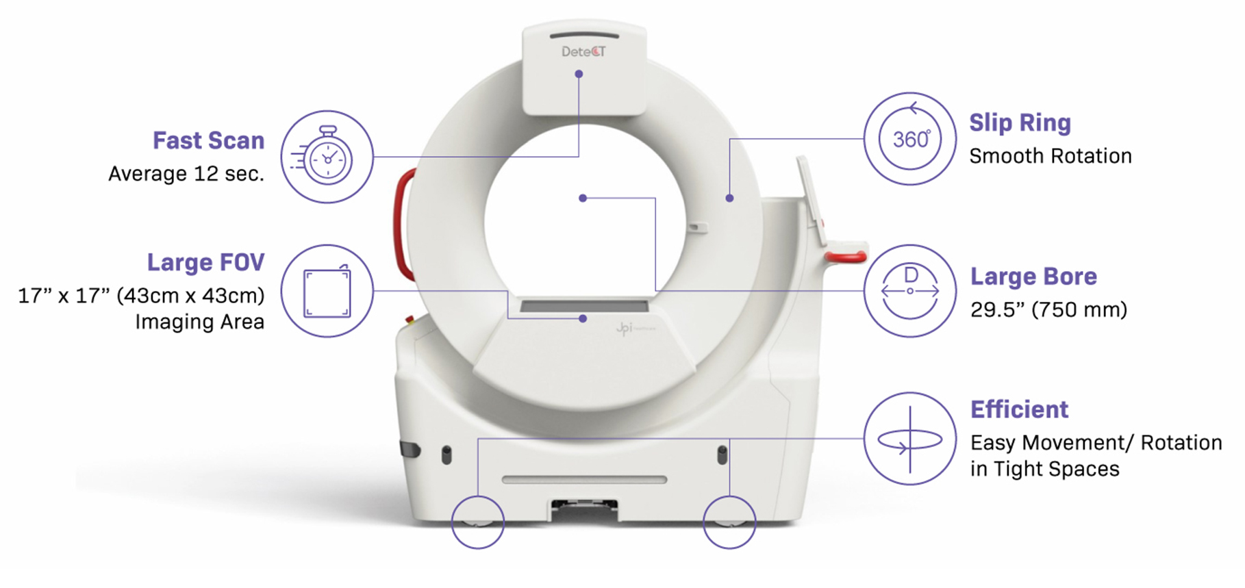
Rapid Scan Times to Support Patient Safety
The system completes scans in a rapid range of 4 to 20 seconds (averaging 12 to 19 seconds depending on the protocol) and finishes image reconstructions in just 25 seconds. Fast processing limits the time veterinary patients spend under anesthesia or sedation, streamlining your daily workflow.
High Patient Accessibility with an Extra-Large Bore
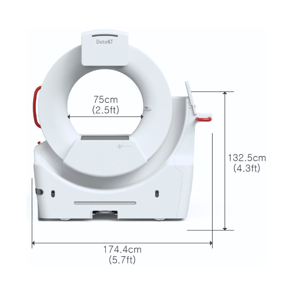
Designed with a spacious 29.5 inch (750 mm) bore diameter and a generous 17 x 17 inch (43 cm x 43 cm) active imaging area. This allows veterinary technicians to easily position a diverse range of patient sizes, from small companion animals to large dog breeds.
True Mobility with Low Infrastructure Demands
The compact, wheeled design with 360-degree slip-ring rotation allows the system to be rolled and maneuvered easily, even in tight rooms. Unlike conventional CT scanners, it operates with lower energy requirements and has no special room shielding or heavy power infrastructure demands.
Three Integrated Imaging Modalities
1. Cone Beam Computed Tomography (CBCT)
-
- Fast Reconstruction: Reconstruction completed in 25 seconds for rapid chair-side decisions.
- High-Definition 3D Scans: Exceptional volumetric imaging quality to locate structural abnormalities.
- Multiplanar Reconstruction (MPR): Built-in MPR viewer lets you inspect pathology from sagittal, coronal, and transverse perspectives.
2. Live Video X-Ray (Fluoroscopy)
-
- High Frame Rate: Delivers smooth, real-time imaging at 30 frames per second.
- Dynamic Visualizations: Features Digital Subtraction Angiography (DSA) and Auto Brightness Control (ABC) for enhanced visualization of moving structures.
- Large Field of View: Utilizes the full 17 x 17 inch active area for wide-area coverage during real-time procedures.
3. High-Definition Digital Radiography
-
- Instant Image Preview: Radiographic previews display in less than 0.8 seconds to immediately verify patient positioning.
- AI-Enhanced Software: ExamVue Duo acquisition software includes powerful measurement tools and advanced image processing filters.
- Anatomical Optimization: Automated thickness-based protocol selection dynamically optimizes exposure settings for consistent image quality.
Real-World Clinical Applications
Dental and Craniofacial Diagnostics
Evaluate complex dental cases with high-resolution 3D visualization. The system provides clear details of tooth roots, periodontal ligament spaces, jawbone structures, and nasal cavities to improve planning for extractions and oral surgeries.
Orthopedic and Joint Assessments
Locate micro-fractures, evaluate joint spacing, and identify free-floating bone fragments. The multi-angle volumetric images assist in planning orthopedic interventions and confirming post-operative hardware placement.
Vascular and Internal Soft-Tissue Evaluations
Utilize dynamic fluoroscopy to observe moving internal processes. Clinicians can track vascular pathways via contrast-enhanced Digital Subtraction Angiography (DSA) and monitor internal organ movement with real-time feedback.
Technical Specifications & Dimensions
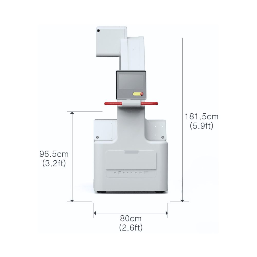
- System Width: 5.72 feet
- System Height: 5.79 feet
- System Depth: 2.69 feet
- Bore Diameter: 29.5 inches (750 mm)
- Active Imaging Area (FOV): 17 x 17 inches (43 cm x 43 cm)
- Scan Time Range: 4 to 20 seconds (Average: 12 to 19 seconds)
- CT Reconstruction Time: 25 seconds
- Fluoroscopy Frame Rate: 30 frames per second
- Radiography Preview Time: less than 0.8 seconds
- Room Requirements: Fits comfortably in standard clinical rooms as small as 8 x 12 feet