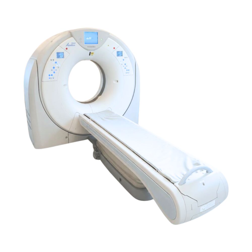
Canon Aquilion PRIME S CT
- Category:
- CT Scanners
- Brand:
- Canon
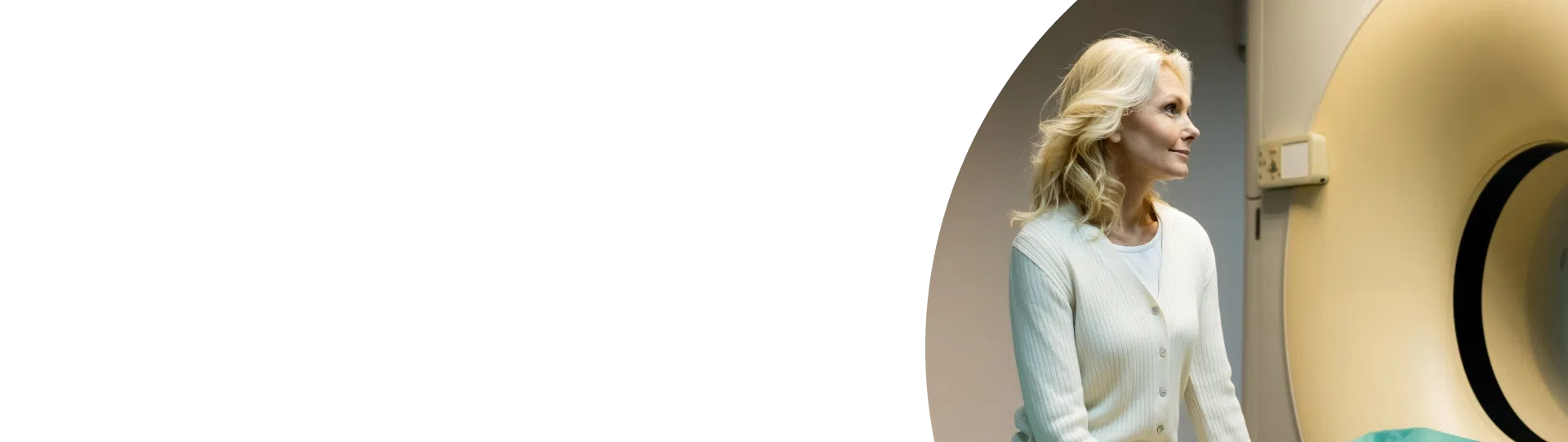
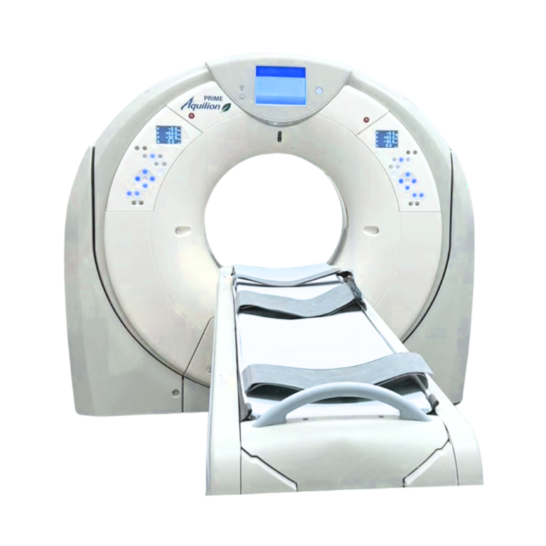
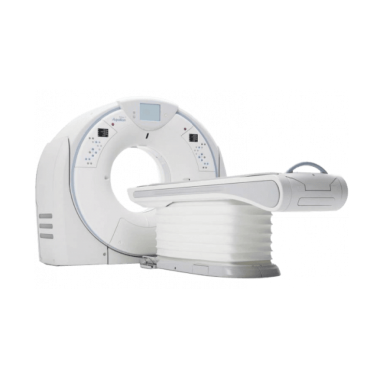
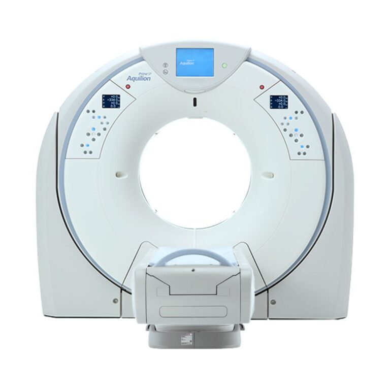
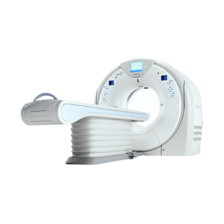
The Canon Aquilion PRIME S is a CT scanner available in 80- and 160-slice configurations for hospitals, imaging centers, specialty practices, and veterinary referral hospitals. Its 0.35-second rotation helps reduce motion, while 40 mm thin-slice coverage and a 78 cm bore support fast imaging and broader patient access.
Aquilion PRIME S Highlights for 80/160-Slice CT
The Canon Aquilion PRIME S CT scanner combines thin-section imaging, fast acquisition, wide patient access, dose-management technology, and configurable cardiac, vascular, interventional, and advanced image-reconstruction capabilities.
PRIME S Performance & Patient Access Benefits
-
- Available 80- and 160-slice configurations
- 80 rows of 0.5 mm detector elements
- 40 mm physical detector coverage per rotation
- Rotation times down to 0.35 seconds
- 78 cm (30.7 in) scanner opening
- Patient table capacity up to 660 lb (300 kg) on applicable configurations
- AIDR 3D reconstruction for image-noise and dose management
- SUREExposure 3D automatic exposure management
- Up to ±30° scanner tilt for positioning flexibility
- Configurable cardiac, vascular, metal artifact reduction, and interventional imaging options
PRIME S Acquisition Architecture & Slice Configurations
80 × 0.5 mm Thin-Section Detector
The Aquilion PRIME S uses 80 rows of 0.5 mm detector elements, providing 40 mm of physical coverage per rotation.
Thin 0.5 mm acquisition provides detailed image data that can be reconstructed in multiple viewing planes and 3D views for routine body CT as well as neurologic, vascular, cardiac, musculoskeletal, oncology, and other advanced examinations.
Aquilion PRIME S 80- & 160-Slice Configurations
The Aquilion PRIME S 80-slice and 160-slice configurations use the same 80-row, 0.5 mm detector architecture. The primary difference is the number of images reconstructed from the acquired data.
The 80-slice configuration produces 80 slices per rotation. Applicable 160-slice systems use Double Slice reconstruction, a processing method that generates 160 reconstructed slices from the 80-row detector.
For a pre-owned system, the active slice configuration, image-reconstruction hardware, software level, and installed licenses should be confirmed on the individual scanner.
0.35-Second Rotation for Motion Control
Rotation times down to 0.35 seconds shorten the time required to collect CT data.
Faster acquisition can reduce the opportunity for patient motion and is particularly useful for chest, emergency, trauma, vascular, cardiac, and other examinations where scan speed directly affects workflow or image consistency.
PRIME S Image Quality & Dose Management
AIDR 3D Iterative Reconstruction
Adaptive Iterative Dose Reduction 3D, or AIDR 3D, reduces image noise during reconstruction while helping preserve clinically useful anatomical detail.
Because AIDR 3D is integrated into the acquisition and reconstruction process, it supports routine dose management without requiring a separate image-processing step.
Automatic Exposure with SUREExposure 3D
SUREExposure 3D automatically adjusts X-ray output according to patient anatomy and the selected examination.
This helps match exposure to the individual patient rather than applying the same X-ray output throughout the scan.
Dose Check & Protocol Management
Applicable PRIME S configurations support CT Dose Check, which alerts technologists when planned exposure exceeds preset reference levels, along with automatic exposure controls and dose reporting.
Adult and pediatric reference protocols can also help standardize scan settings for different patient groups.
Installed dose-management features and software versions should be verified on the individual scanner.
SEMAR for Imaging Around Metal
Single Energy Metal Artifact Reduction, or SEMAR, is an available reconstruction option that reduces streaking and distortion caused by metallic implants.
When installed, SEMAR can improve visualization of bone and soft tissue surrounding orthopedic hardware and other metal-containing anatomy.
PRIME S Wide-Bore Patient Access & Positioning
78 cm Gantry Opening & 47 cm Couch
The scanner opening, also called the gantry, measures 78 cm (30.7 in), making the PRIME S a wide-bore CT scanner with additional room for patient positioning. The patient couch is approximately 47 cm (18.5 in) wide.
The additional clearance supports positioning across a broad patient population, including pediatric, bariatric, mobility-limited, emergency, and interventional patients.
High-Capacity Patient Support
Applicable PRIME S table configurations support patient weights up to approximately 660 lb (300 kg).
A low couch height and wide patient-support surface can make transfers and positioning easier for larger patients and patients with limited mobility.
Lateral Table Positioning
Available Tech Assist Lateral Slide moves the tabletop from side to side so the anatomy of interest can be centered without repeatedly repositioning the patient.
This can simplify setup while reducing physical handling for both technologists and patients.
Tilted Helical Acquisition
The gantry can support up to ±30° of physical tilt on applicable configurations, including helical acquisition, which moves the patient continuously through the scanner while data is collected in a spiral path.
Gantry tilt provides additional positioning flexibility and can help adapt scan geometry when anatomy or patient condition makes standard positioning difficult.
PRIME S Clinical Environments & Applications
The Aquilion PRIME S supports routine and advanced CT across a broad range of clinical environments. Exact capability depends on the active slice configuration, software, reconstruction hardware, patient table, and installed clinical applications.
Care Settings for PRIME S
-
- Hospitals and health systems
- Community hospitals
- Outpatient imaging centers
- Independent diagnostic imaging facilities
- Emergency and trauma departments
- Cardiac and vascular imaging programs
- Oncology and interventional imaging departments
- Physician-owned and specialty practices with dedicated CT
- Veterinary referral and specialty hospitals
Clinical Specialties
-
- Diagnostic Radiology
- Emergency Medicine & Trauma
- Cardiology
- Vascular Medicine & Surgery
- Neurology & Neurovascular Imaging
- Oncology
- Orthopedics & Musculoskeletal Imaging
- Interventional Radiology
- Pediatric Radiology
- Veterinary Medicine
Common PRIME S CT Applications
-
- Brain, head, and neck CT
- Chest and lung CT
- Abdominal and pelvic CT
- Emergency and trauma imaging
- Stroke and neurologic imaging
- CT angiography
- Cardiac CT and coronary CT angiography on appropriately configured systems
- Oncology imaging and treatment follow-up
- Musculoskeletal and orthopedic CT
- Pediatric CT using age- and size-appropriate protocols
- CT-guided biopsy and intervention
- Veterinary diagnostic CT in specialty and referral settings
Where PRIME S Fits in the Aquilion PRIME Family
The Aquilion PRIME S is a later-generation system within the original Aquilion PRIME family. Earlier PRIME 80/160 systems and PRIME S systems can share similar slice counts and external design cues, so the exact generation and installed configuration should be identified when comparing pre-owned units.
PRIME S vs. Earlier PRIME 80/160 Systems
PRIME S represents a later generation of the original PRIME platform while retaining the 80-row, 0.5 mm detector architecture used across the family.
Compared with earlier PRIME systems, individual PRIME S units may incorporate later scanner hardware, detector-signal processing components, image-reconstruction systems, software versions, and workflow options. Slice count alone therefore does not identify the scanner’s generation or complete capabilities.
PRIME S vs. Prime SP
The Aquilion PRIME S and Prime SP can have similar slice counts, but they belong to different generations of the Aquilion platform.
The PRIME S is the earlier generation, technically identified as the TSX-303A platform. Prime SP is the later TSX-303B generation, with updated imaging hardware, workflow capabilities, and support for newer reconstruction technologies.
Prime SP systems can include later-generation features such as PUREViSION Optics, which improves how efficiently the scanner uses the available X-ray signal, and AiCE, a deep-learning reconstruction technology designed to separate useful image information from noise. These technologies should not be assumed on a PRIME S simply because both scanners may be available in 80- or 160-slice configurations.
When comparing pre-owned systems, the more important differences are platform generation, installed reconstruction technology, software, and clinical options rather than slice count alone.
PRIME S Cardiac, Vascular & Interventional Imaging
Advanced applications depend on installed software and should be confirmed on the individual PRIME S.
Cardiac CT & ECG-Gated Acquisition
Appropriately configured PRIME S systems can support cardiac CT and coronary CT angiography.
Available SURECardio Prospective coordinates scanning with the patient’s heartbeat. This electrocardiogram, or ECG-gated, approach times image acquisition to selected portions of the cardiac cycle and can adjust acquisition when irregular heart rhythms are detected during applicable examinations.
Variable Helical Pitch for Combined Acquisitions
Available Variable Helical Pitch changes table speed during a continuous spiral CT scan.
This allows different portions of an examination to use different acquisition settings, supporting applications such as cardiac and aortic imaging without requiring completely separate scan sequences.
Vascular Subtraction & Perfusion Options
Available SURESubtraction digitally removes bone and calcium from applicable contrast-enhanced vascular images to provide a clearer view inside blood vessels.
Depending on installed software, PRIME S systems may also support perfusion imaging, which evaluates blood flow through tissue over time, for neurologic, vascular, and other advanced examinations.
Dual Energy Imaging
Available Dual Energy imaging collects CT data at two X-ray energy levels to provide additional information about tissue and material composition.
Dual Energy capability is configuration-dependent and should not be assumed on every pre-owned PRIME S.
CT Fluoroscopy & Image-Guided Procedures
Available CT fluoroscopy provides rapidly updated CT images during image-guided procedures.
When equipped with the required interventional software and accessories, PRIME S can support biopsy, drainage, needle placement, and other CT-guided procedures.
PRIME S Workflow from Patient Setup to Image Review
In-Room Setup & Scan Controls
Gantry-mounted controls, patient-positioning tools, and dual scanograms, which are planning images taken from two directions, can help technologists prepare the examination before scanning.
Available iStation functionality provides additional room-side workflow and positioning controls so the technologist can remain closer to the patient during final setup.
Rapid Reconstruction & Immediate Review
Applicable PRIME S configurations can reconstruct full-resolution images at speeds up to approximately 60 images per second.
InstaView provides images during the scanning workflow so technologists can begin reviewing acquired data before the examination is complete.
DICOM, PACS & Advanced Visualization
The PRIME S supports Digital Imaging and Communications in Medicine, or DICOM, the standard used to exchange medical images and study information.
DICOM enables integration with compatible picture archiving and communication systems, or PACS, radiology worklists, and other clinical imaging systems.
Depending on configuration, available workstations can add multiplanar reconstruction, 3D visualization, cardiovascular, neurovascular, oncology, and other post-processing capabilities.
Pre-Owned Canon Aquilion PRIME S Configurations
A pre-owned Canon Aquilion PRIME S can vary significantly by model year, 80- or 160-slice configuration, tube history, detector and data-acquisition hardware, software level, reconstruction system, patient table, workstations, clinical applications, and previous upgrades.
80- or 160-Slice Configuration
Confirm whether the scanner is actively configured for 80- or 160-slice reconstruction.
Important items include:
-
- Active slice configuration
- Image-reconstruction hardware
- Software version
- Double Slice reconstruction capability
- Previous upgrades
- Installed advanced imaging licenses
Detector, Data Acquisition & Hardware Configuration
PRIME S systems can differ in detector and data-acquisition hardware depending on model year and installed configuration.
The data-acquisition system is the electronics that convert signals from the CT detector into digital information used to create the images.
Important items to verify include:
-
- Detector configuration
- Data-acquisition system
- Gantry hardware generation
- Image-reconstruction system
- Patient table configuration
- Positioning accessories
- Tech Assist Lateral Slide availability
- X-Ray Tube & Generator Condition
- Important system details include:
- Model year
- X-ray tube model
- Tube installation date and usage history
- Generator configuration
- Tube and generator service history
- Current hardware condition
Installed Clinical Software
A pre-owned PRIME S may include different combinations of:
-
- Cardiac CT applications
- SURECardio Prospective
- Variable Helical Pitch
- SURESubtraction
- Dual Energy
- SEMAR
- CT fluoroscopy
- Perfusion applications
- Advanced 3D visualization
- DICOM workflow options
The installed software package should be matched to the facility’s actual examination mix.
Toshiba Aquilion PRIME S & PRIME S FAST Branding
Many earlier systems carry Toshiba Aquilion PRIME S branding because the product generation predates the transition from Toshiba Medical Systems to Canon Medical Systems branding.
Some pre-owned 80-slice systems may also be listed as the Aquilion PRIME S FAST. The exact model designation, active slice configuration, hardware, software, and installed options should be confirmed when comparing units.
Branding alone does not determine clinical capability. Slice configuration, model year, tube history, detector and data-acquisition hardware, software version, reconstruction system, and installed options are more important.
Canon Aquilion PRIME S Price & Availability
Canon Aquilion PRIME S pricing varies by model year, 80- or 160-slice configuration, tube history, detector and data-acquisition hardware, reconstruction system, software licenses, patient table, workstations, accessories, and overall project scope.
MXR can help identify or source a pre-owned Canon Aquilion PRIME S CT scanner aligned with your clinical workload, advanced imaging requirements, facility needs, and equipment budget.
For buyers evaluating certified pre-owned CT options, select systems may also be available through MXR Platinum Certified CT & MRI Systems.
Aquilion PRIME S Installation & Site Requirements
The Aquilion PRIME S can accommodate a minimum equipment-room footprint of approximately 14.8 m² (159.3 sq ft) with the short couch configuration.
Actual room requirements depend on the installed patient table, electrical service, cooling, shielding, accessories, equipment-access path, and control-room layout.
A documented PRIME S configuration uses a 72 kW X-ray generator, while facility power requirements vary with the exact system and installation.
MXR can support site planning, equipment coordination, and installation through Turnkey Imaging Project Management.
Canon Aquilion PRIME S Specifications
Specifications vary by model year, active slice configuration, detector and data-acquisition hardware, software level, patient table, and installed options.
80/160-Slice Acquisition & Detector
-
- System configurations: 80- or 160-slice CT
- Physical detector rows: 80 on documented configurations
- Detector element thickness: 0.5 mm
- Physical detector coverage: 40 mm per rotation
- 80-slice configuration: 80 slices per rotation
- 160-slice configuration: up to 160 reconstructed slices per rotation on applicable systems
- Minimum rotation time: 0.35 seconds
- Maximum scan field of view: 50 cm (19.7 in)
- Whole-body CT capability
- Detector and data-acquisition hardware: configuration-dependent
Gantry, Couch & Positioning
-
- Gantry aperture: 78 cm (30.7 in)
- Gantry tilt: up to ±30°
- Couch width: approximately 47 cm (18.5 in)
- Patient capacity: up to approximately 660 lb (300 kg) on applicable configurations
- Tech Assist Lateral Slide: available on applicable configurations
- In-room gantry controls: configuration-dependent
X-Ray Tube & Generator
-
- X-ray tube heat capacity: 7.5 MHU (million heat units) on documented PRIME S configurations
- Generator output: 72 kW on documented configurations
- Tube-voltage selections: 80, 100, 120, and 135 kV on applicable configurations
- X-ray tube model and usage history: configuration-dependent
- Generator and tube specifications should be verified on the individual scanner
Reconstruction, Dose & Clinical Software
-
- AIDR 3D iterative reconstruction
- Reconstruction speed: up to approximately 60 images per second on applicable configurations
- SUREExposure 3D automatic exposure management
- CT Dose Check on applicable software configurations
- SURECardio Prospective: available
- Variable Helical Pitch: available
- SURESubtraction: available
- SEMAR: available
- Dual Energy: available
- CT fluoroscopy: available
- Perfusion and advanced clinical applications: configuration-dependent
Connectivity & Installation
-
- Minimum documented installation area: approximately 14.8 m² (159.3 sq ft) with short couch
- DICOM connectivity
- PACS and radiology worklist integration
- Electrical, cooling, shielding, and room requirements vary by configuration
- Final site planning should be based on the exact PRIME S being installed