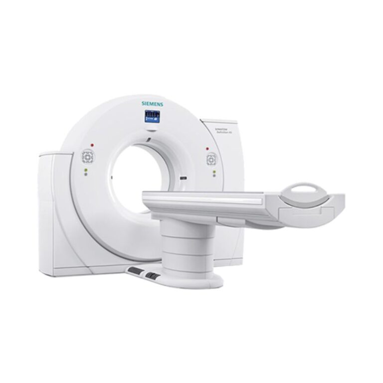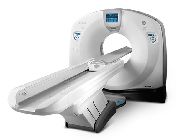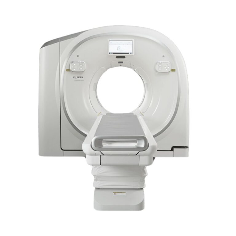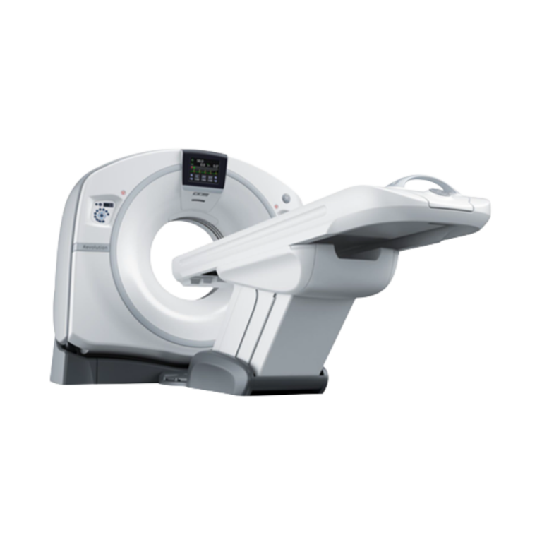
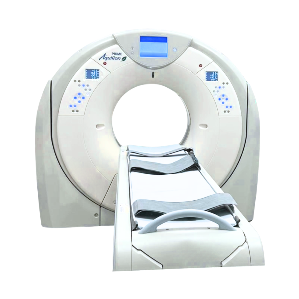
Canon Aquilion PRIME 160-Slice CT
- Category:
- CT Scanners
- Brand:
- Canon
The Canon Aquilion PRIME 160 is an 80-detector-row CT scanner that generates 160 reconstructed slices per rotation. It is well suited for hospitals, imaging centers, specialty practices, and veterinary referral hospitals needing fast, wide-bore CT with 0.35-second rotation, a 78 cm bore, and configurable cardiac, vascular, and interventional imaging.
Cannon Aquilion PRIME 160 CT Highlights & Benefits
Also found in the pre-owned market as the Toshiba Aquilion PRIME 160, the system combines 160-slice reconstruction with a wide-bore patient platform, fast acquisition, and advanced imaging options for routine and specialized CT.
PRIME 160 CT Key Benefits
-
- 160 reconstructed slices per rotation from an 80-row detector
- 0.5 mm detector elements with 40 mm physical coverage
- Fast 0.35-second gantry rotation
- 78 cm (30.7 in) gantry opening
- Patient table capacity up to 660 lb (300 kg), depending on installed couch
- AIDR 3D reconstruction for image-noise and radiation dose management
- Up to ±30° gantry tilt for additional positioning flexibility
- Reconstruction speeds up to 60 images per second on applicable configurations
Aquilion PRIME Platform Video
See the Aquilion PRIME platform, patient positioning, and imaging workflow.
Aquilion PRIME 160-Slice Imaging Performance
Detector & Slice Configuration
The Aquilion PRIME 160 is an 80-detector-row CT scanner that generates 160 reconstructed slices per rotation. Its 80 × 0.5 mm detector provides 40 mm of physical coverage, while Double Slice and coneXact reconstruction create the 160-slice dataset.
The additional reconstructed sampling supports detailed multiplanar and 3D imaging while retaining 40 mm of physical detector coverage. This distinction is important when comparing the PRIME 160 with scanners that use a different number of physical detector rows.
Scan Speed & Anatomical Coverage
Rotation times down to 0.35 seconds support rapid chest, abdominal, vascular, trauma, and other examinations where shorter acquisition can help reduce the effects of patient motion.
The 80 × 0.5 mm detector configuration provides 40 mm of anatomical coverage per rotation while maintaining thin-section acquisition for detailed reconstruction.
Image Reconstruction Speed
Applicable Aquilion PRIME configurations support full-resolution reconstruction at speeds up to 60 images per second.
Faster reconstruction helps move acquired studies into review without creating an unnecessary processing delay, particularly during emergency, trauma, vascular, and higher-volume CT workflows.
Image Quality & Radiation Dose Management
Image Noise Reduction & Reconstruction
Adaptive Iterative Dose Reduction 3D, or AIDR 3D, is Canon/Toshiba’s iterative reconstruction technology for reducing image noise while maintaining clinically useful image detail.
AIDR 3D works within the acquisition and reconstruction process to help balance image quality with radiation exposure rather than relying only on conventional filtered reconstruction.
Automatic Exposure & Dose Controls
Available PRIME dose-management tools include SUREExposure 3D Adaptive, automatic tube-current modulation, active collimation, and Dose Check.
SUREExposure 3D Adaptive adjusts X-ray exposure based on the patient and selected examination. Active collimation limits unnecessary exposure at the beginning and end of applicable helical acquisitions, while Dose Check improves operator awareness of planned radiation levels.
Installed dose-management software should be verified on the individual scanner.
Detector & System Configuration
Aquilion PRIME 160 systems were produced across multiple hardware and software generations. Detector hardware, data-acquisition components, reconstruction systems, and software levels can therefore vary among pre-owned units.
These components should be reviewed as part of the individual scanner configuration rather than assumed from the model name alone.
Patient Access & Positioning
Wide-Bore Patient Access
The Aquilion PRIME uses a 78 cm (30.7 in) gantry opening. The wider bore provides additional patient clearance and can make positioning easier for larger patients, trauma cases, and examinations requiring greater procedural access.
The gantry supports up to ±30° physical tilt for applicable acquisitions, providing additional positioning flexibility.
Patient Table Capacity & Accessibility
Applicable PRIME patient tables support up to 660 lb (300 kg). Documented configurations include a couch width of approximately 47 cm (18.5 in), a minimum couch height of approximately 33 cm (13 in), and scan ranges up to approximately 180 cm (70.9 in).
The combination of a wide bore, high-capacity couch, low table height, and long scan range supports imaging across a broad patient population.
Patient Positioning & In-Room Controls
Available Tech Assist Lateral Slide allows the couch top to move from side to side, helping technologists center anatomy without repeatedly repositioning the patient manually.
Applicable systems also include gantry-mounted controls and iStation in-room guidance features that can support positioning, patient communication, and workflow from inside the scan room.
Installed table and positioning features should be confirmed on the individual system.
Clinical Environments & Applications
The Canon Aquilion PRIME 160 supports routine and advanced CT across a wide range of clinical settings. Its suitability depends on clinical requirements and installed capabilities rather than the size of the organization operating it.
Clinical Environments
-
- Hospitals and health systems
- Outpatient imaging centers
- Independent diagnostic imaging centers
- Emergency and trauma departments
- Physician-owned and specialty practices with dedicated CT capabilities
- Veterinary referral and specialty hospitals
Clinical Specialties
-
- Diagnostic Radiology
- Emergency Medicine & Trauma
- Cardiology
- Vascular Medicine & Surgery
- Oncology
- Neurology & Neurosurgery
- Orthopedics & Musculoskeletal Imaging
- Interventional Radiology
- Veterinary Medicine
Common CT Applications
-
- Brain, head, and neck CT
- Chest CT and clinically appropriate lung screening protocols
- Abdominal and pelvic CT, including CT colonography
- Emergency and trauma imaging
- CT angiography and vascular imaging
- Cardiac CT and coronary CT angiography on appropriately configured systems
- Oncology, musculoskeletal, and orthopedic imaging
- CT-guided procedures and veterinary diagnostic CT in appropriately configured settings
Advanced Imaging Capabilities
Advanced imaging capabilities can vary significantly among pre-owned Canon Aquilion PRIME 160 systems. Hardware, software licenses, and installed application packages should be reviewed on the individual scanner.
Cardiac CT Imaging
Appropriately configured Aquilion PRIME 160 systems support ECG-gated cardiac CT and coronary CT angiography.
SURECardio Prospective is an available cardiac acquisition package designed to coordinate imaging with the patient’s heart rhythm. Applicable configurations can also include tools for managing irregular heartbeats during cardiac examinations.
CT Angiography, Perfusion & Vascular Imaging
Thin-section acquisition and fast rotation support CT angiography of the head, neck, chest, abdomen, aorta, and peripheral vascular system.
Available SURESubtraction technology can separate bone or calcium from applicable contrast-enhanced datasets to improve vascular visualization. Variable Helical Pitch can change table speed during a continuous acquisition, while applicable systems may also include Shuttle Helical capabilities for extended dynamic and perfusion imaging.
Advanced Tissue & Metal Implant Imaging
Available Dual Energy acquisition can support material characterization and iodine-based analysis on appropriately configured systems.
Single Energy Metal Artifact Reduction, or SEMAR, is designed to reduce image artifacts caused by metal. This can improve visualization around orthopedic hardware, prostheses, and adjacent bone or soft tissue.
CT-Guided Procedures
Applicable PRIME configurations support CT fluoroscopy, which provides rapidly updated CT images during image-guided procedures.
Available interventional tools can support needle visualization and procedural planning for biopsy, drainage, and other CT-guided procedures.
Oncology Imaging & Treatment Follow-Up
Thin-section whole-body imaging supports lesion assessment, chest, abdominal and pelvic imaging, treatment follow-up, and other oncology CT examinations.
Multiphase acquisition, advanced visualization, subtraction, and other software capabilities may provide additional oncology imaging options depending on the installed configuration.
CT Workflow & Image Management
Scan Setup & Immediate Image Review
The Aquilion PRIME workflow can include real-time dual scanograms, scan planning tools, gantry controls, rapid reconstruction, and InstaView.
InstaView provides immediate image reconstruction and review during the scanning workflow so technologists can begin evaluating acquired images without waiting for the complete study to finish processing.
Image Review & Storage Efficiency
HybridView combines fine lung detail and soft-tissue visualization into a single reconstructed image series.
For applicable examinations, this can reduce the need to create and review separate reconstruction series while also reducing image-storage requirements.
PACS, Worklist & Network Integration
The Aquilion PRIME supports DICOM connectivity for integration with PACS, radiology worklists, and other compatible healthcare information systems.
DICOM is the standard used to exchange medical images and associated study information between imaging equipment and clinical systems.
Depending on the installed workstation and software, PRIME systems may also include Vital Images or Vitrea tools for cardiovascular, neurovascular, oncology, multiplanar, and 3D image processing.
Available Configurations & Installed Options
The Canon Aquilion PRIME 160 was sold across multiple hardware and software generations. Pre-owned systems can therefore differ substantially even when they share the same model designation.
Scanner Hardware & Installed Equipment
Important configuration details include:
-
- Year of manufacture
- Detector and data-acquisition hardware
- X-ray tube model and tube usage history
- Generator configuration
- Patient table and weight capacity
- Reconstruction hardware
- Gantry and table accessories
- Included injector and positioning accessories
Image Reconstruction & Clinical Software
Available systems may differ in their reconstruction software, dose-management tools, cardiac packages, vascular applications, metal artifact reduction, Dual Energy, perfusion capabilities, and CT fluoroscopy options.
Installed licenses should be evaluated against the facility’s actual examination mix rather than assuming every PRIME 160 includes the complete advanced application package.
Workstations & Image Processing
The operator console, reconstruction hardware, Vital Images or Vitrea workstation, active software licenses, and DICOM functionality should also be evaluated as part of the complete system.
An MXR representative can help identify an available configuration that aligns with the facility’s clinical applications, patient-capacity needs, workflow, and advanced imaging requirements.
Select CT systems may also be available through MXR’s Platinum Certified CT & MRI Systems.
Aquilion PRIME 160 Price & Availability
Canon Aquilion PRIME 160 pricing can vary based on model year, tube history, hardware configuration, installed software, patient table, workstations, accessories, service requirements, and overall project scope.
MXR can help identify or source an Aquilion PRIME 160 configuration that aligns with your clinical requirements, facility needs, and equipment budget. Service, parts support, installation planning, and project requirements can also be reviewed as part of the purchasing process.
See CT maintenance and repair support.
Site Planning & Installation Requirements
The Aquilion PRIME has a documented minimum installation footprint of approximately 14.8 m² (159.3 sq ft) when configured with the short patient couch.
Actual room requirements depend on the individual scanner configuration, patient table, generator, electrical service, cooling, shielding, accessories, equipment access path, and control-room layout.
Final site planning should therefore be based on the exact Canon Aquilion PRIME 160 being installed rather than a generic model specification.
MXR can support equipment planning and installation through Turnkey Imaging Project Management.
Canon Aquilion PRIME 160 Specifications
Specifications can vary by model year, installed hardware, software version, and optional equipment.
CT Acquisition & Detector
-
- System configuration: 160-slice CT scanner
- Physical detector rows: 80
- Reconstructed slices: 160 per rotation
- Detector element thickness: 0.5 mm
- Detector coverage: 40 mm per rotation
- Minimum rotation time: 0.35 seconds
- Whole-body CT capability
Gantry & Patient Table
-
- Gantry aperture: 78 cm (30.7 in)
- Gantry tilt: up to ±30°
- Patient table capacity: up to 660 lb (300 kg), depending on installed couch
- Couch width: approximately 47 cm (18.5 in)
- Minimum couch height: approximately 33 cm (13 in)
- Maximum documented scan range: up to 180 cm (70.9 in)
- Tech Assist Lateral Slide: available on applicable configurations
X-Ray Generator & Tube
-
- X-ray tube heat capacity: 7.5 MHU
- Generator output: 60 kW and 72 kW configurations documented within the platform
- Generator and tube configuration should be verified on the individual scanner
Image Reconstruction & Performance
-
- Double Slice and coneXact reconstruction supporting 160 reconstructed slices
- AIDR 3D iterative reconstruction
- Reconstruction speeds up to 60 images per second on applicable configurations
- Thin-section acquisition: 0.5 mm
- Multiplanar and 3D reconstruction support
- InstaView immediate image reconstruction and review on applicable configurations
- HybridView image reconstruction on applicable configurations
System & Site Requirements
-
- Minimum documented installation area: approximately 14.8 m² (159.3 sq ft) with short couch
- DICOM connectivity
- Electrical, cooling, shielding, and room requirements depend on the individual system configuration
- Final site requirements should be verified against the exact scanner being installed