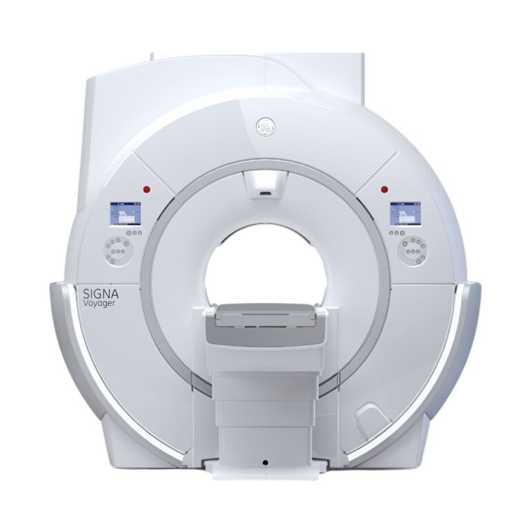


Siemens MAGNETOM Espree 1.5T MRI
- Category:
- MRI Systems
- Brand:
- Siemens
The Siemens MAGNETOM Espree is a 1.5T MRI scanner distinguished by its 70 cm opening and unusually short 125 cm housing. It suits hospitals, imaging centers, and specialty practices serving larger or claustrophobic patients, while Siemens Tim coils support efficient examinations across multiple anatomical regions.
MAGNETOM Espree MRI Highlights & Benefits
The Siemens MAGNETOM Espree combines high-field imaging with one of the shortest wide-bore designs of its generation. Its 70 cm opening, flexible patient positioning, and Siemens Tim technology help facilities accommodate more patients while performing routine and specialized MRI examinations.
Key Features & Benefits
-
- 70 cm Wide Bore: Provides more room around patients than conventional 60 cm scanners, helping accommodate larger patients and those uncomfortable in enclosed spaces.
- 125 cm Short System: Allows head-out and feet-first positioning for many examinations, reducing the sensation of enclosure.
- Up to 550 lb Patient Capacity: Compatible patient tables support up to 250 kg (550 lb), helping facilities accommodate larger patients.
- Low Patient Table: Available configurations lower to approximately 47 cm (18.5 in), making transfers easier for patients with limited mobility.
- 1.5T High-Field Imaging: Supports detailed neurological, orthopedic, abdominal, breast, cardiac, and vascular examinations with compatible equipment.
- Siemens Tim Technology: Connects multiple lightweight coils to reduce repositioning and coil changes during extended examinations.
- Up to 32 Receiver Channels: Available 8-, 18-, and 32-channel configurations support different imaging requirements and compatible accelerated scans.
- Up to 205 cm Imaging Coverage: Compatible Tim equipment supports extended spine, body, and vascular examinations.
- Automated MRI Workflows: AutoAlign, Phoenix, and Inline Composing simplify compatible scan planning, protocol management, and image processing.
- Zero Helium Boil-Off: Limits helium evaporation during normal operation, reducing routine replenishment requirements.
Espree 70 cm Wide-Bore Patient Access & Positioning
The Espree’s defining advantage is its combination of a 70 cm bore and 125 cm housing. This design helps facilities accommodate patients who may have difficulty tolerating longer or narrower MRI scanners.
70 cm Bore & 125 cm Short Housing
The Espree features a 70 cm (27.6 in) bore and a 125 cm (49.2 in) system length, measured from cover to cover.
Its wide opening provides additional space around patients, while the short housing reduces how much of the body is surrounded by the scanner.
Key positioning advantages include:
-
- Head-Out Imaging: Keeps the patient’s head outside the magnet during many suitable examinations, helping reduce claustrophobia and anxiety.
- Feet-First Scanning: Allows patients to enter feet-first during appropriate examinations, avoiding head-first positioning.
- Greater Facial Clearance: Provides additional space around the face during examinations requiring head-in positioning.
- Flexible Joint Positioning: Allows selected wrist, elbow, and other extremity examinations with the limb alongside the body rather than overhead.
Siemens originally estimated that approximately 60% of routine Espree examinations could be performed with the patient’s head outside the magnet. Actual positioning depends on the anatomy, imaging coils, and examination requirements.
The Espree pioneered 1.5T Open Bore MRI. Although described as an Open Bore scanner, it is a short, wide cylindrical MRI rather than a fully open-sided system.
Up to 550 lb Capacity & Low Patient Table
The Espree supports patient-table configurations rated for up to 250 kg (550 lb).
Compatible tables also provide a minimum height of approximately 47 cm (18.5 in), helping patients transfer onto and off the scanner.
These features are particularly useful for:
-
- Larger Patients: Additional bore clearance and compatible higher-capacity tables accommodate a broader range of body sizes.
- Patients with Claustrophobia: The short housing and head-out positioning options reduce the feeling of enclosure.
- Patients with Limited Mobility: A low table helps simplify transfers and examination preparation.
- Orthopedic Patients: Additional positioning space accommodates selected painful joints and restricted movements.
Patient fit depends on body dimensions, bore clearance, coil placement, and examination requirements.
Some Espree configurations have lower patient-table weight limits. MXR Imaging confirms the installed table’s rated capacity and positioning capabilities during equipment selection.
Flexible Wrist, Elbow & Joint Positioning
The 70 cm bore provides additional room for joint examinations that may be uncomfortable in narrower scanners.
For example, certain wrist and elbow examinations can be performed with the arm alongside the body rather than raised overhead.
This is useful for patients with shoulder restrictions, joint pain, or difficulty maintaining conventional imaging positions. Compatible lightweight Tim coils provide additional positioning flexibility.
Espree 1.5T Imaging & Gradient Performance
The Siemens MAGNETOM Espree combines a superconducting 1.5T magnet, Siemens gradient technology, and Tim receiving coils to support routine and advanced MRI.
1.5T Magnet for Detailed Anatomical Imaging
The 1.5 Tesla magnetic field provides the signal strength needed for detailed brain, spine, joint, abdominal, and pelvic examinations.
With compatible software, the scanner also supports specialized techniques for evaluating tissue characteristics, blood vessels, and selected functional processes.
Its combination of high-field imaging and wide-bore access allows facilities to accommodate patients who may struggle with conventional scanners without moving to a lower-field open-sided MRI system.
Z-Engine vs DZ-Engine: Gradient Speed & Performance
MRI gradients create additional magnetic fields that help the scanner determine where imaging signals originate within the patient’s anatomy.
Documented Espree configurations include:
-
- Z-Engine: Provides 33 mT/m maximum gradient amplitude and 100 T/m/s slew rate, supporting routine imaging and compatible accelerated acquisitions.
- DZ-Engine: Provides the same 33 mT/m gradient amplitude with a faster 170 T/m/s slew rate, supporting more rapid gradient changes during selected advanced sequences.
Gradient amplitude describes the strength of the additional magnetic fields used during imaging. Slew rate describes how quickly those fields change.
The DZ-Engine can benefit examinations requiring faster gradient switching. Actual performance depends on the imaging sequence, software, and installed hardware.
Espree Tim Coils & Extended Imaging Coverage
The Espree uses Siemens Tim (Total imaging matrix) technology to collect MRI signals from multiple lightweight coils positioned around the patient.
Instead of repeatedly changing coils between anatomical regions, compatible Tim configurations allow several coils to remain connected while the scanner selects the receiving elements needed for each acquisition.
This reduces preparation interruptions and supports longer examinations with less patient repositioning.
Up to 10 Tim Coils with Fewer Coil Changes
Compatible Siemens MAGNETOM Espree configurations support combinations of up to ten imaging coils.
Technologists can position coils around multiple anatomical regions during preparation, allowing the scanner to select the appropriate receiving elements as the examination progresses.
This provides three practical benefits:
-
- Fewer Coil Changes: Connected coils can remain in place during compatible multiregion examinations.
- Less Patient Repositioning: Different anatomical regions can be examined without repeatedly moving the patient to replace coils.
- Flexible Anatomical Coverage: Lightweight coils accommodate different patient sizes and examination requirements.
The Siemens Body Matrix coil weighs approximately 950 g (2.1 lb), making it easier to position over the abdomen and other body regions.
Tim technology can reduce examination time through fewer coil changes and compatible accelerated acquisitions. Actual time savings depend on the examination and installed equipment.
8-, 18- & 32-Channel Tim Configurations
The Siemens MAGNETOM Espree supports three documented Tim configurations:
-
- Tim [32 × 8]: Connects up to 32 coil elements with eight independent receiver channels, supporting routine MRI and compatible parallel imaging.
- Tim [76 × 18]: Connects up to 76 coil elements with 18 receiver channels, providing additional receiving capability for compatible advanced and extended examinations.
- Tim [76 × 32]: Connects up to 76 coil elements with 32 independent receiver channels, supporting more simultaneous signal information from compatible coils.
The first number identifies the maximum connected coil elements. The second identifies the independent receiver channels available during acquisition.
Higher-channel configurations support more demanding coil arrangements and compatible accelerated imaging techniques.
All three are configurations of the same MAGNETOM Espree model.
Up to 205 cm of Whole-Body Imaging Coverage
Compatible Tim Whole Body equipment supports imaging coverage of up to approximately 205 cm (80.7 in).
The scanner acquires information across different table positions while compatible coils remain connected.
This supports:
-
- Whole-Spine MRI: Extended imaging of the cervical, thoracic, and lumbar spine.
- Peripheral Angiography: Imaging of blood vessels across larger anatomical regions.
- Multiregion Body MRI: Examination of several body regions with fewer coil changes.
- Oncology Imaging: Selected extended-coverage tumor assessments and follow-up examinations.
The primary benefit is reduced interruption for coil replacement and patient repositioning during longer studies.
iPAT Parallel Imaging for Shorter Scans
Siemens iPAT (integrated Parallel Acquisition Technique) uses information collected by multiple imaging coils to reconstruct images from fewer measurements.
The scanner uses differences between coil signals to reconstruct information that would otherwise require additional acquisition time.
This can shorten compatible scans or support greater image detail within an appropriate examination period.
On the Espree, iPAT complements Tim technology by combining flexible coil coverage with accelerated image acquisition.
Espree Exam Setup & Automated Imaging Workflow
The Espree supports Siemens syngo MR software and workflow tools that simplify examination planning, protocol management, and image processing. These technologies reduce manual steps and help technologists maintain consistent examinations.
AutoAlign & Phoenix for Consistent Scan Setup
-
- AutoAlign: Identifies anatomical reference points and automatically positions imaging slices during compatible brain and spine examinations. This reduces manual planning and helps maintain consistent anatomical coverage between initial and follow-up studies.
- Phoenix & PhoenixZIP: Store, retrieve, and transfer MRI examination protocols, allowing technologists to reuse established scanning settings. This reduces setup work and helps standardize examinations across operators.
TimCT & Inline Processing for Extended Exams
-
- Inline Composing: Combines images acquired at different table positions into one continuous anatomical view. This is useful for whole-spine and vascular examinations, reducing the need to manually assemble images.
- syngo Inline Processing: Automatically performs selected reconstruction tasks, including compatible subtraction images and vascular projections. This reduces post-processing work after scanning.
- syngo TimCT: Coordinates continuous patient-table movement with image acquisition during compatible extended examinations. This reduces interruptions between scanning stations during selected whole-body oncology and vascular studies.
syngo TimCT requires compatible software, receiving equipment, and patient-table capabilities.
The Espree also supports DICOM connectivity for transferring examinations to compatible hospital imaging and picture archiving and communication systems (PACS).
Espree Motion Correction & Metal Implant Imaging
The Siemens MAGNETOM Espree supports imaging techniques that address two common challenges during MRI examinations: patient movement and distortion around metal implants.
- syngo BLADE: Collects overlapping imaging information to reduce motion-related artifacts during reconstruction. This helps improve image clarity during compatible brain, spine, joint, and body examinations, particularly when patients have difficulty remaining still. It can also reduce the need to repeat some movement-affected scans.
- syngo WARP: Uses optimized acquisition settings and View Angle Tilting (VAT) to reduce distortion caused by compatible metal implants. VAT helps correct the apparent displacement of anatomy near metal, improving visibility of surrounding tissues during selected postoperative orthopedic examinations.
Some motion and metal-related artifacts may remain. Patients with implants require appropriate MRI safety screening, and scanning must follow the implant manufacturer’s MR-conditional requirements.
Espree 3D Imaging & Tissue Measurements
Compatible Espree software provides additional ways to visualize anatomy and measure tissue characteristics beyond conventional MRI images.
- SPACE: Collects detailed three-dimensional imaging information that can be reconstructed into different viewing planes. This helps radiologists evaluate complex anatomy without acquiring separate scans for every orientation. Compatible SPACE DIR techniques also help distinguish selected neurological abnormalities by suppressing signals from certain tissues.
- DIXON: Separates fat and water signals to produce more consistent fat suppression. This improves the visibility of selected soft-tissue structures during orthopedic and body examinations.
- syngo MapIt: Produces quantitative tissue measurements using compatible T1, T2, and T2* mapping techniques. These measurements provide additional information about tissue characteristics, including cartilage assessment during specialized orthopedic examinations.
These capabilities require compatible scanning sequences, software, and clinical protocols.
Espree Advanced Brain, Vascular & Body Imaging
The Siemens MAGNETOM Espree supports specialized neurological, vascular, and body imaging techniques with appropriate software and hardware. These applications provide additional information beyond conventional anatomical MRI.
Nerve Pathway & Brain Blood Flow Imaging
-
- Diffusion Tensor Imaging (DTI): Measures the direction of water movement within tissue to provide information about nerve pathways. This supports specialized neurological examinations and selected presurgical assessments.
- Susceptibility-Weighted Imaging (SWI): Uses differences in magnetic susceptibility to help visualize small hemorrhages and other blood-related tissue characteristics that may be less apparent on conventional MRI.
- Arterial Spin Labeling (ASL): Magnetically labels flowing blood to estimate tissue perfusion without injected contrast, supporting selected neurological assessments.
Blood Vessel, Tumor & Liver Imaging
-
- syngo NATIVE: Uses differences between flowing blood and surrounding tissue to produce vascular images without conventional contrast injection. This provides an additional option for selected thoracic, abdominal, and peripheral vascular examinations.
- syngo REVEAL: Uses diffusion-weighted MRI to evaluate differences in water movement within body tissues. This supports selected oncology examinations, including tumor assessment and follow-up imaging, with compatible techniques to manage breathing-related motion.
- MR Elastography: Uses externally generated mechanical vibrations and MRI measurements to estimate tissue stiffness. These measurements provide additional information during selected liver assessments and require specialized hardware and software.
Available clinical applications depend on the installed software, licenses, imaging coils, and compatible examination protocols.
Clinical Environments & Applications
The Espree’s wide bore, short housing, and Tim technology support facilities serving patients who need additional space, flexible positioning, or specialized MRI capabilities.
Clinical Environments
-
- Hospitals & Radiology Departments: Wide-bore imaging for patients with varied body sizes, mobility limitations, and complex examination needs.
- Outpatient Diagnostic Imaging Centers: Routine and specialty 1.5T MRI with a short-bore design that can help patients who find conventional scanners uncomfortable.
- Orthopedic & Sports Medicine Centers: Joint and spine imaging with additional room for positioning painful or restricted extremities.
- Neurology & Neurosurgery Departments: Brain and spine examinations with compatible diffusion, susceptibility, and perfusion applications.
- Cardiac & Vascular Imaging Departments: Specialized examinations using compatible cardiac software and angiography techniques.
- Breast Imaging Centers: Diagnostic breast MRI using dedicated coils and appropriate clinical applications.
- Oncology Imaging Departments: Regional and extended-coverage imaging using compatible Tim equipment.
- Specialized Equine Veterinary Facilities: High-field MRI in purpose-built facilities equipped for large-animal handling and positioning.
Clinical Specialties & Applications
-
- Neurology & Neurosurgery: Brain and spine MRI, including compatible diffusion imaging, nerve pathway assessment, and small-hemorrhage visualization.
- Orthopedics & Sports Medicine: Joint, cartilage, ligament, tendon, and soft-tissue examinations, including selected postoperative imaging using syngo WARP.
- Spine Care: Cervical, thoracic, lumbar, and whole-spine imaging with compatible extended coverage and Inline Composing.
- Abdominal Imaging: Liver, pancreatic, abdominal, and pelvic MRI using appropriate body coils and motion-reduction techniques.
- Cardiology: Cardiac anatomy, function, and selected flow studies using compatible cardiac software.
- Vascular Medicine: Peripheral and abdominal angiography, including selected non-contrast examinations using syngo NATIVE.
- Breast Radiology: Diagnostic breast MRI, cancer evaluation, implant assessment, and compatible MRI-guided procedures.
- Oncology: Tumor assessment, treatment follow-up, and selected extended-coverage examinations using compatible diffusion imaging.
- Pediatric Radiology: Appropriate brain, spine, joint, and body examinations using patient-specific positioning and scanning protocols.
- Equine Veterinary Imaging: Selected orthopedic, soft-tissue, and head or brain examinations in specially configured large-animal MRI facilities.
Equine MRI is a documented real-world Espree application. These installations require specialized patient handling, anesthesia, positioning, and facility planning.
Clinical capabilities depend on the installed coils, software, and examination requirements.
Espree Configurations, Coils & Available Options
Individual Siemens MAGNETOM Espree systems can differ in receiver channels, gradients, imaging coils, software, and patient-table equipment.
MXR Imaging helps customers select configurations based on clinical applications, patient requirements, and available equipment.
Receiver Channels, Gradients & Table Options
-
- Receiver Channels: Tim [32 × 8], [76 × 18], or [76 × 32], affecting compatible coil arrangements and accelerated imaging.
- Gradient Hardware: Z-Engine or DZ-Engine, affecting gradient switching performance during supported examinations.
- Tim Whole Body Equipment: Compatible coils and patient-table arrangements for extended anatomical coverage.
- Patient Table: Installed movement capabilities, height adjustment, and rated capacity.
- Software & Clinical Options: Installed syngo MR generation and licensed specialized imaging applications.
Tim Coil Options & Anatomical Coverage
Compatible coil options include:
-
- Head & Neck Matrix Coils: Support neurological, head, neck, and cervical spine examinations.
- Spine Matrix Coils: Provide regional and extended spinal coverage with fewer coil changes.
- Body Matrix Coils: Support abdominal and pelvic imaging through flexible anatomical coverage.
- Shoulder & Extremity Coils: Collect signals close to joints for detailed orthopedic imaging.
- Flexible Coils: Accommodate anatomical areas that are difficult to position within rigid coil arrangements.
- Breast Coils: Support diagnostic breast MRI and compatible procedures.
- Peripheral Angiography Coils: Provide extended coverage for selected vascular examinations.
Coil compatibility and included accessories depend on the selected system.
MXR Imaging also provides MRI coil repair for supported equipment.
Software & Installed Clinical Options
Espree systems can include different syngo MR software generations, including compatible B19 configurations.
Installed options may include:
-
- Scan Planning & Workflow: AutoAlign, Phoenix, and PhoenixZIP.
- Accelerated Imaging: iPAT parallel imaging.
- Image Processing: Inline Composing and syngo Inline Processing.
- Extended Imaging: Compatible syngo TimCT applications.
- Artifact Reduction: syngo BLADE and syngo WARP.
- Specialized Image Quality: SPACE, DIXON, and MapIt.
- Neurological Imaging: DTI, SWI, and ASL.
- Vascular & Body Imaging: syngo NATIVE, syngo REVEAL, and MR elastography.
Software availability depends on installed licenses, software generation, and hardware compatibility.
MXR Imaging helps customers identify configurations equipped for their required examinations.
Espree Magnet, Cooling & Site Requirements
The Espree’s short magnet and compact equipment arrangement can simplify certain MRI installations while helping facilities manage operating requirements.
Final installation specifications depend on the selected equipment and approved site plan.
Zero Helium Boil-Off & Magnet Cooling
The Espree uses liquid helium to cool its superconducting magnet.
Its zero helium boil-off refrigeration system limits helium evaporation during normal operation, reducing routine replenishment requirements.
The magnet still requires refrigeration maintenance, monitoring, and emergency helium venting.
MXR Imaging provides MRI refrigeration and helium services for supported equipment.
Compact Installation & MRI Shielding
The Espree supports a representative minimum installation area of approximately 27 m² (290 sq ft) under specified planning conditions.
This figure represents the equipment installation area for a suitable layout, not a universal minimum examination-room size.
Site planning must address:
-
- Equipment Delivery: Adequate access for transporting the magnet, patient table, and supporting equipment.
- Structural Support: Floor capacity for the installed magnet and concentrated equipment loads.
- RF Shielding: Specialized shielding that prevents external radiofrequency interference from degrading images.
- Magnetic Field Boundaries: Appropriate safety zones and access controls around the magnet.
- Quench Venting: Emergency infrastructure for directing released helium safely outside.
- Service Clearances: Access for maintenance, inspections, and repairs.
MXR Imaging provides custom MRI shielding solutions and turnkey imaging project management for new installations, replacements, and facility upgrades.
These services also support qualifying Platinum Certified equipment projects.
Electrical Power & Cooling
The Espree requires dedicated electrical service and appropriate equipment cooling.
Requirements depend on the installed gradients, refrigeration system, power distribution equipment, and site configuration.
MXR Imaging coordinates installation planning and technical requirements for the selected system.
Espree vs Siemens MAGNETOM Aera
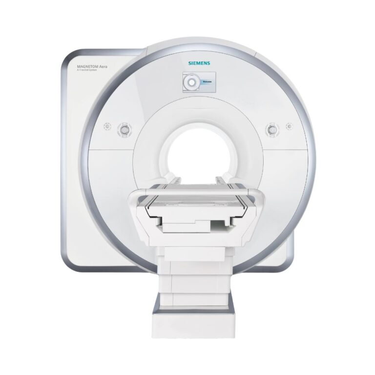
The Espree and Siemens MAGNETOM Aera 1.5T both offer a 70 cm wide bore, but they belong to different Siemens MRI generations.
Espree emphasizes patient access through its exceptionally short 125 cm housing and original Tim coil technology. Aera uses a 145 cm system design with newer Tim 4G coils and Dot guided scanning workflows.
Facilities prioritizing a particularly short magnet may prefer Espree, while those seeking newer coil technology and automated workflows may find Aera better suited to their needs.
MXR Imaging helps customers compare clinical applications, installed options, equipment condition, and installation requirements.
Pre-Owned Espree Systems: Condition & Installed Options
MXR Imaging helps healthcare facilities evaluate pre-owned Siemens MAGNETOM Espree systems based on clinical requirements, equipment condition, and installation needs.
Available units can differ in:
-
- Receiver Channels & Gradients: Eight-, 18-, or 32-channel configurations and installed gradient hardware.
- Software & Licenses: syngo MR generation and available specialized applications.
- Imaging Coils: Included neurological, body, orthopedic, breast, and vascular coils.
- Patient Table: Installed configuration, height adjustment, movement capabilities, and rated capacity.
- Magnet & Refrigeration: Helium condition, cooling performance, and service history.
- Equipment History: Previous maintenance, repairs, upgrades, and included accessories.
MXR Imaging works with customers to identify suitable equipment and coordinate system evaluation, installation planning, and long-term MRI support.
MAGNETOM Espree MRI Price & Availability
Siemens MAGNETOM Espree pricing depends on equipment condition, receiver-channel capacity, gradient hardware, software, imaging coils, service history, and installation requirements.
MXR Imaging offers pre-owned MRI systems, including Platinum Certified equipment where applicable.
Platinum Certified Reconditioning & Warranty
Qualifying Platinum Certified systems undergo MXR Imaging’s 100-point refurbishment process under its ISO 13485:2016-certified quality management system.
MXR Imaging also provides turnkey imaging project management, MRI maintenance and repair, coil repair, helium services, replacement parts, and ongoing equipment support.
Contact MXR Imaging to discuss an Espree configuration suited to your facility and request a quote.
Siemens MAGNETOM Espree 1.5T Specifications
Magnet System
-
- Field Strength: 1.5T
- Magnet Type: Superconducting, actively shielded
- Magnet Design: Siemens OR122
- Bore Diameter: 70 cm (27.6 in)
- Magnet Length: Approximately 120 cm (47.2 in)
- System Length Including Covers: Approximately 125 cm (49.2 in)
- Helium Consumption: Zero helium boil-off under normal operating conditions
Gradient System
-
- Documented Configurations: Z-Engine and DZ-Engine
- Z-Engine Maximum Gradient Amplitude: 33 mT/m
- Z-Engine Maximum Slew Rate: 100 T/m/s
- DZ-Engine Maximum Gradient Amplitude: 33 mT/m
- DZ-Engine Maximum Slew Rate: 170 T/m/s
- Installed Gradient Hardware: Configuration-dependent
RF System
-
- RF Technology: Siemens Tim (Total imaging matrix)
- Receiver Configurations: Tim [32 × 8], Tim [76 × 18], and Tim [76 × 32]
- Independent Receiver Channels: 8, 18, or 32
- Maximum Connected Coil Elements: Up to 76
- Compatible Coil Combinations: Up to ten coils, depending on configuration
- Extended Imaging Coverage: Up to approximately 205 cm (80.7 in)
- Parallel Imaging: Compatible Siemens iPAT
Bore & Patient Table
-
- Bore Diameter: 70 cm (27.6 in)
- Maximum Patient Weight: Up to 250 kg (550 lb) with an appropriately rated table; other configurations may have lower limits
- Representative Minimum Table Height: 47 cm (18.5 in)
- Representative Maximum Table Height: 89 cm (35 in)
- Representative Tabletop Width: 54 cm (21.3 in)
- Positioning: Head-first and feet-first for appropriate examinations
- Patient Comfort: Short magnet housing, wide opening, and lightweight coils
Coils & Accessories
-
- Coil Technology: Siemens Tim Matrix
- Neurological Coils: Compatible head and neck coils
- Spine Coils: Compatible Spine Matrix arrays
- Body Coils: Compatible Body Matrix arrays
- Flexible Coils: Supported flexible surface coils
- Orthopedic Coils: Compatible shoulder and extremity coils
- Breast Coils: Compatible diagnostic breast coils
- Vascular Coils: Compatible peripheral angiography coils
- Included Accessories: Determined by the selected equipment package
Workflow & Imaging Software
-
- Software Platform: Siemens syngo MR
- Documented Software Generations: Earlier releases and compatible B19 configurations
- Exam Planning: Compatible AutoAlign
- Protocol Management: Phoenix and PhoenixZIP
- Image Processing: Compatible syngo Inline and Inline Composing
- Extended Imaging: Compatible syngo TimCT
- Parallel Imaging: Siemens iPAT
- Motion Reduction: Compatible syngo BLADE
- Metal-Artifact Reduction: Compatible syngo WARP
- Advanced Imaging: Configuration-dependent SPACE, DIXON, MapIt, DTI, SWI, ASL, NATIVE, REVEAL, and MR elastography
Siting & Utilities
-
- Representative Minimum Installation Area: Approximately 27 m² (290 sq ft), subject to the approved layout
- RF Shielding: Required
- Magnetic-Field Safety Planning: Required
- Emergency Helium Venting: Required
- Electrical Service: Configuration-specific dedicated service
- Equipment Cooling: Configuration-specific refrigeration and cooling infrastructure
- Final Site Requirements: Determined by the selected system and approved planning documentation
System Dimensions & Weight
-
- Bore Diameter: 70 cm (27.6 in)
- Magnet Length: Approximately 120 cm (47.2 in)
- System Length Including Covers: Approximately 125 cm (49.2 in)
- Representative Operating System Weight: Approximately 5.1 metric tons (11,244 lb)
- Patient Table Capacity: Configuration-dependent, up to 250 kg (550 lb) with the specified table
- Installation Dimensions: Depend on the patient table, equipment arrangement, shielding, and service clearances