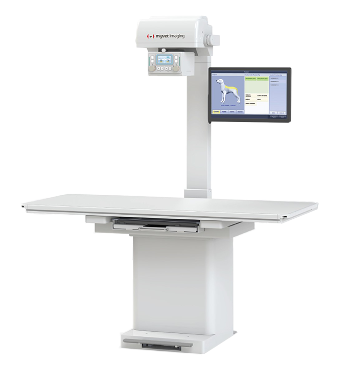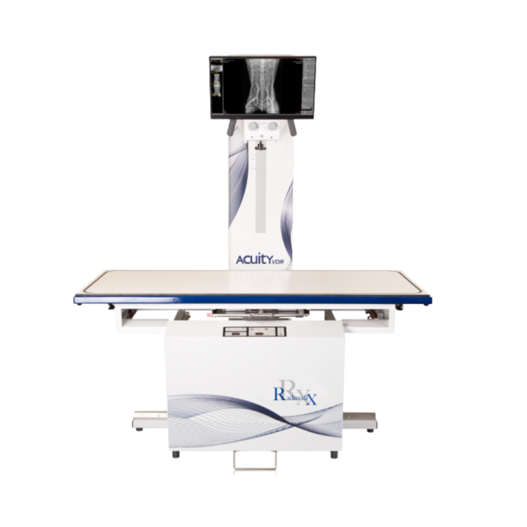
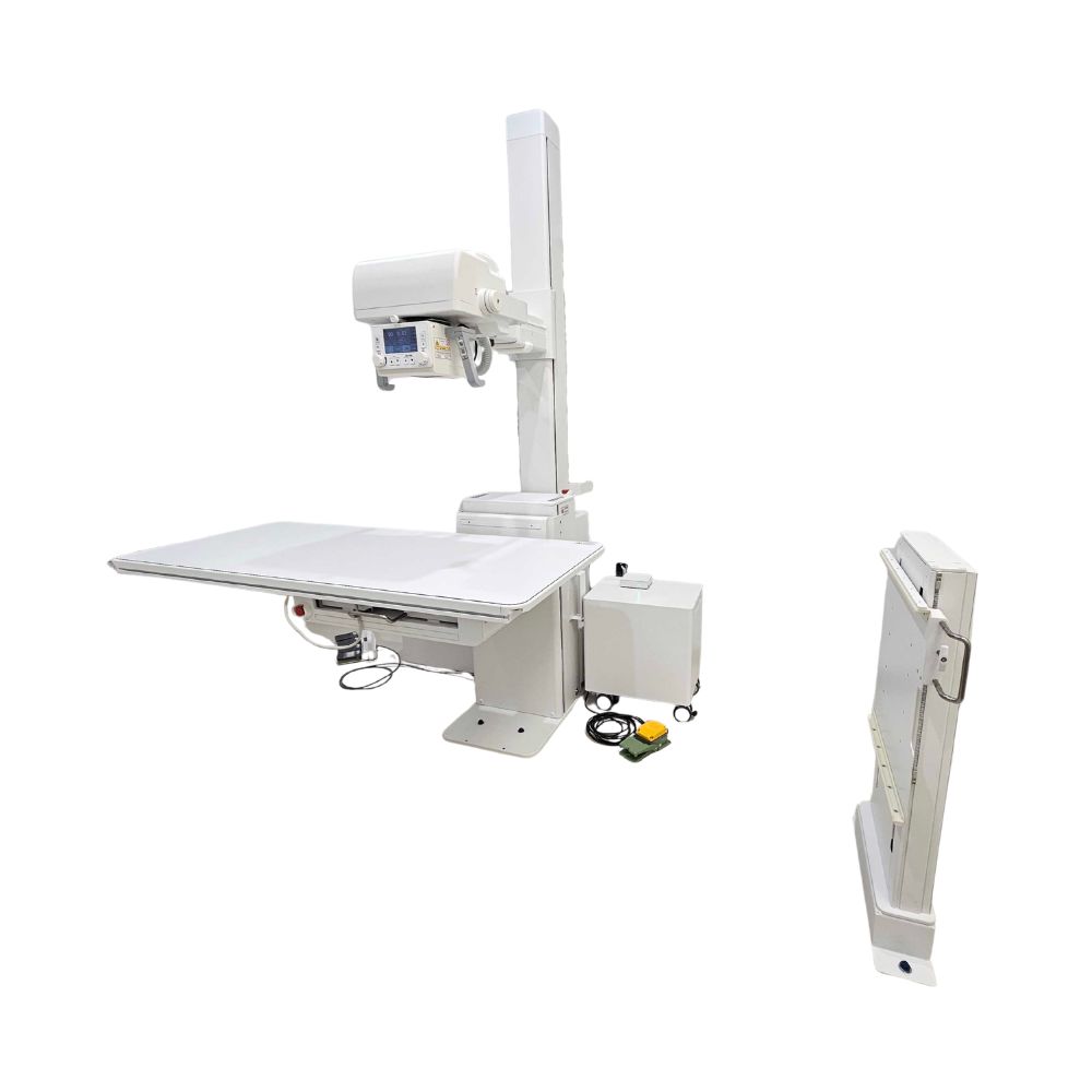
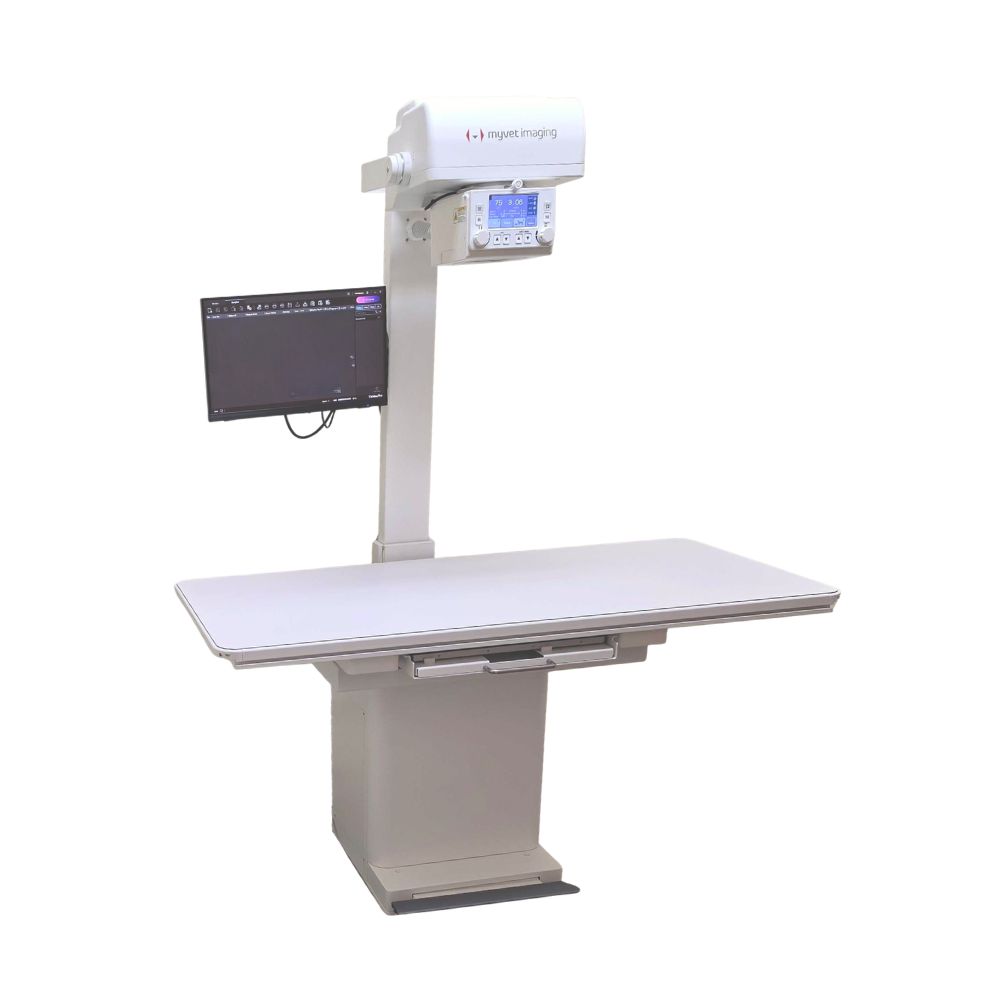
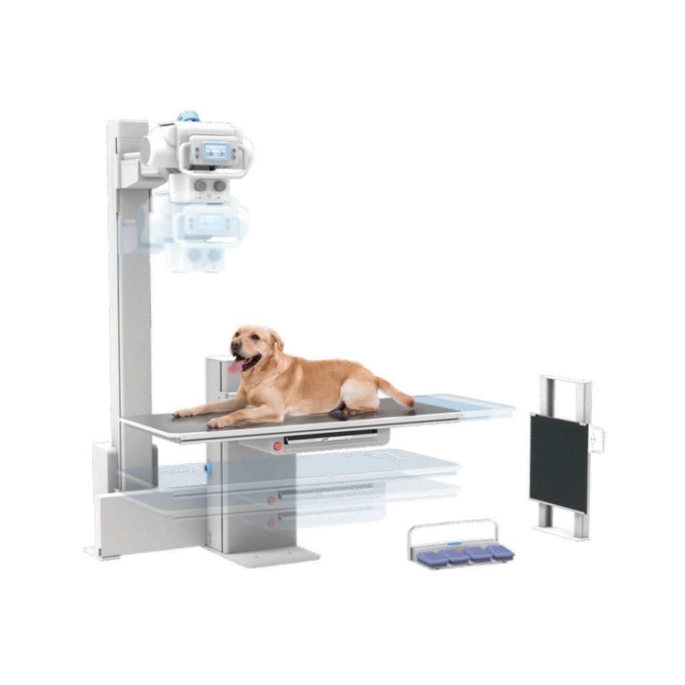
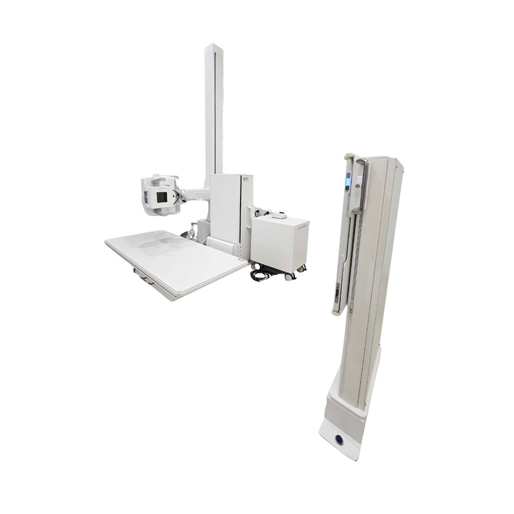
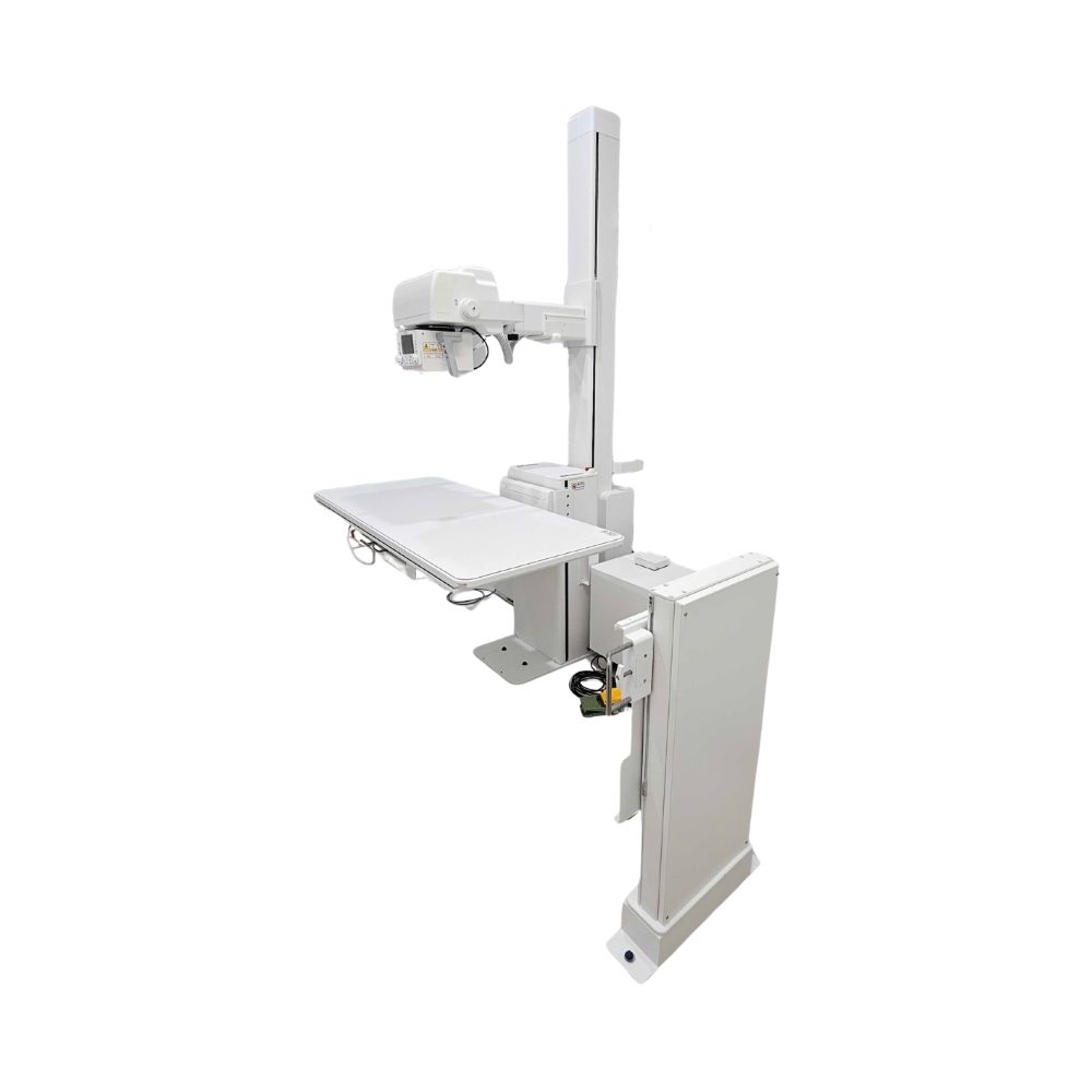
MyVet i72W X-ray Table
- Category:
- X-Ray Systems
- Brand:
- MyVet
The MyVet i72W X-ray is a power height-adjustable veterinary X-ray table best suited to small-animal practices, hospitals, and specialty facilities. A 10.5-inch low tabletop, quiet powered elevation, 40- to 72-inch imaging distance, and upright imaging provide flexible patient positioning in one integrated digital radiography system.
MyVet i72W X-Ray Positioning & Digital Imaging Highlights
The MyVet i72W combines an elevating radiographic table, adjustable tube positioning, digital imaging, and upright radiography in one veterinary X-ray system. Its positioning range is designed to reduce unnecessary patient lifting and repeated equipment adjustments during small-animal exams.
Key Benefits
-
- 40- to 72-inch (101.6-182.9 cm) source-to-image distance
- Powered height-adjustable table with a 10.5-inch (26.7 cm) minimum height
- 90° X-ray tube rotation for upright imaging
- Quiet powered table movement
- 40-inch synchronized tube and table positioning
- 17 × 17-inch (43.2 × 43.2 cm) wireless digital detector
- Automatic stitching of up to three images
- Magnetic in-table detector charging
Rounded tabletop corners, foot-switch table control, and sensor-assisted tube positioning further simplify routine radiography.
Low Table Height, 40-72-Inch Imaging Distance & Upright Positioning
The i72W supports a source-to-image distance, or SID, from 40 to 72 inches (101.6 to 182.9 cm). Combined with table elevation and tube movement, this allows the system to adapt to different patient sizes and radiographic projections without relying on one fixed imaging position.
Power Height Adjustment with a 10.5-Inch Low Position
The tabletop lowers to approximately 10.5 inches (26.7 cm) from the floor. This can reduce lifting for larger ambulatory dogs and other patients able to step onto the table.
Powered elevation then raises the patient to a practical working height, while quiet motor movement limits unnecessary noise during positioning.
40-Inch Tube & Table Position Sync
At the 40-inch (101.6 cm) imaging distance, the tube and table can move in synchronization.
This maintains their imaging relationship while table height changes, reducing the need to repeatedly realign the X-ray tube during patient positioning.
72-Inch Vertical & Horizontal Imaging
The i72W extends to a 72-inch (182.9 cm) source-to-image distance in both vertical and horizontal imaging arrangements.
This provides additional positioning flexibility when a projection requires greater distance between the X-ray source and detector than the standard 40-inch position.
Upright Imaging with 90° Tube Rotation
The X-ray tube rotates 90° to support upright and horizontal-beam imaging.
This allows selected examinations to be performed with the detector positioned vertically rather than requiring every projection to be completed on the tabletop.
Proximity-Sensor Tube Release & Foot Controls
An infrared proximity sensor is built into the tube-head handle. Placing a hand beneath the sensor releases the positioning locks so the tube head can be moved vertically or horizontally.
Foot-switch controls operate table elevation, allowing the technologist to adjust table height while remaining focused on patient positioning.
MyVet i72W Table Positioning Demo
See how the elevating table, tube movement, adjustable imaging distance, and upright positioning work together during veterinary radiography.
MyVet i72W Veterinary X-Ray Applications
The i72W is an elevating X-ray table for veterinary practices that need flexible table height, tube positioning, digital imaging, and upright radiography for small-animal exams.
Veterinary Practice Settings
The system is best suited to:
-
- Small-animal veterinary practices
- Veterinary hospitals
- Emergency and critical-care facilities
- Orthopedic and surgical practices
- Specialty and referral centers
Its configuration is centered on small-animal radiography rather than dedicated equine or large-animal imaging.
Tabletop, Upright & Extended-Field Imaging
The combination of tabletop positioning, upright detector positioning, variable imaging distance, and image stitching supports a range of veterinary X-ray applications, including:
-
- Skeletal and orthopedic imaging
- Thoracic radiography
- Abdominal radiography
- Upright or weight-bearing views when clinically appropriate
- Horizontal-beam imaging, including air/fluid level views when indicated
- Longer anatomical studies requiring multiple images
The positioning flexibility allows the imaging setup to be adjusted around the patient and required projection rather than forcing every study into the same table position.
17 × 17-Inch Wireless Digital Detector
The MyVet i72W uses a 17 × 17-inch (43.2 × 43.2 cm) wireless flat panel detector for digital X-ray imaging.
Square Detector Coverage
The square 17 × 17-inch imaging area provides coverage in either orientation without requiring the detector to be rotated between portrait and landscape positions.
Compared with a rectangular 14 × 17-inch detector, the square format can simplify positioning when anatomy extends in different directions.
Wireless CsI Detector
The detector uses cesium iodide, or CsI, as the image-capture layer that converts incoming X-rays into light before the digital image is produced.
Its documented imaging specifications include a 3268 × 3268 pixel matrix, 140 µm pixel pitch, and maximum resolution of 3.9 line pairs/mm.
The detector also supports wireless image transfer up to 1.3 Gbps and can store up to 200 images internally.
Magnetic In-Table Detector Charging
The detector tray uses magnetic charging contacts to recharge the wireless panel while it remains positioned in the table.
This reduces the need to remove the detector solely for routine charging and helps keep the panel ready for continued use.
Automatic Three-Image Stitching
The i72W can automatically combine up to three sequential X-ray exposures into one longer composite image.
During the stitching sequence, the table moves automatically between exposures while the system displays a stitching-process indicator. This reduces manual detector repositioning when the anatomy extends beyond a single 17 × 17-inch image field.
MyVet i72W X-Ray Table Specifications
X-Ray Generator & Tube
-
- Standard generator: 32 kW, 400 mA
- X-ray tube heat capacity: 200 KHU
- Focal spots: 0.6 / 1.5 mm
- Electrical requirement: 280/230 VAC, single phase
Table & Positioning
-
- Source-to-image distance: 40-72 in (101.6-182.9 cm)
- Minimum tabletop height: 10.5 in (26.7 cm)
- Tabletop size: 59.1 × 25.2 in (150 × 64 cm)
- Stand height: 6.8 ft (approximately 2.07 m)
- Tube rotation: 90°
- Automatic positioning to 40- and 72-inch imaging distances
- 40-inch synchronized tube and table positioning
- Vertical and horizontal imaging capability
- Quiet powered table movement
- Rounded tabletop corners
- Foot-switch table operation
- Infrared proximity-sensor tube release
Wireless DR Detector
-
- Detection area: 17 × 17 in (43.2 × 43.2 cm)
- Detector type: Wireless flat panel digital radiography detector
- Sensor: Amorphous silicon with thin-film transistor technology
- Scintillator: Cesium iodide
- Pixel matrix: 3268 × 3268
- Pixel pitch: 140 µm
- Resolution: Up to 3.9 lp/mm
- Wireless transfer speed: Up to 1.3 Gbps
- Wireless standards: 802.11a/g/n/ac
- Internal image storage: Up to 200 images
- Typical battery operation: Approximately 4 hours
- Detector weight: 7.7 lb (3.5 kg), including battery
- Construction: Carbon fiber
- Water resistance: IPX6
- Magnetic battery charging
Image Stitching
-
- Up to three-image stitching
- Automatic table movement during stitching
- Stitching-process indicator
- Available Equipment Options
Available configurations include:
-
- 40 kW, 500 mA generator
- 17 × 17-inch (43.2 × 43.2 cm) tethered flat panel detector, i72T
Generator, detector, and system configuration should be confirmed for the specific i72W installation
For additional information and specifications, access the PDFs provided below.