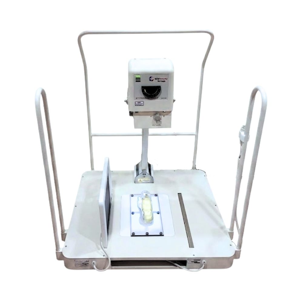
20/20 Imaging HF PXS-710 Podiatry X-Ray
- Category:
- X-Ray Systems
The 20/20 Imaging PXS-710D is a high-frequency digital podiatry X-ray system for podiatry and orthopedic foot and ankle practices. Its 3.5-inch weight-bearing platform, 450 lb patient capacity, dedicated foot and ankle positioning, and configurable DR options support compact, specialty-focused radiography.
PXS-710 High-Frequency X-Ray System & Weight-Bearing Design
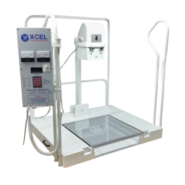
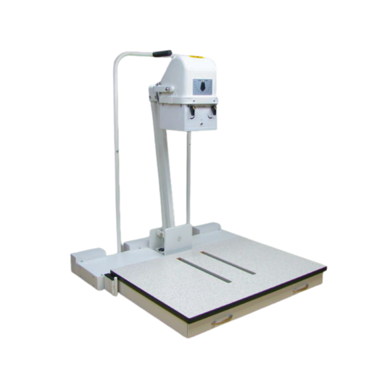
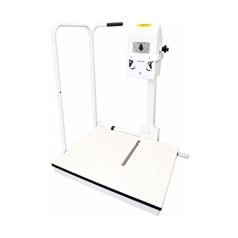
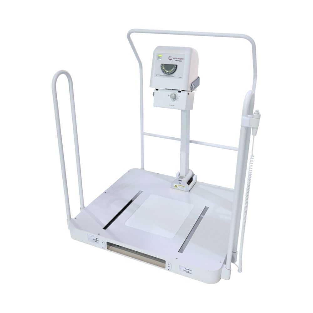
The 20/20 Imaging PXS-710D podiatry X-ray system combines a high-frequency X-ray unit with a 0.5 mm focal spot and positioning features built specifically around foot and ankle radiography. The PXS-710 generator delivers 70 kV at 10 mA, while the low-profile platform, support rails, adjustable tube head, and detector positioning support common weight-bearing exams.
Key Benefits
-
- High-frequency X-ray output at 70 kV and 10 mA
- 0.5 mm focal spot for detailed foot and ankle imaging
- 3.5 in (8.9 cm) low-profile weight-bearing base
- UL-certified patient weight limit up to 450 lb (204 kg)
- Patient support rails with front and side access
- Bi-directional tube movement
- Tilting tube head for AP/DP positioning
- 29 in (73.7 cm) source-to-image distance for AP and oblique views
- 36 in (91.4 cm) source-to-image distance for lateral views
- Integrated cable management for compatible DR configurations
- Built in the USA
These features keep the positioning requirements of routine podiatric radiography within one dedicated system rather than requiring the practice to adapt a general-purpose X-ray configuration around standing foot and ankle exams.
PXS-710 Weight-Bearing Positioning for Foot & Ankle Exams
The physical design of the PXS-710 is centered on positioning patients, the X-ray tube, and the detector for the projections commonly performed in podiatry.
3.5-Inch PXS-710 Weight-Bearing Platform
The 3.5 in (8.9 cm) base keeps the step-up height low for standing exams. The platform supports patients up to 450 lb (204 kg), while the integrated support rail gives patients a stable handhold during positioning.
The rail system can be accessed from the front or side, giving the operator additional flexibility when moving patients onto and around the platform.
PXS-710 Tube Positioning for AP/DP, Oblique & Lateral Views
The tilting tube head and bi-directional movement support common foot and ankle projections, including AP/DP, oblique, and lateral views. AP/DP refers to anteroposterior or dorsoplantar positioning used for routine foot radiography.
The PXS-710 provides a 29 in (73.7 cm) source-to-image distance for AP and oblique views and a 36 in (91.4 cm) distance for lateral views.
PXS-710 Detector Positioning & Integrated Cable Management
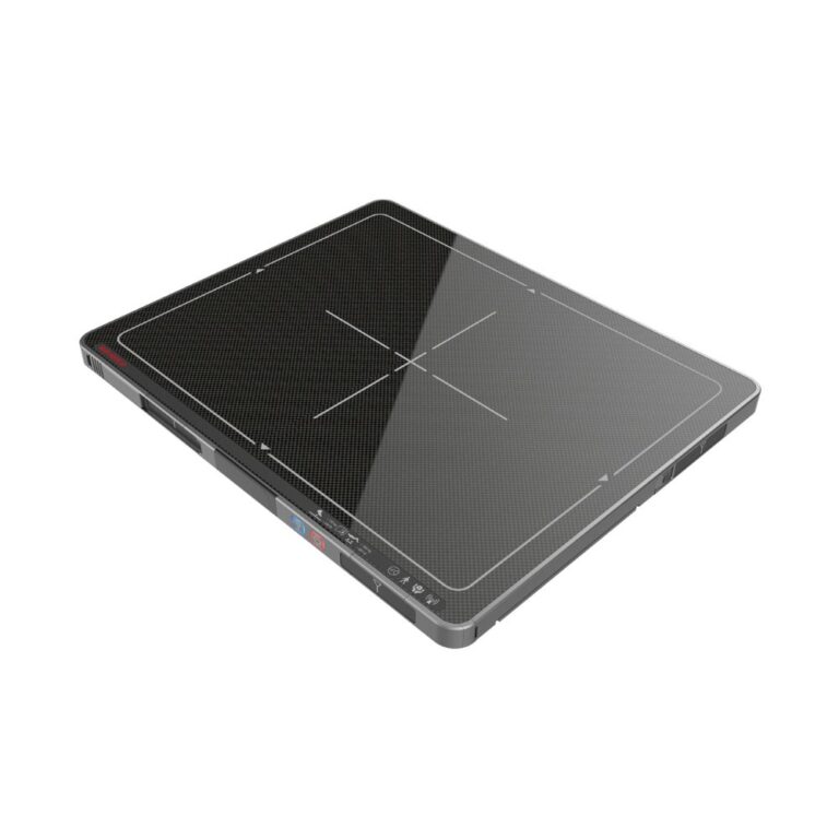
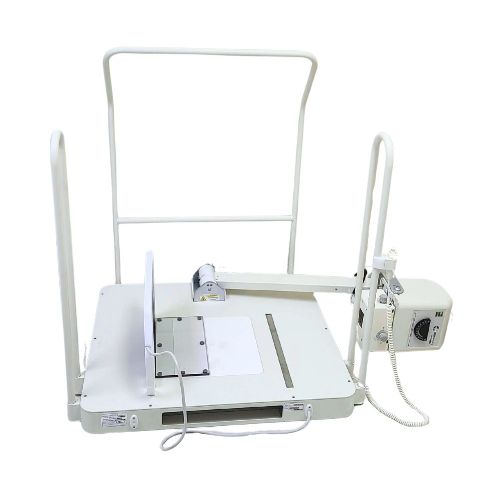
Compatible detector holders are designed to position the DR panel around the PXS-710 platform for different foot and ankle projections.
With tethered DR configurations, detector cables can be routed inside the system base to reduce loose cabling around the patient platform. Wireless detector options are also available for practices that prefer cable-free detector handling.
20/20 Imaging PXS-710D System Demonstration
See how the weight-bearing platform, X-ray tube positioning, detector placement, and digital components of the PXS-710D work together in a dedicated podiatry imaging system.
Clinical Applications for the 20/20 Imaging PXS-710
The 20/20 Imaging PXS-710D is designed for podiatric radiography and orthopedic foot and ankle applications, particularly when standing and weight-bearing positioning are part of the imaging workflow.
PXS-710 for Podiatry & Orthopedic Foot/Ankle Practices
Relevant clinical environments include:
-
- Podiatry practices
- Foot and ankle specialty clinics
- Orthopedic practices performing foot and ankle radiography
Its specialty-focused design makes the system particularly relevant for practices that regularly perform foot and ankle X-rays and want a dedicated positioning platform instead of a general-purpose radiography setup.
PXS-710 Weight-Bearing & Routine Foot/Ankle Exams
The platform, tube head, and detector positioning support applications including:
-
- Weight-bearing foot radiography
- Weight-bearing ankle radiography
- AP/DP foot views
- Oblique foot and ankle views
- Lateral foot and ankle views
- General podiatric radiographic examinations
- Foot and ankle imaging used in surgical planning
- Podiatric imaging used within diabetic foot care workflows
The selected DR detector and software configuration determine how images are acquired, processed, measured, reviewed, and stored after exposure.
PXS-710D Digital Acquisition & Imaging Workflow
The digital PXS-710D configuration connects the X-ray unit with DR acquisition, exam selection, image processing, review, and storage at the workstation.
PXS-710D Workstation-Based Exam Selection & Generator Integration
Anatomy-based program selection and preset X-ray techniques can be accessed directly from the acquisition workstation. Preconfigured exams can also be customized, allowing users to add or remove views as needed.
This keeps technique selection and digital image acquisition within the same workstation-based workflow instead of requiring separate setup for each step.
PXS-710D Automated Processing, Cropping & Image Saving
After exposure, the digital workflow includes automated functions designed to reduce repetitive image-processing steps.
Capabilities include:
-
- Automatic contrast processing
- Automatic image shuttering and cropping
- Quick image viewing
- Customizable image sharpness
- Automatic image saving during acquisition
- Under 6-second image processing
Acquired images can be saved to the imaging server for access within the connected imaging environment.
On-Site PXS-710D Installation & Training
Current complete-system configurations include on-site installation and training. System setup is performed by a certified installer, followed by training covering X-ray acquisition, detector positioning, software basics, and image viewing tools.
This helps the practice transition from installation to routine use with both the equipment and software configured as one system.
DR Panel Options for the PXS-710D
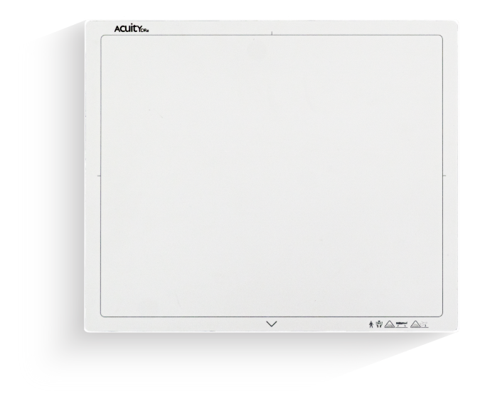
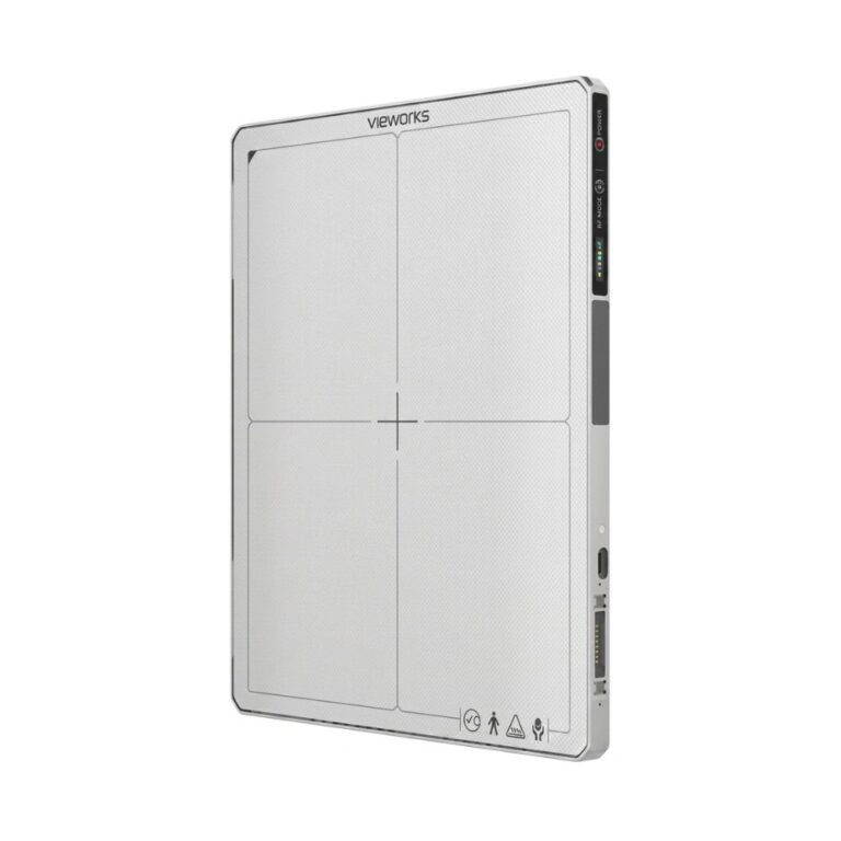
The PXS-710D can be configured with multiple 20/20 Imaging DR detectors, allowing practices to choose the detector size and connection type that best fit their podiatry workflow.
Current detector choices range from compact 10 × 12 in (254 × 305 mm) formats to a larger 14 × 17 in (356 × 432 mm) option. The available detectors use cesium iodide imaging technology and are paired with holders designed for integration with the PXS-710.
Wireless & Tethered PXS-710D Detector Configurations
-
- UniversalDR 14 × 17 Wireless: Larger-format wireless detector with a 13.75 × 16.75 in (349 × 425 mm) active imaging area for broader anatomical coverage.
- UniversalDR 10 × 13 Wireless: Compact wireless option with a true 10 × 13 in (254 × 330 mm) active imaging area.
- UniversalDR 1012V Tethered: Compact wired detector with a 9.34 × 11.8 in (237 × 300 mm) active imaging area for practices that prefer a permanent tethered connection.
- Momentum DR 10 × 12 Wireless: Wireless detector with rapid-charge capacitor technology and REALISM image processing.
Detector choice allows the PXS-710D to be configured around imaging area, wireless versus tethered handling, and the practice’s preferred digital workflow.
The 20/20 Imaging 1012V Tethered DR Detector and 20/20 Imaging Momentum Wireless DR Panel should be internally linked to their dedicated MXR product pages for buyers who need complete detector specifications.
Symmetry-Sync Software Integration with the PXS-710D
Symmetry-Sync brings image acquisition, review, study management, and storage into the digital PXS-710D workflow. Its podiatry-specific tools extend the system beyond image capture by supporting measurements commonly used in foot and ankle evaluation.
Symmetry-Sync Acquisition, Viewing & PACS Workflow
Symmetry-Sync combines acquisition, image viewing, study management, and PACS, or picture archiving and communication system, functionality within one software environment.
Workflow capabilities include:
-
- Customizable users and groups
- Patient and study search
- Image import and export
- Integrated image viewing
- Customizable workflow settings
- PACS functionality
- Electronic health record (EHR) integration readiness
Additional fees may apply for EHR integration.
Symmetry-Sync Podiatry Measurement Tools
The included podiatry toolset provides measurements designed for foot and ankle evaluation and surgical planning, including:
-
- Hallux interphalangeus and hallux abductus angles
- First intermetatarsal angle
- P.A.S.A. (proximal articular set angle) and D.A.S.A. (distal articular set angle)
- Metatarsus adductus and total adductory angle
- Metatarsal parabola, metatarsal protrusion, and talo-calcaneal measurements
Keeping these measurements inside the imaging software allows clinicians to perform common podiatric analysis within the same digital study used for image review.
Optional Symmetry-Sync Zero-Footprint Web Viewer
An optional zero-footprint web viewer extends study access to compatible web-connected devices. “Zero-footprint” means the viewer operates through a web browser without requiring a separate viewer application to be installed on the device.
Available functions include:
-
- Patient and study search
- Standard annotations
- Touch-based image navigation
- Access to the study list and image viewer
- Browser-based viewing on compatible tablets, phones, and computers
The zero-footprint viewer is an add-on option and should not be assumed to be included with every PXS-710D configuration.
20/20 Imaging PXS-710D x-Ray System Specifications
PXS-710 High-Frequency Generator & X-Ray Tube
System type: High-frequency podiatry X-ray system
Generator output: 70 kV at 10 mA
Focal spot: 0.5 mm
X-ray tube: HF-series proprietary tube
Exposure control: Low-voltage exposure switch
Tube head: Tilting design
Built in the USA
PXS-710 Platform & Positioning Specifications
-
- Base height: 3.5 in (8.9 cm)
- Patient weight limit: Up to 450 lb (204 kg)
- AP/oblique source-to-image distance: 29 in (73.7 cm)
- Lateral source-to-image distance: 36 in (91.4 cm)
- Tube movement: Bi-directional
- Patient support rails: Front and side access
- Detector positioning: Multiple foot and ankle positions
- DR cable management: Integrated into the base for compatible tethered configurations
PXS-710D System Dimensions
-
- Approximate overall width: 68.95 in (175.1 cm)
- X-ray unit depth: 33.75 in (85.7 cm)
- Detector holder depth: 20.19 in (51.3 cm)
- Approximate total system depth: 53.94 in (137 cm)
Actual room planning requirements should be confirmed for the selected system configuration before installation.
PXS-710D Digital System Configuration
-
- High-frequency PXS-710 X-ray unit
- Choice of compatible DR detector
- Generator-integrated digital acquisition
- Anatomy-based exam selection
- Preset and customizable X-ray techniques
- Under 6-second image processing
- Automatic contrast processing
- Automatic image shuttering and cropping
- Automatic image saving during acquisition
- Symmetry-Sync imaging software
- Podiatry-specific measurement toolset
- PACS functionality
- EHR integration readiness
- Optional zero-footprint web viewer
PXS-710D Power & Connectivity Requirements
-
- X-ray unit power: 120 V, 60 Hz, 20 A grounded outlet
- Dedicated X-ray outlet recommended
- Acquisition computer: Standard grounded 120 V outlet
- Network connection: Minimum 1 Gbps
- Network cabling: Cat 5e or better
- Static IP address required for the acquisition computer at installation
Internet connection: 10 Mbps or greater
PXS-710D Compliance & Warranty
-
- FDA 510(k) cleared for general-purpose podiatry radiographic applications
- UL and CUL listed
- PXS-710 mechanical warranty: 5 years
- X-ray tube and electrical warranty: 3 years
- DR detector warranty: 3 to 5 years depending on detector
- Extended DR detector warranties available on eligible configurations
Current PC components: 3-year on-site warranty including parts
Actual radiation dose and efficiency vary based on patient size, exposure settings, imaging technique, and equipment configuration. Current OEM materials likewise note that dose reduction and efficiency gains vary with patient size, technique, settings, and equipment configuration.