Mindray Animal Vetus EQ Portable Equine Ultrasound
- Category:
- Veterinary Ultrasound Products
- Brand:
- Mindray Animal Medical
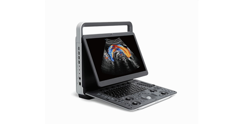
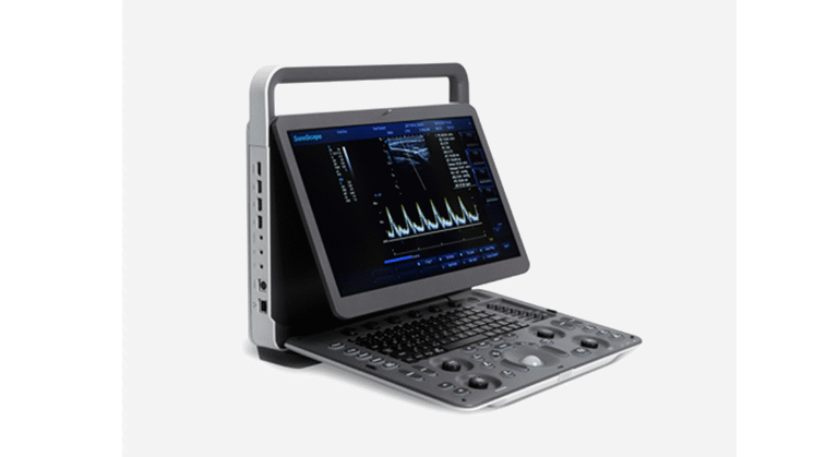
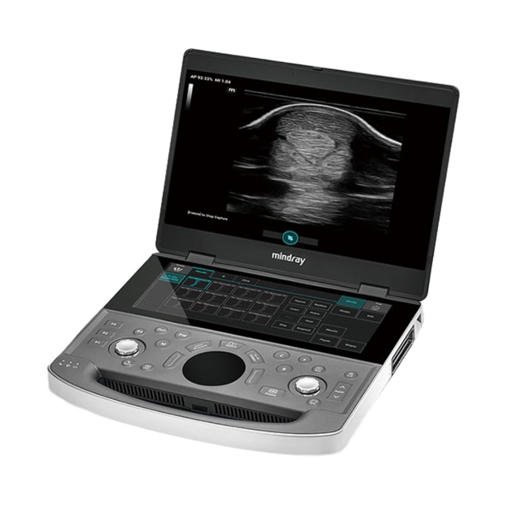
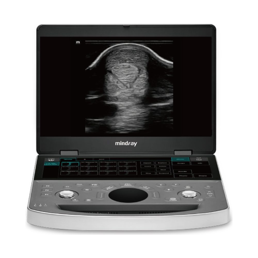
The Mindray Animal Medical Vetus EQ is a portable equine ultrasound system designed for field and clinic use. Built specifically for equine practitioners, it combines ZST+ imaging, dedicated equine presets, a 15.6-inch high resolution color LED monitor, and a lightweight design to support fast, consistent imaging in barn-side and clinical environments across reproduction, tendon, abdominal, and cardiac exams.
Portable Equine Ultrasound System Built for Barn-Side & Clinical Workflows
The Mindray Animal Medical Vetus EQ is designed specifically for equine imaging, combining portability with workflow tools and imaging performance tailored to large animal exams. It allows practitioners to perform diagnostic imaging in barns, clinics, and field environments without sacrificing consistency or control.
Key Highlights
-
- ZST+ imaging platform for faster image acquisition and improved clarity
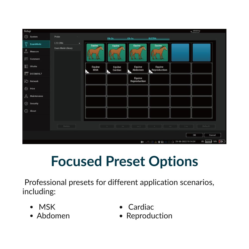
- Dedicated equine presets for reproduction, tendon, abdominal, and cardiac
- workflows
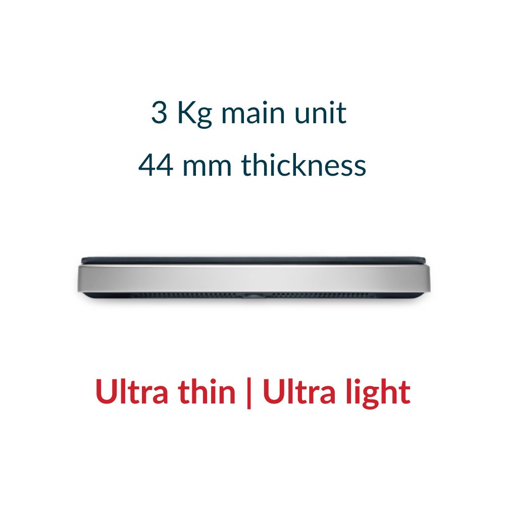
- Lightweight ~3.0 kg system designed for field mobility
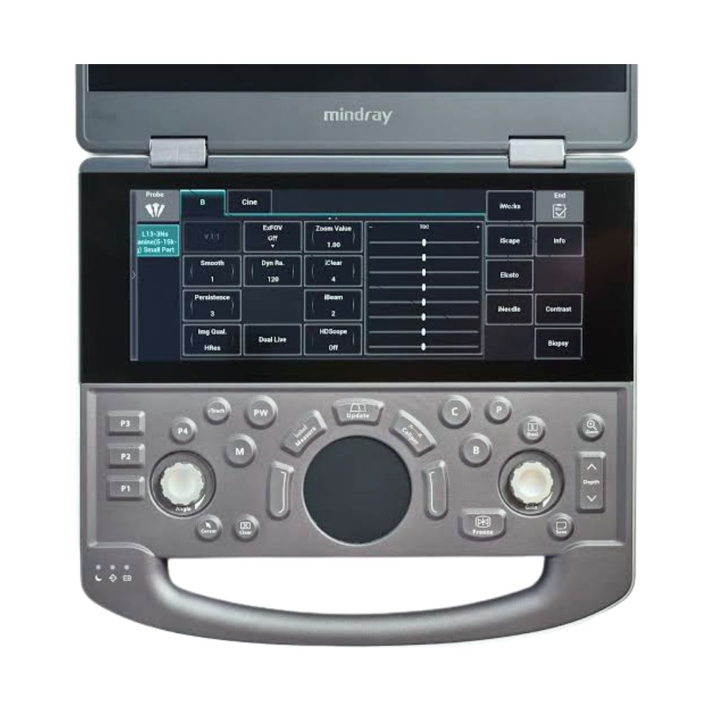
- 15.6-inch main display with 12.3-inch touchscreen control interface
- Battery-powered operation for mobile equine imaging
- iWorks workflow automation to reduce exam time and keystrokes
The Vetus EQ is built to reduce setup time, improve repeatability, and maintain diagnostic confidence across equine workflows.
Mindray Animal Medical Vetus EQ Platform & Equine Imaging Workflow Overview
This video highlights the Vetus ultrasound platform, including equine-focused workflow design, imaging performance, and how the system is used in real-world veterinary environments.
What Types of Equine Exams Can the Mindaray Vetus EQ Support?
The Mindray Animal Medical Vetus EQ is designed around the core imaging needs of equine practitioners, with transducers and presets aligned to reproduction, tendon, abdominal, and cardiac workflows.
Reproduction & Intrarectal Imaging
-
- Intrarectal imaging with dedicated equine probe design
- Follicle tracking and reproductive monitoring
The 6LE5Vs intrarectal probe is purpose-built for equine reproductive exams.
Tendon & Musculoskeletal Imaging
-
- High-frequency imaging for tendon and ligament evaluation
- Injury assessment and follow-up imaging
The L13-3Ns linear transducer supports detailed imaging of equine tendon structures.
Abdominal Imaging
-
- Large animal abdominal imaging with appropriate depth penetration
The C5-1s convex probe supports equine abdominal exams.
Cardiac Imaging
-
- Echocardiography and flow assessment
The P4-2s phased array probe supports equine cardiac imaging.
How the Mindray Animal Medical Vetus EQ Improves Equine Ultrasound Workflow Efficiency
The Mindray Animal Medical Vetus EQ is designed to reduce exam time and improve consistency through equine-specific workflow tools and imaging architecture.
iWorks Workflow Automation for Standardized Exams
iWorks is a preset-driven workflow tool that structures equine ultrasound exams into guided steps based on the selected application.
When a preset is selected (such as reproduction, tendon, or cardiac), the system:
-
- loads optimized imaging parameters for that exam type
- guides the user through required views and scan positions
- prompts for measurements and annotations at the appropriate stage
- organizes captured images into a structured exam sequence
This reduces the need to manually adjust settings, remember exam protocols, or manage image organization during the scan.
In equine workflows, this is particularly valuable when working in time-sensitive or less controlled environments, where maintaining consistency across exams and operators can otherwise be difficult.
ZST+ Imaging Platform for Faster, More Stable Real-Time Imaging
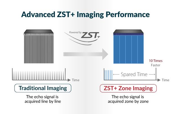
ZST+ changes how ultrasound data is captured and processed by acquiring signals in zones rather than line-by-line, allowing the system to process more imaging data at once during live scanning.
In practice, this means:
- fewer delays when moving or repositioning the probe
- less need for the system to refocus or rebuild the image after each adjustment
- smoother real-time imaging when scanning across larger anatomical areas
Instead of rebuilding the image with each small movement, the Vetus EQ maintains a more continuous image as the probe moves, which is especially important during equine exams.
In equine workflows, this helps:
- maintain image clarity when the horse shifts or moves
- reduce interruptions when scanning tendons, abdomen, or cardiac structures
- capture diagnostic images more consistently in barn-side and field conditions
Designed for Equine Field Use, Mobility, & Durability
The Mindray Animal Medical Vetus EQ is built for equine practitioners who routinely work across barns, clinics, and mobile environments.
Lightweight System for Transport Between Exam Locations
-
- Approximately 3.0 kg system weight (6 lbs 9.8 oz)
- Designed for movement between barns, stalls, and exam areas
- Supports mobile workflows without requiring fixed installation
Built for Barn-Side & Clinical Environments
-
- Sealed interface helps reduce contamination from hair and debris
- Resistant to cleaning agents used in veterinary environments
- Protective accessories available for transport and durability
Battery Operation for Mobile Equine Imaging
-
- Built-in battery supports field imaging
- Approximately 1.5 hours of continuous scanning per battery
Optional battery configurations support extended use in mobile settings.
Equine Imaging Tools & Procedural Support
The Mindray Animal Medical Vetus EQ includes imaging tools designed to support both diagnostic imaging and guided procedures in equine practice.
iNeedle™, iScape™ & Imaging Tools
-
- iNeedle™ improves needle visualization during guided procedures
- iScape™ panoramic imaging supports extended tendon evaluation
- Imaging tools support both diagnostic and interventional workflows
How the Mindray Animal Medical Vetus EQ Streamlines Equine Exams
The Mindray Animal Medical Vetus EQ is designed to reduce setup time, guide exam execution, and simplify image review in equine workflows where efficiency and consistency directly impact throughput.
Equine Presets That Load Imaging Parameters Instantly
When an equine preset is selected, the system automatically loads imaging parameters such as depth, frequency range, gain, and focal zones optimized for that exam type.
This allows practitioners to:
-
- begin scanning without manual setup
- maintain consistent imaging conditions across exams
- reduce time spent adjusting settings between patients
Touchscreen Controls That Allow Adjustments Without Breaking Probe Contact
The touchscreen allows users to adjust key imaging parameters such as gain, depth, and measurement tools directly on-screen while scanning.
This allows the operator to:
-
- maintain probe position during adjustments
- avoid reaching for external controls
- make quick changes without interrupting the scan
This is particularly important in equine exams where consistent probe positioning directly impacts image quality.
Exam-Based Image Organization That Reduces Post-Scan Sorting
Captured images and cine loops are automatically grouped based on the selected exam workflow.
This allows practitioners to:
-
- keep studies organized during acquisition
- reduce time spent sorting images after the exam
- move directly into review or export without restructuring files
How the Mindray Animal Medical Vetus EQ Stores, Transfers & Organizes Equine Studies
The Mindray Animal Medical Vetus EQ supports local storage, structured study management, and flexible data transfer between barn-side and clinic environments.
Internal Storage for On-System Study Management
The system includes integrated solid-state storage (SSD) for saving ultrasound images, cine loops, and exam data directly during use.
This allows practitioners to:
-
- store multiple exams locally without interruption
- review studies immediately after scanning
- continue working without requiring immediate export
For most equine workflows, this supports a full day of imaging, including multi-image studies and cine loops, before requiring export or archiving.
Export Options for Moving Studies from Field to Clinic Systems
The Mindray Animal Medical Vetus EQ supports multiple methods for transferring studies depending on whether imaging is performed in the field or within a clinic.
Studies can be transferred using:
-
- USB export to save images and cine loops directly to a flash drive after the exam
- Wired network connection (LAN) to send studies to a workstation or server when connected in a clinic
- Wireless transfer (Wi-Fi) to move images to connected devices or network locations when available
In a typical equine workflow, studies are captured in the field, stored locally on the system, and then exported via USB or transferred over a network once back in a clinic setting. This allows images to move between environments without requiring real-time connectivity during the exam.
DICOM Integration for Centralized Image Management
The Mindray Animal Medical Vetus EQ supports DICOM for integration with PACS and other imaging systems used in veterinary practices.
This allows studies to be:
-
- sent directly to a PACS server for long-term storage
- accessed from other workstations within the practice
- reviewed alongside other imaging modalities such as radiography
This is most relevant for practices that want ultrasound studies stored within an existing imaging system rather than managed as standalone files.
Equine-Specific Transducers for Reproduction, Tendon, Abdominal & Cardiac Imaging
The Mindray Animal Medical Vetus EQ is built around transducers designed specifically for equine imaging, allowing each exam type to be performed with the appropriate probe.
Transducers Matched to Equine Exam Types
-
- 6LE5Vs intrarectal probe for reproductive imaging
- L13-3Ns linear probe for tendon and musculoskeletal exams
- C5-1s convex probe for abdominal imaging in large animals
- P4-2s phased array probe for cardiac imaging
Each probe is designed for a specific equine application, allowing practitioners to move between exam types without switching systems.
Needle Visualization for Ultrasound-Guided Procedures
The Mindray Animal Medical Vetus EQ includes iNeedle, which enhances needle visibility by improving alignment between the needle and the ultrasound beam.
This allows practitioners to:
-
- better visualize needle trajectory during procedures
- maintain alignment between the needle and imaging plane
- improve accuracy during injections and aspirations
Panoramic Imaging for Tendon Evaluation
iScape panoramic imaging creates extended field-of-view images by stitching together multiple frames during probe movement.
This allows practitioners to:
-
- capture the full length of tendons in a single image
- assess the extent of injuries across a larger area
- compare affected and unaffected regions more easily
Portable System Design for Barn-Side Imaging & Transport
The Mindray Animal Medical Vetus EQ is designed for equine practitioners who work across barns, clinics, and mobile environments.
Lightweight System for Transport Between Locations
-
- approximately 3.0 kg system weight
- can be carried between barns, stalls, and exam areas without carts or mounting systems
- allows imaging in locations where fixed systems are not practical
Sealed & Durable Design for Hair, Debris & Cleaning Exposure
-
- sealed interface helps prevent contamination from hair and barn debris
- designed to withstand frequent cleaning with veterinary disinfectants
- protective accessories help reduce damage during transport
This supports consistent system performance in environments where dust, moisture, and repeated cleaning are common.
Battery Operation for Barn-Side Imaging
-
- built-in battery supports imaging without external power
- approximately 1.5 hours of continuous scanning per battery
In practice, this supports multiple exams in a barn or field setting before requiring recharge or battery swap.
Mindray Animal Medical Vetus EQ Specifications
System
-
- Display: 15.6-inch high-resolution monitor
- Touchscreen: 12.3-inch anti-glare interface
- Weight: ~3.0 kg main unit (6.6 lbs)
- Thickness: 44 mm main unit (1.73 inches)
- Design: portable system for field and clinic use
Imaging Modes
-
- B-mode
- M-mode
- Color Doppler
- Power Doppler
- Pulsed wave Doppler
Imaging Technologies
-
- ZST+ imaging platform
- iWorks workflow automation
- iClear speckle reduction
- iBeam spatial compounding
- THI harmonic imaging
- HR Flow
Storage & Connectivity
-
- Internal SSD storage for images and cine loops
- USB export capability
- Wired LAN connectivity
- Wi-Fi wireless transfer
- DICOM support
Transducer Support
-
- Linear transducers
- Convex transducers
- Phased array transducers
- Intrarectal equine-specific probe
Power & Mobility
-
- Battery-powered operation
- ~1.5 hours continuous scanning per battery
- Optional extended battery configurations