Mindray Animal Vetus 9 Veterinary Ultrasound
- Category:
- Veterinary Ultrasound Products
- Brand:
- Mindray Animal Medical
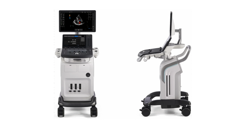
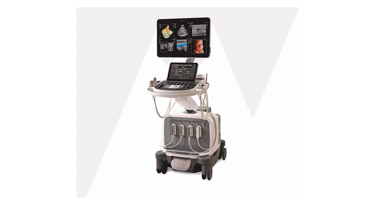
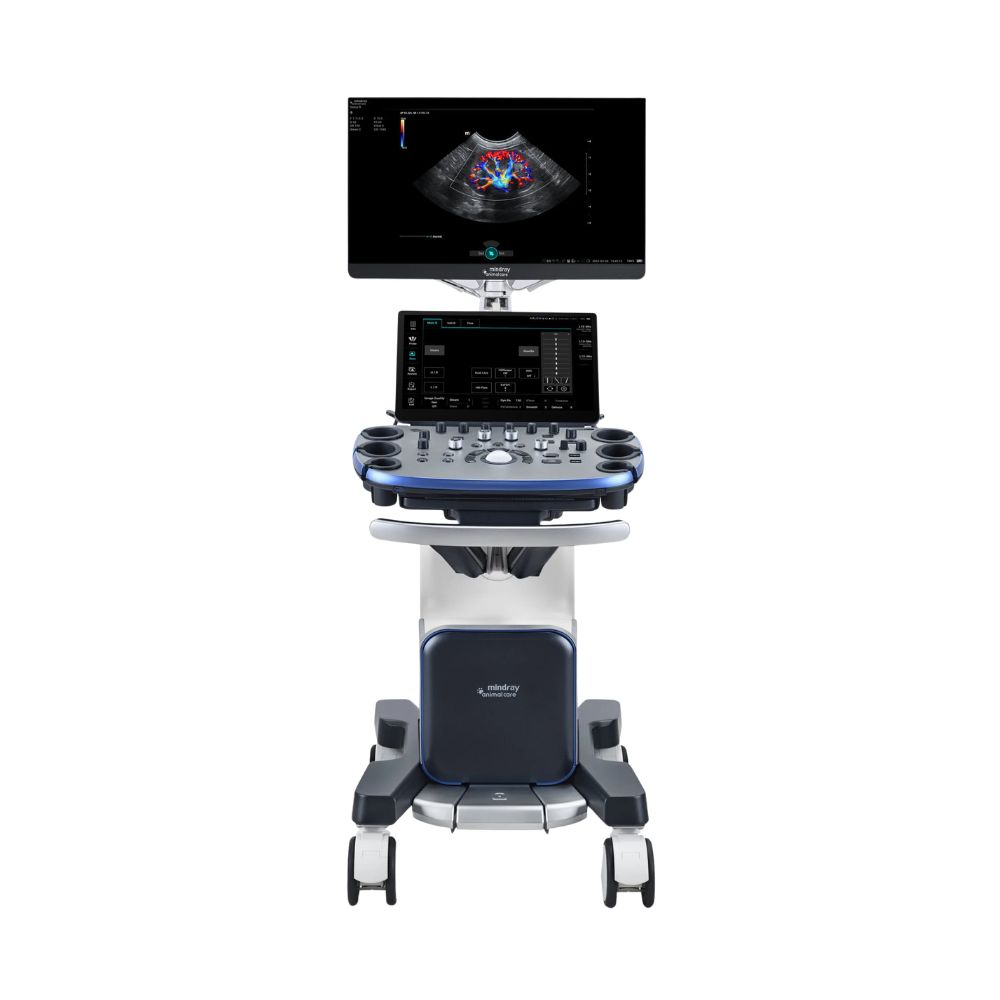
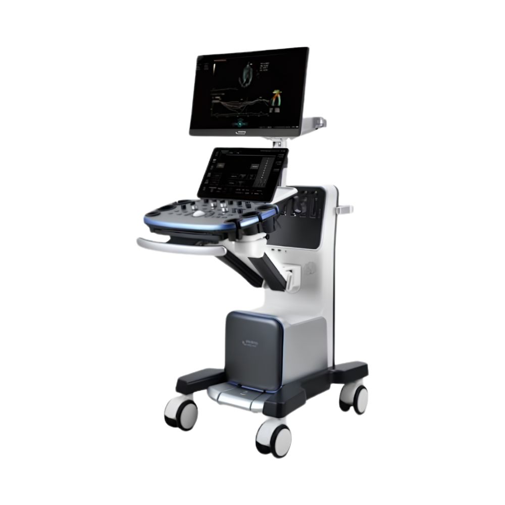
The Mindray Animal Medical Vetus 9 is a premium cart-based veterinary ultrasound system for advanced companion, equine, farm-animal, and exotic-animal imaging. Powered by ZST+, it combines high-speed imaging, advanced abdominal and cardiac tools, five active transducer ports, and intelligent workflow features for demanding veterinary hospitals and specialty practices.
Premium Mindray Animal Medical Vetus 9 Imaging Powered by ZST+
The Mindray Animal Vetus 9 is built for veterinary hospitals, specialty practices, and advanced imaging services managing challenging cases and heavier ultrasound workloads. Its ZST+ platform combines high-speed imaging and dynamic focusing with advanced cardiac analysis, tissue stiffness assessment, contrast imaging, five active transducer ports, and a dual-screen workflow.
Key Benefits
-
- ZST+ Imaging Platform: Uses zone-based image acquisition, dynamic focusing, and stored acoustic data to support fast imaging, consistent image quality, and image adjustments after acquisition.
- Advanced Abdominal & Tissue Assessment: Supports tissue stiffness measurement, contrast-enhanced imaging, perfusion assessment, and focal-lesion evaluation for more complex abdominal studies.
- Advanced Veterinary Cardiology: Provides tools for heart-muscle motion analysis, ventricular function, Doppler measurements, myocardial tracking, and advanced echocardiography.
- Five Active Transducer Ports: Keeps multiple probes connected for faster transitions between abdominal, cardiac, reproductive, superficial, ocular, and musculoskeletal exams.
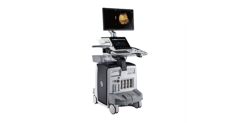
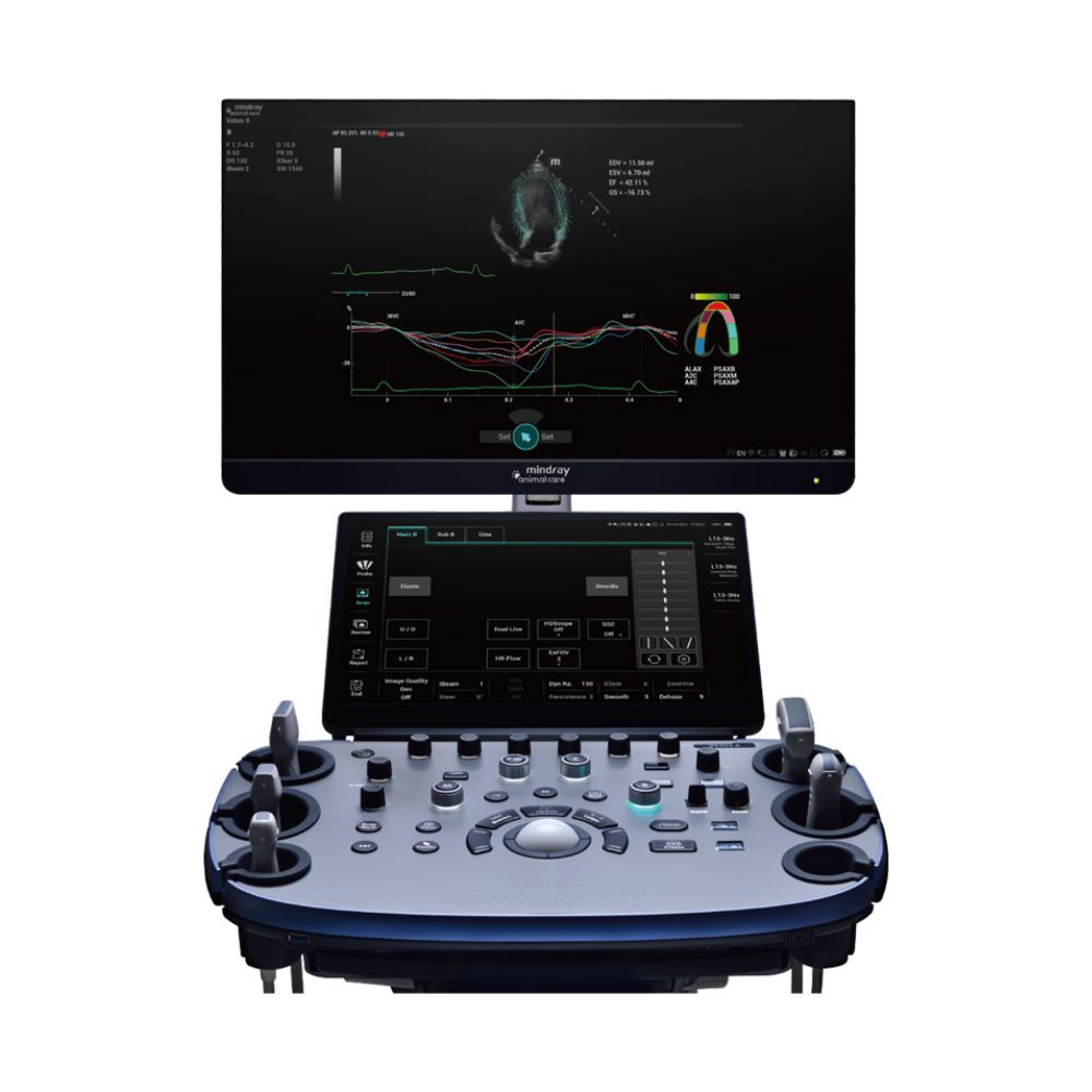
- Dual Full-HD Displays: Combines a 23.8-inch main monitor with a 15.6-inch anti-glare touch screen for image review and direct exam control.
- Veterinary Workflow Automation: Provides guided exam protocols, scanning references, voice control, study management, reporting, measurement assistance, and remote image-access tools.
The Vetus 9 is best suited for veterinary hospitals, specialty practices, and advanced imaging services that need advanced cardiac and abdominal imaging, elastography, and deeper workflow automation from a cart-based system.
Mindray Animal Medical Vetus 9 Imaging, Workflow, & System Design
This Mindray Animal Medical video highlights the Vetus 9, including its ZST+ imaging platform, advanced diagnostic tools, intelligent workflow features, and veterinary-focused cart design. It provides a closer look at how these capabilities come together within the Vetus 9 system.
How ZST+ Improves Mindray Animal Medical Vetus 9 Image Quality
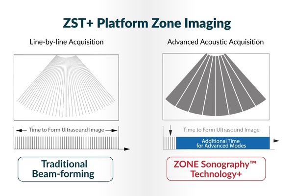
ZST+ is one of the primary distinctions of the Vetus 9. Instead of relying only on conventional line-by-line beamforming, the platform acquires larger imaging zones and processes the underlying acoustic data before the final image is formed. This approach is designed to improve image detail, capture fast-moving anatomy, and maintain more consistent image quality across the field of view.
Zone Imaging for Fast-Moving Anatomy
Zone Imaging transmits and receives a smaller number of larger imaging zones rather than building the image line by line.
Because animal hearts can beat rapidly, cardiac ultrasound requires the system to capture motion quickly and clearly. Zone Imaging supports high-speed real-time visualization of cardiac anatomy and blood flow.
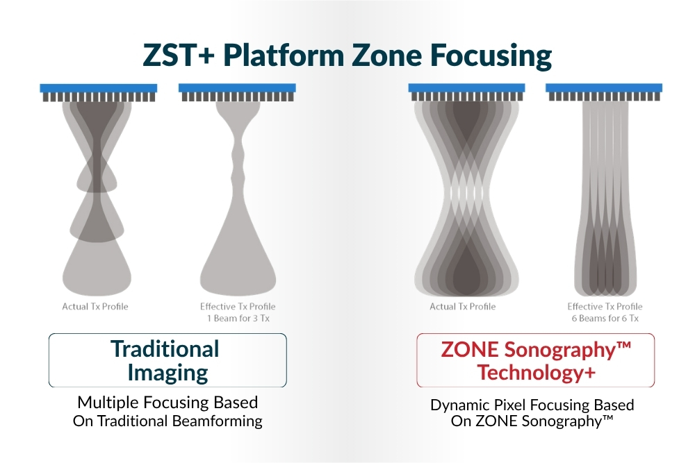
Zone Focusing Across the Full Field of View
Animal size and body shape can vary substantially between patients. Zone Focusing uses Dynamic Pixel Focusing, a ZST+ technique that maintains focus throughout the image rather than relying on only a limited number of conventional focal zones.
This supports more consistent visualization from superficial tissue to deeper anatomy across different patient sizes.
Zone Processing & Total Recall Imaging
ZST+ stores the underlying acoustic data collected during the scan, not only the final displayed image. Zone Processing allows supported image settings to be adjusted after acquisition using that stored data.
Total Recall Imaging (TRI) extends this capability so supported image optimization can continue after the scan rather than requiring every setting to be finalized while the animal is still being imaged.
Mindray Animal Vetus 9 Veterinary Applications Across Animal Types
The Mindray Animal Care Vetus 9 supports abdominal, cardiac, reproductive, small-parts, vascular, musculoskeletal, and ocular imaging across companion animals, large animals, and supported wild and exotic species. Curved, linear, and phased-array transducers help the system adapt to substantial differences in patient size and anatomy.
Vetus 9 Abdomen & Reproduction
Curved and micro-convex transducers support companion- and large-animal abdominal and reproductive imaging.
Relevant applications include:
-
- liver, gallbladder, spleen, kidney, urinary bladder, gastrointestinal, and other abdominal structures
- abdominal blood-flow and tissue perfusion assessment
- uterine and ovarian imaging
- pregnancy and gestational assessment
- canine and feline reproduction
- equine, bovine, and ovine reproduction
The reproduction measurement package adds species-specific tools for canine, feline, equine, bovine, and ovine exams, including gestational, ovarian, uterine, follicular, prostate, and testicular measurements.
Vetus 9 Veterinary Cardiology
Dedicated phased-array transducers support structural imaging, Doppler, heart-muscle motion analysis, ventricular-function assessment, and advanced cardiac measurements across different patient sizes.
Smart VTI supports VTI tracing and LVOT diameter measurement for assessing blood flow and cardiac function. Additional advanced cardiac tools are detailed below.
Vetus 9 Small Parts, Ocular, MSK & Vascular Imaging
High-frequency linear transducers expand the Vetus 9 into superficial, ocular, musculoskeletal, and vascular applications.
Supported workflows include:
-
- small-parts and superficial tissue evaluation
- ocular imaging
- musculoskeletal imaging
- vascular Doppler assessment
- IMT, or intima-media thickness, measurement
- peripheral arterial measurements
- tissue stiffness assessment with supported elastography modes
- panoramic imaging with iScape View
- ultrasound-guided procedures with iNeedle
Vetus 9 Wild & Exotic Animal Imaging
Mindray Animal Medical specifically includes the Vetus 9 within its wild and exotic animal ultrasound solutions. The manufacturer highlights its advanced transducers and imaging capabilities for the diagnostic challenges presented by species with very different sizes, anatomy, and physiology. Mindray’s broader exotic-animal solution covers animals ranging from small mammals and birds to reptiles and larger exotic species.
Depending on the patient, transducer, and exam, Vetus 9 ultrasound capabilities can support:
-
- abdominal and soft-tissue imaging
- cardiac assessment
- reproductive imaging
- superficial and small-parts evaluation
- musculoskeletal imaging
- vascular and blood-flow assessment
This broader species coverage makes the Vetus 9 relevant not only to companion- and mixed-animal practices, but also to specialty, zoo, wildlife, and exotic-animal imaging environments where patient size and anatomy can vary substantially.
Mindray Animal Vetus 9 Advanced Abdominal Imaging & Tissue Assessment
The Mindray Animal Medical Vetus 9 goes beyond routine abdominal B-mode and Doppler imaging with tools for tissue stiffness, lesion perfusion, contrast analysis, and quantitative assessment.
HiFR STE for Tissue Stiffness Assessment
High Frame Rate Sound Touch Elastography (HiFR STE) measures tissue stiffness using shear waves while the ZST+ platform supports a faster, smoother real-time elastography display.
Motion stability and reliability tools help clinicians assess shear-wave image quality before interpreting tissue stiffness measurements.
UWN+ Contrast Imaging for Lesion Perfusion
UWN+ Contrast Imaging supports focal-lesion and tissue perfusion assessment by improving visualization of contrast-agent signals and small-vessel blood flow.
The technology is designed to detect weaker contrast-agent signals more effectively, while Micro Flow Enhancement provides additional visualization of small-vessel perfusion. Contrast Imaging QA supports time-intensity curve analysis, which helps quantify how contrast moves through selected tissue regions over time.
Contrast imaging availability depends on applicable regulatory approval, authorized markets, compatible probes, and approved contrast agents.
Natural Touch Elastography, STE & STQ for Tissue Assessment
The Mindray Animal Medical Vetus 9 provides multiple approaches to tissue stiffness assessment:
-
- Natural Touch Elastography: Evaluates how tissue deforms under pressure, with strain ratio and histogram measurements.
- Sound Touch Elastography (STE): Uses shear waves for real-time two-dimensional tissue stiffness imaging.
- Sound Touch Quantification (STQ): Provides quantitative stiffness measurements within a selected area, or region of interest.
- HiFR STE: Increases shear-wave elastography frame rate for smoother real-time visualization.
Mindray Animal Vetus 9 Advanced Veterinary Cardiology & Cardiac Analysis
The Mindray Animal Medical Vetus 9 provides dedicated cardiac analysis tools for practices performing advanced echocardiography, including heart-muscle motion and timing analysis, ventricular-function assessment, and Doppler measurements.
TDI QA for Myocardial Motion & Synchronization
TDI QA, or Tissue Doppler Imaging Quantitative Analysis, supports TVI, TEI, TVD, and TVM modes and provides quantitative analysis of myocardial, or heart-muscle, velocity, strain, strain rate, and synchronization.
Up to eight regions of interest can be analyzed during myocardial assessment.
TTQA for Angle-Independent Myocardial Tracking
Tissue Tracking Quantitative Analysis (TTQA) tracks two-dimensional speckle patterns within the ultrasound image to evaluate myocardial movement without relying on Doppler angle.
Supported analysis includes myocardial velocity, displacement, strain, strain rate, rotation, and other quantitative motion measurements across multiple cardiac views.
Auto EF for Left Ventricular Function
Auto EF analyzes two-dimensional echo clips, identifies end-diastolic and end-systolic frames, and calculates measurements used to evaluate left ventricular function.
Supported outputs include EDV, ESV, ejection fraction, stroke volume, and cardiac output using the Simpson method.
Mindray Animal Vetus 9 Workflow Tools for High-Volume Veterinary Imaging
The Mindray Animal Medical Vetus 9 combines exam guidance, study management, measurement support, voice control, reporting, and image transfer tools so advanced imaging capability does not automatically create a more cumbersome workflow.
iWorks, iScanHelper & iVocal for Guided Scanning
iWorks provides configurable step-by-step exam guidance with support for annotations, body marks, measurements, and predefined workflow steps. Mindray Animal Medical states that its standardized workflows can reduce exam time and keystrokes.
iScanHelper provides scanning reference images and instructional guidance to help operators confirm probe positioning and exam technique during supported studies.
iVocal allows supported system functions to be controlled through voice commands. This can be particularly useful when the operator needs to maintain probe position or assist with animal handling while changing supported system functions.
The workflow suite also includes iMeasure, which uses supported measurements to help indicate potential or suspected clinical findings during applicable workflows.
iStation, iStorage & iReport for Study Management
iStation provides veterinary exam management, including the ability to search, retrieve, and review current and prior studies and perform measurements on archived images.
iStorage provides direct network storage between the Vetus 9 and a compatible computer.
iReport supports application-specific and customizable report templates, selectable images, editable report modules, and report export.
MedSight, MedTouch & DICOM Connectivity
The Mindray Animal Medical Vetus 9 supports:
-
- MedSight for wireless image and cine transfer to compatible smart devices
- MedTouch for compatible remote system control and workflow functions
- DICOM Storage, Worklist, Query/Retrieve, MPPS, Storage Commitment, and structured DICOM
- reporting for supported applications
- built-in wired and wireless network connectivity
This gives veterinary hospitals and imaging services multiple ways to review, transfer, archive, and manage studies beyond the ultrasound cart itself.
Mindray Animal Vetus 9 iConsole & Dual-Screen Workflow
The Mindray Animal Medical Vetus 9 combines a large imaging monitor, Full HD touch screen, floating control panel, and exam-specific controls to reduce unnecessary navigation during more involved studies.
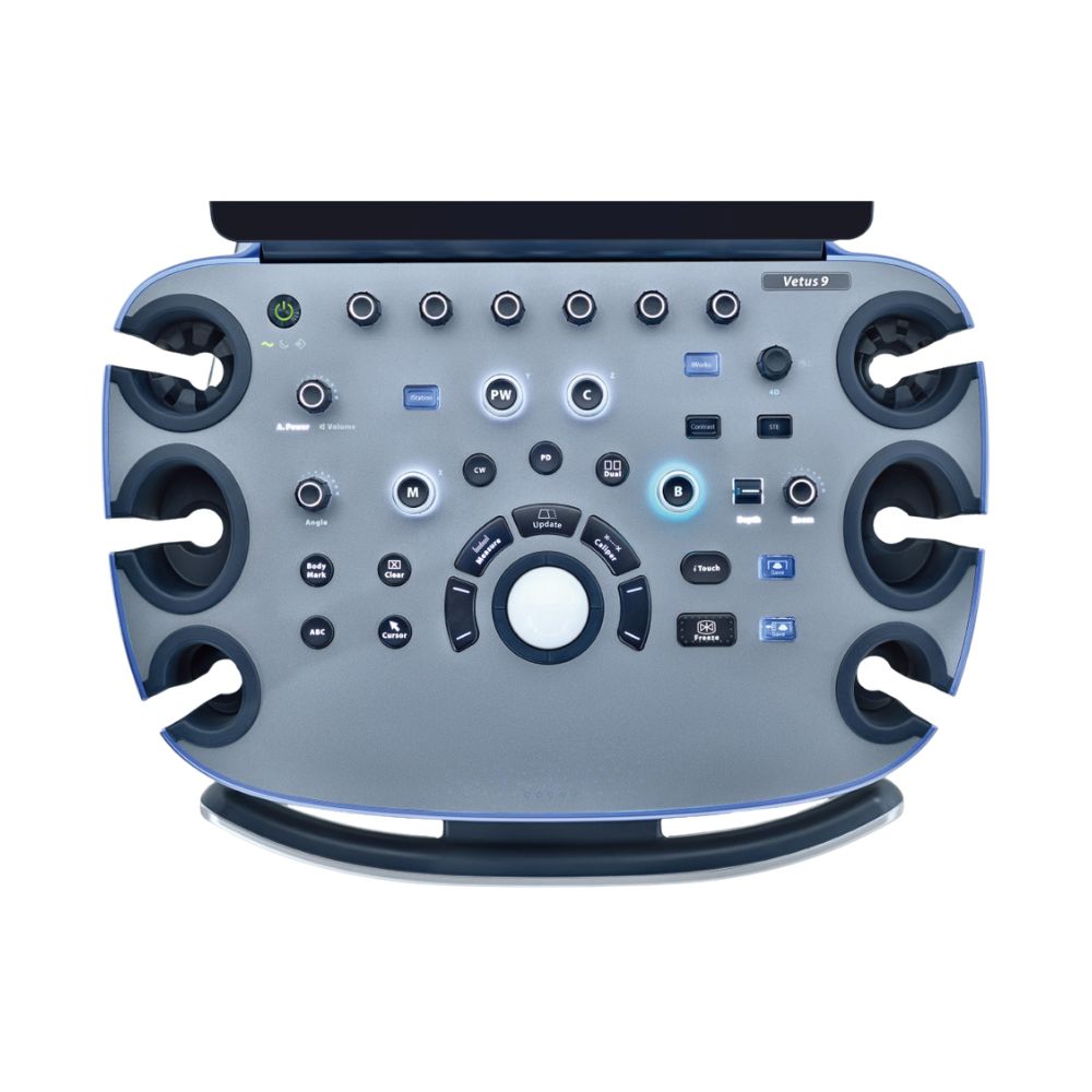
iConsole Adaptive E-Ink Controls
iConsole uses six programmable keys with small E-ink digital displays that change based on the exam being performed. The control layout can adapt to workflows such as abdomen, cardiology, small parts, and MSK.
This keeps frequently needed functions closer to the operator during different exam types instead of relying on the same fixed control layout for every specialty.
23.8-Inch Monitor & 15.6-Inch Touch Screen
The Mindray Animal Medical Vetus 9 includes:
-
- 23.8-inch bezel-less 1920 × 1080 main monitor
- 178° viewing angle
- adaptive monitor brightness
- independently adjustable tilt and rotation
- 15.6-inch 1920 × 1080 anti-glare touch screen
- touch operation with gloved hands
- digital Time Gain Compensation (TGC)
- customizable touch gestures
- shortcuts for recently used probes and exams
Five Active Transducer Ports for Faster Exam Changes
Five active, dust-protected transducer ports allow multiple probes to remain connected simultaneously, with hot-plug support and active indicator lights.
For practices performing abdominal, cardiac, superficial, reproductive, ocular, and MSK studies on the same system, this reduces repeated probe changes between exam types.
Mindray Animal Medical Vetus 9 Cart Design & Veterinary Ergonomics
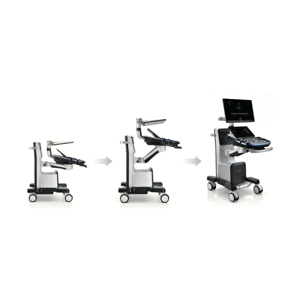
Despite its advanced imaging capabilities, the Mindray Animal Vetus 9 is designed to remain adjustable and movable within hospitals, specialty practices, and imaging departments.
Floating Controls for Flexible Scan Positioning
The floating control panel supports extensive left/right, vertical, and front/back adjustment and can be fixed at the desired scanning position.
The dual-wing monitor arm provides additional positioning flexibility so displays and controls can be adjusted around different animal sizes, tables, and scanning approaches.
Quiet Operation for Veterinary Exam Rooms
System noise is rated at ≤26 dB at 25°C. The quiet operating design helps limit unnecessary environmental noise around nervous, reactive, or sensitive animals during ultrasound exams.
Fast Startup & Battery-Supported Operation
The Vetus 9 boots from shutdown in under 30 seconds and recovers from standby in under 5 seconds.
An optional replaceable rechargeable battery supports continued system operation when the cart is moved between clinical areas or temporarily disconnected from AC power. Available battery configurations can support extended scanning away from a wall outlet depending on system configuration.
Mindray Animal Medical Vetus 9 Multi-Species Transducers
The Mindray Animal Medical Vetus 9 supports curved, linear, and phased-array transducers for companion, equine, farm-animal, and supported wild and exotic animal imaging across cardiac, abdominal, reproductive, ocular, small-parts, and musculoskeletal applications. Mindray Animal Medical specifically identifies the Vetus 9 and its advanced transducers as part of its dedicated exotic-animal ultrasound solution.
Representative options include:
- SC6-1s Convex: Abdomen and reproduction in supported companion and large-animal workflows.
- C11-3s Micro-Convex: Companion-animal abdominal and reproductive applications.
- L13-3Ns Linear: Superficial, small-parts, and supported musculoskeletal applications.
- SP5-1s Phased Array: Cardiology across companion and supported large-animal species.
- P8-2s Phased Array: Cardiac imaging in small- and medium-sized patients.
- P10-4s Phased Array: Higher-frequency cardiac imaging for smaller patients.
The broad probe range allows the Vetus 9 to be configured around advanced cardiology, abdominal imaging, mixed-animal practice, exotic-animal imaging, or multidisciplinary veterinary imaging without limiting the system to a single application.
Mindray Animal Medical Vetus 9 Veterinary Ultrasound Specifications
System & Physical Design
-
- System type: Cart-based veterinary diagnostic ultrasound system designed for advanced multi-application imaging.
- Dimensions: 1020 ± 20 mm D × 550 ± 10 mm W × 1000 ± 20 mm H with the monitor and control panel in their lowest positions.
- Net weight: 105 ± 4 kg in the standard configuration, excluding transducers.
- Operating noise: ≤26 dB at 25°C for quieter use around animal patients.
- Electrical input: 100–240 VAC, 50/60 Hz.
- Power consumption: 650 VA.
Displays & Operator Controls
-
- Primary monitor: 23.8-inch bezel-less Full HD display with 1920 × 1080 resolution and 178° viewing angle.
- Monitor positioning: Independent tilt and rotation help adapt the display to different operator positions and animal sizes.
- Touch screen: 15.6-inch anti-glare Full HD display with 1920 × 1080 resolution and 170° viewing angle.
- Touch operation: Supports handwriting, gloved-hand operation, image adjustment, measurements, zoom, and customizable gestures.
- iConsole: Six programmable E-ink keys can change displayed functions based on the clinical exam and support user-defined settings.
- Keyboard and gain controls: Full-size backlit QWERTY keyboard with digital Time Gain Compensation (TGC) on the touch screen.
Transducer Access & Cart Design
-
- Active transducer ports: Five dust-protected ports allow multiple probes to remain connected for faster switching between exam types.
- Probe handling: Hot-plug support allows supported probes to be connected without shutting down the system, while active indicator lights help identify connected transducers.
- Transducer holders: Seven holders, including one designed for a hockey-style probe.
- Floating control panel: Adjustable vertically, laterally, and front-to-back so controls can be positioned around the patient and scanning approach.
- Mobility: 125 mm wheels support movement between clinical areas, with a central brake available depending on configuration.
Startup, Battery & Storage
-
- Startup: Boots from shutdown in under 30 seconds and resumes from standby in under 5 seconds.
- Battery: Optional replaceable rechargeable lithium battery supports continued operation when the cart is moved or temporarily disconnected from AC power.
- Internal storage: 1 TB hard drive for exam and image storage, with a 128 GB system SSD.
Imaging Modes & Performance
-
- Core imaging: B-mode, M-mode, Color M-mode, Color Doppler, Power Doppler, Pulsed Wave Doppler, and Continuous Wave Doppler.
- Flow imaging: HR Flow and Glazing Flow support additional visualization of vascular and blood-flow information.
- Cardiac imaging: Free Xros M, Free Xros CM, and Tissue Doppler Imaging support anatomical M-mode and evaluation of heart-muscle motion.
- Advanced imaging: UWN+ Contrast Imaging, Natural Touch Elastography, STE, STQ, HiFR STE, Smart 3D, and iScape View extend the system into perfusion, tissue stiffness, 3D, and panoramic imaging.
- Maximum frame rates: Up to 1701 fps in B-mode, 790 fps in Color Doppler, and 3049 fps in supported TDI/TVI imaging.
- Imaging depth: 1.5–40 cm.
- Dynamic range: Up to 30–260 dB depending on the transducer.
Advanced Imaging & Analysis Tools
-
- ZST+ platform: Uses Advanced Acoustic Acquisition, Dynamic Pixel Focusing, Sound Speed Compensation, processing of underlying acoustic data, and Total Recall Imaging to support high-speed acquisition and image optimization after acquisition.
- iClear / iClear+: Speckle-suppression processing helps improve contrast and tissue-border definition.
iTouch: Automatically optimizes supported image parameters to reduce manual adjustment during scanning. - Echo Boost: Improves image uniformity and noise control in supported cardiac exams.
TDI QA and TTQA: Support quantitative analysis of heart-muscle motion, strain, strain rate, and timing for advanced cardiac assessment. - Auto EF: Calculates left ventricular measurements including EDV, ESV, ejection fraction, stroke volume, and cardiac output.
- Smart VTI: Supports VTI tracing and LVOT diameter measurement for assessing blood flow and cardiac function.
- Contrast Imaging QA: Provides time-intensity curve analysis to quantify contrast movement through selected tissue regions.
- iNeedle and Smart Trace: Support enhanced needle visualization and semi-automated measurement workflows.
Connectivity & Study Management
-
- Network connectivity: Built-in wireless adapter and Ethernet support connection to compatible practice and imaging networks.
- DICOM: Supports Storage, Worklist, Query/Retrieve, MPPS, Storage Commitment, and application-specific structured reporting.
- iStation: Manages veterinary exams, including searching, retrieving, and reviewing current and prior studies.
- iStorage: Supports direct network storage between the Vetus 9 and a compatible computer.
MedSight and MedTouch: Support compatible smart-device image sharing, remote control, and workflow functions. - USB connectivity: Six ports total, including five USB 3.0 ports and one USB Type-C port.
Additional interfaces: HDMI, VGA, S-Video, ECG, and audio connections.
Representative Transducer Options
-
- SC6-1s convex array: 1.0–6.0 MHz for supported abdominal and reproductive imaging.
- C11-3s micro-convex array: 2.6–12.8 MHz for companion-animal abdominal and reproductive applications.
- L13-3Ns linear array: High-frequency option for superficial, small-parts, and supported musculoskeletal imaging.
- SP5-1s phased array: Single-crystal cardiac transducer for supported companion- and large-animal cardiology.
- P8-2s phased array: Mid/high-frequency cardiac transducer for small- and medium-sized patients.
- P10-4s phased array: Higher-frequency cardiac transducer for smaller patients.
Feature, transducer, battery, contrast-imaging, and accessory availability may vary by configuration, mode, market, and regulatory approval.