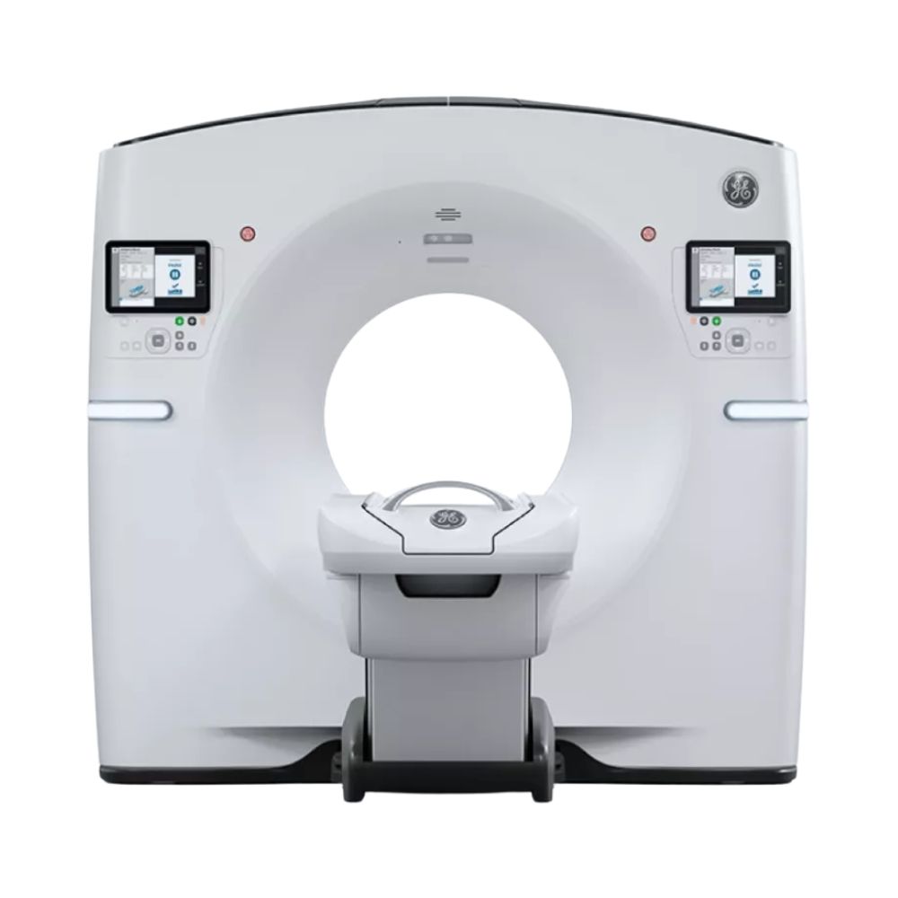
GE Revolution Maxima Select 64-Slice CT
- Category:
- CT Scanners
- Brand:
- GE
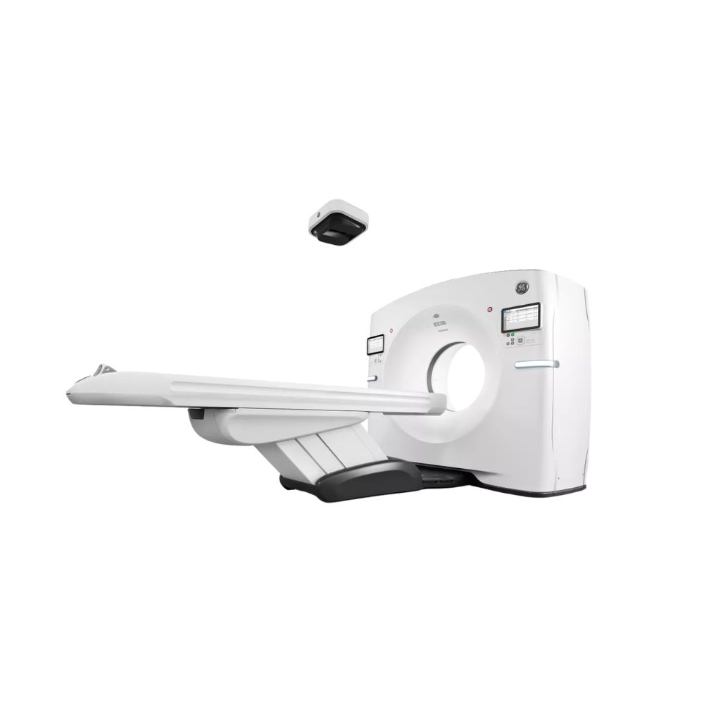
The GE Revolution Maxima Select 64 is a 64-slice CT scanner well suited for hospitals, imaging centers, and specialty practices. Its Clarity Imaging Chain delivers detailed images, while flexible detector coverage, advanced image processing, and optional AI Auto Positioning support routine and specialized cardiac, vascular, and neurological examinations.
GE Revolution Maxima Select 64 Highlights & Benefits
The GE Revolution Maxima Select 64 combines detailed diagnostic imaging with efficient CT scanning and configurable clinical technology. This 64-slice Revolution Maxima system offers two detector-coverage arrangements, advanced image-processing options, and an upgrade pathway for facilities seeking additional imaging and workflow capabilities.
Key Features & Benefits
-
- 64-Slice CT Imaging: Provides 64-slice imaging capabilities using documented 20 mm or 40 mm detector configurations.
- Clarity Imaging Chain: Supports spatial resolution as fine as 0.28 mm (0.011 in), helping reveal detailed bone, vascular, and soft-tissue anatomy.
- Flexible Detector Coverage: Offers 20 mm coverage with 0.625 mm acquired sections or 40 mm coverage with 1.25 mm acquired sections, allowing facilities to balance fine detail and scanning coverage.
- Efficient Scanning Performance: Features 0.5-second standard gantry rotation, with faster performance available through compatible upgrades.
- ASiR & ASiR-V Image Processing: Available technologies help produce clearer images with less visual noise while supporting patient-specific radiation-dose management.
- Advanced Clinical Applications: Compatible cardiac, vascular, and neurological software expands the scanner’s diagnostic capabilities.
- Optional AI Auto Positioning: Eligible systems can add a camera-and-software package that automates patient centering.
- Upgradeable Platform: Supports eligible modernization options, including 128-slice reconstruction and additional imaging technologies.
Revolution Maxima Select 64 Detector Coverage & Image Quality
The Revolution Maxima Select 64 uses GE’s Clarity Imaging Chain and configurable scanning settings to balance anatomical detail with scanning efficiency. Detector coverage determines how much anatomy the scanner captures during each rotation, while section thickness and image reconstruction influence the level of detail available for clinical review.
64-Slice Imaging with 20 mm & 40 mm Coverage
The GE Revolution Maxima Select 64 supports two documented detector configurations, providing different combinations of anatomical coverage and acquired section thickness.
-
- 20 mm Coverage at 0.625 mm: Uses a 32 × 0.625 mm acquisition arrangement to capture thinner sections, providing detailed views of smaller anatomical structures.
- 40 mm Coverage at 1.25 mm: Uses a 32 × 1.25 mm acquisition arrangement to capture a wider area per rotation, supporting efficient body and vascular examinations.
Both configurations support the Maxima Select’s 64-slice imaging capabilities. Their physical detector coverage and acquired section thickness differ, making these specifications important when comparing equipment.
Image reconstruction processes the acquired scanning data into cross-sectional images that clinicians can review from different angles. Compatible higher-tier upgrades support 40 mm coverage with 0.625 mm sections and 128-slice overlapping reconstruction.
Clarity Imaging Chain for Detailed CT Images
GE’s Clarity Imaging Chain brings together the scanner’s X-ray detector, tube, data-processing electronics, and image-reconstruction software to produce detailed CT images.
Its principal technologies include:
-
- Clarity Detector: Measures X-rays passing through the patient while minimizing electronic interference, helping preserve fine anatomical detail.
- Clarity Data Acquisition System: Converts detector measurements into digital information used to create CT images.
- Performix 40 Plus X-Ray Tube: Generates the X-rays needed for scanning, with heat-management capabilities that support repeated examinations.
- High-Resolution Reconstruction: Produces images using up to a 1024 × 1024 pixel matrix on compatible systems, allowing clinicians to review smaller anatomical details.
The platform supports spatial resolution as fine as 0.28 mm (0.011 in), helping clinicians evaluate small vessels, bone structures, and other detailed anatomy.
Fast Gantry Rotation & Helical Scanning
The Revolution Maxima Select 64 supports 0.5-second standard gantry rotation, with faster rotation available through compatible upgrades.
The gantry is the circular scanner housing that rotates the X-ray tube and detector around the patient.
During helical scanning, the patient table moves continuously while the gantry rotates, allowing the scanner to collect images along the body in a spiral pattern.
The system supports helical pitch values up to 1.531 on compatible configurations. Pitch describes how far the table advances during each rotation relative to the detector’s scanning width. Higher pitch settings can help cover larger anatomical regions more quickly when appropriate for the examination.
These capabilities benefit routine body imaging, CT angiography, and selected examinations where acquisition speed or breath-hold duration matters.
Revolution Maxima Select 64 Reconstruction & Dose Management
The scanner offers image-processing and exposure-control technologies that help balance image detail, clarity, and patient-specific radiation-dose requirements.
ASiR & ASiR-V Image Reconstruction
The GE Revolution Maxima Select 64 supports ASiR and ASiR-V, two image-processing technologies that use a method called iterative reconstruction. This method repeatedly refines CT images to reduce visual noise.
The Revolution Maxima Select offers two reconstruction technologies:
-
- ASiR: Adaptive Statistical Iterative Reconstruction processes CT data to reduce image noise, helping maintain useful anatomical detail while supporting appropriately selected radiation-dose settings.
- ASiR-V: An advanced version that uses more detailed calculations to reduce noise and improve low-contrast visibility. This helps clinicians distinguish structures with subtle differences in tissue density.
- 1024 × 1024 Reconstruction: Produces higher-resolution images with more pixels, allowing finer anatomical details to be reviewed on compatible examinations.
ASiR-V has demonstrated radiation-dose reductions of up to 82% under specified comparative testing conditions. Actual results depend on patient characteristics, examination settings, and the clinical task.
Automated Exposure Control & Dose Monitoring
Compatible Maxima Select configurations provide technologies for adjusting radiation exposure according to the patient and examination.
-
- AutomA & SmartmA: Automatically adjust X-ray tube current as the scanner moves through areas of different body thickness, helping deliver appropriate exposure for each anatomical region.
- Organ Dose Modulation: Adjusts X-ray exposure during selected examinations to help reduce unnecessary radiation reaching sensitive organs.
- Dose Check: Alerts technologists when planned examination settings approach or exceed configured radiation-dose thresholds, supporting protocol review before scanning.
- Dose Reporting: Records examination radiation-dose information so facilities can review exposure levels, track protocols, and support quality-management programs.
These technologies work alongside image reconstruction to support consistent imaging protocols and appropriate exposure management.
Smart MAR for Imaging Around Metal Implants
Metallic implants can create bright streaks and distortions in CT images, making nearby bone and soft tissue harder to evaluate.
-
- Smart Metal Artifact Reduction (Smart MAR): Available reconstruction software that reduces image distortions caused by joint replacements, spinal hardware, and other metallic objects, helping clinicians see surrounding structures more clearly.
Smart MAR can support postoperative and orthopedic examinations where metal implants would otherwise interfere with image interpretation. Availability depends on the installed software configuration.
Revolution Maxima Select 64 Patient Access & Positioning
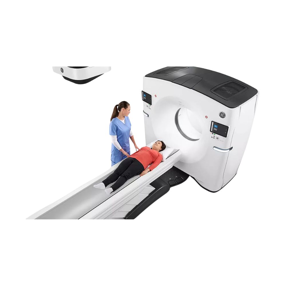
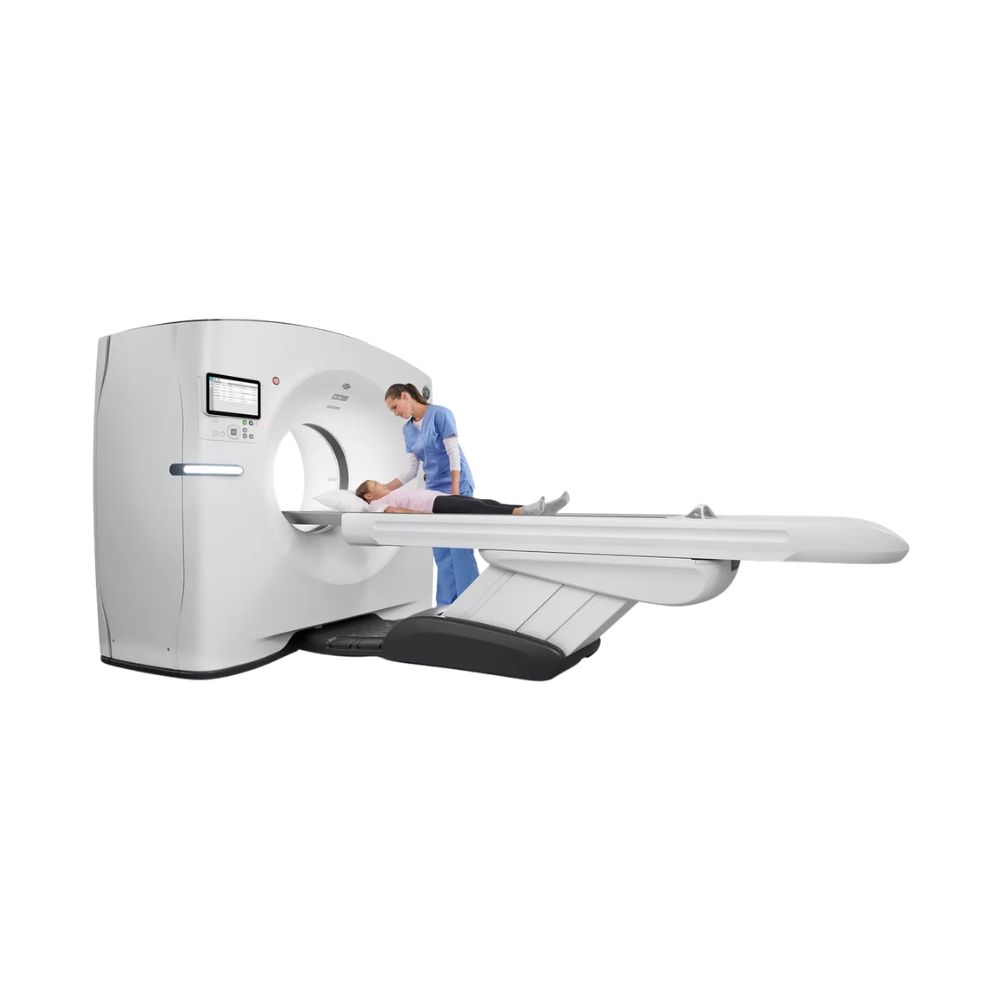
The Revolution Maxima Select 64 combines a 70 cm gantry opening with motorized patient-table configurations and accessible examination controls. These capabilities support patient preparation across routine and specialty imaging workflows.
70 cm Gantry & Digital Tilt
The scanner features:
-
- 70 cm (27.6 in) Gantry Opening: Provides space for positioning patients within the scanner’s circular opening.
- 50 cm (19.7 in) Maximum Scan Field of View: Defines the maximum diameter of the area that can be reconstructed into a CT image.
- Digital Tilt: Creates images at selected angles of up to ±30° without physically tilting the scanner, allowing clinicians to review anatomy from the required orientation.
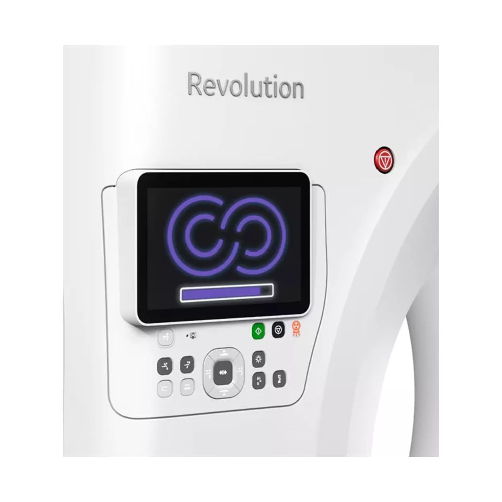
- Dual Xtream Touchscreens: Integrated displays positioned on each side of the gantry provide patient information, positioning controls, and examination status.
Digital Tilt can be useful when a particular viewing angle is needed without additional mechanical positioning.
Patient Table Options & Scanning Range
The Revolution Maxima platform supports different motorized patient-table configurations. Their specifications can vary according to the installed system.
Important considerations include:
-
- Patient Weight Capacity: Determined by the installed table and approved accessories.
- Longitudinal Scan Range: The distance along the patient’s body that the scanner can cover during an examination, depending on table and scanning configuration.
- Motorized Positioning: Allows controlled table movement to help position patients accurately for scanning.
- Positioning Accessories: Compatible headrests, supports, and other accessories help accommodate different patient needs and examinations.
MXR Imaging helps facilities select a configuration based on their patient population, imaging applications, and room requirements.
Revolution Maxima Select 64 Cardiac, Vascular, & Neurological Imaging
The Revolution Maxima Select 64 supports routine diagnostic CT and selected advanced applications through compatible scanning and image-processing software. These technologies help clinicians examine the heart, blood vessels, and brain in greater detail.
Cardiac CT & Coronary Imaging
Appropriately equipped systems support examinations that help clinicians evaluate coronary artery disease, including calcium scoring and cardiac CT angiography.
Available capabilities include:
-
- Cardiac Calcium Scoring: Uses CT images to detect and measure calcium deposits within the coronary arteries, which supply blood to the heart muscle. The software calculates a calcium score that helps clinicians assess the amount of hardened plaque present and evaluate the patient’s risk of coronary artery disease.
- Cardiac CT Angiography: Uses an injected contrast agent to make the coronary arteries more visible on CT images. This allows clinicians to examine the arteries for narrowing, plaque buildup, and other abnormalities that may affect blood flow to the heart.
- ECG-Synchronized Acquisition: Uses an electrocardiogram (ECG), which records the heart’s electrical activity, to coordinate scanning with the heartbeat. This helps reduce motion-related image distortion during cardiac examinations.
- Cardiac Reconstruction Software: Processes the acquired scan data into additional views of the heart and coronary arteries, helping clinicians examine vessel anatomy and assess areas of concern from different angles.
Advanced cardiac technologies such as SnapShot Freeze motion correction, which helps reduce image blurring caused by heart movement, are associated with compatible higher-tier configurations or upgrades rather than standard Maxima Select 64 equipment.
CT Angiography & Vascular Visualization
CT angiography uses injected contrast material to make blood vessels easier to distinguish from surrounding tissues. Compatible Maxima Select configurations support vascular examinations and software that helps clinicians review blood vessel anatomy.
-
- CT Angiography: Produces detailed images of arteries and other blood vessels in the head, neck, chest, abdomen, and extremities. These images help clinicians assess narrowing, blockages, aneurysms, and other vascular abnormalities.
- High-Pitch Helical Scanning: Moves the table through the scanner efficiently during continuous imaging, helping capture larger vascular regions within a shorter scanning period when appropriate.
- Multiplanar Reconstruction: Reconstructs the original CT data into views from the front, side, or other angles, allowing clinicians to follow blood vessels and examine structures that are difficult to assess in a single viewing direction.
- 3D Volume Visualization: Combines CT data into three-dimensional images that help clinicians understand vessel pathways, branching anatomy, and the relationship between blood vessels and surrounding structures.
Available image-processing functions depend on the installed software and workstation configuration.
Brain Perfusion & Neurovascular Imaging
Compatible Maxima Select systems support selected neurological examinations that help clinicians examine brain anatomy, cerebral blood vessels, and blood flow through brain tissue.
-
- Volume Shuttle: Moves the patient table between two scanning positions during repeated imaging, allowing a larger area of the brain to be examined over time without requiring a detector that physically covers the entire region at once.
- Brain Perfusion Analysis: Uses a series of CT images captured as contrast material passes through the brain to measure how blood reaches and moves through different areas of brain tissue. These measurements can help clinicians evaluate blood-flow abnormalities during selected neurological examinations.
- Neurovascular Imaging: Produces contrast-enhanced images of blood vessels in the brain and neck, helping clinicians examine narrowing, blockages, aneurysms, and other abnormalities that may affect cerebral circulation.
Compatible Maxima Select configurations support whole-brain perfusion applications with 8 cm (3.15 in) anatomical coverage through specialized acquisition techniques rather than an 8 cm physical detector.
Revolution Maxima Select 64 Workflow & AI Technology
The Revolution Maxima platform offers technologies that streamline patient setup, scanning protocols, image processing, and examination delivery. Eligible Select 64 configurations can also support optional AI-based positioning and deep-learning reconstruction technologies.
Gantry Controls & Examination Preparation
Available workflow capabilities include:
-
- Gantry-Mounted Interface: Provides selected scanning and positioning controls directly at the scanner, allowing technologists to prepare patients without returning to the main console for every adjustment.
- Protocol Management: Helps technologists select consistent, predefined scanning settings for different examinations.
- Automated Reconstruction: Processes acquired scanning data into predefined image sets and viewing angles, reducing repetitive manual image-processing steps.
- Operator Console Applications: Allow selected clinical image review and processing directly on the scanner’s computer rather than requiring a separate workstation for every examination.
These features help simplify repetitive steps across routine and advanced CT workflows.
AI Auto Positioning & Deep-Learning Upgrades
AI Auto Positioning is an optional upgrade for the GE Revolution Maxima Select 64. Equipped systems include an overhead Xtream depth-sensing camera and compatible software that work together to automate patient centering and examination setup.
The available AI technologies serve different purposes:
-
- AI Auto Positioning: Uses the overhead camera to create a three-dimensional representation of the patient. AI-powered positioning software calculates how to center the patient within the scanner’s opening, helping technologists achieve consistent positioning with fewer manual adjustments.
- TrueFidelity DL: A separate deep-learning reconstruction upgrade that uses artificial intelligence trained on CT image data to reduce noise while preserving anatomical detail and a natural image appearance.
- Remote Control Suite: Supports selected scanning and monitoring functions from compatible remote workstations, giving facilities additional options for managing CT workflows.
- Advanced Protocol Automation: Helps automate selected examination-planning and preparation steps through compatible workflow software.
The camera and positioning software are components of the optional AI Auto Positioning system. TrueFidelity DL is a separate capability and is not automatically included with the camera-based positioning upgrade.
MXR Imaging helps facilities identify available GE Revolution Maxima configurations with the imaging and automation technologies suited to their clinical and operational requirements.
Revolution Maxima AI Auto Positioning Video
See how GE’s optional AI Auto Positioning technology supports patient setup in clinical practice. In this GE HealthCare customer video, Dr. Lombard of Jacques Callot Imaging Medical Center in France discusses the use of automated positioning on the Revolution Maxima platform.
Contrast Timing & DICOM Connectivity
-
- SmartPrep: Tracks how contrast material reaches a selected blood vessel or anatomical region, helping technologists begin scanning when the contrast is in the appropriate location for the examination.
- DICOM Connectivity: Uses the standard medical imaging file format to transfer CT images to compatible picture archiving and communication systems (PACS), where clinical teams store and review scans.
- Clinical Worklist Integration: Exchanges patient and examination information with compatible hospital or imaging information systems, reducing repetitive data entry and supporting more organized workflows.
Connectivity and automation depend on the installed console software and facility network configuration.
Clinical Environments & Applications
The GE Revolution Maxima Select 64 supports routine diagnostic CT and selected advanced clinical applications. Facilities can select scanning and software configurations appropriate to their examination requirements.
Clinical Environments
-
- Hospitals and diagnostic imaging departments
- Outpatient CT and diagnostic imaging centers
- Emergency and trauma departments
- Cardiac and vascular imaging centers
- Neurology and stroke centers
- Oncology and cancer care facilities
- Multispecialty medical practices
- Academic medical centers and teaching hospitals
- Veterinary specialty hospitals and advanced diagnostic imaging centers with appropriately configured equipment
Clinical Specialties
-
- Radiology: Routine head-to-body CT and detailed anatomical imaging.
- Cardiology: Coronary calcium scoring and CT angiography on appropriately equipped systems.
- Vascular Medicine: CT angiography and assessment of blood vessel anatomy.
- Neurology & Neurosurgery: Brain imaging, neurovascular evaluation, and selected blood-flow examinations.
- Orthopedics: Fracture assessment, complex bone anatomy, and postoperative imaging.
- Oncology: Tumor evaluation, disease staging, and treatment follow-up.
- Pulmonology: Chest and lung imaging, including compatible vascular studies.
- Urology: Kidney and urinary tract imaging.
- Emergency Medicine: Acute diagnostic and trauma CT.
- Veterinary Diagnostic Imaging: Selected neurological, orthopedic, and soft-tissue examinations where the system configuration, positioning accessories, and clinical protocols support animal imaging.
Revolution Maxima Select 64 Configurations & Installed Options
GE Revolution Maxima Select 64 CT scanners can differ in their scanning hardware, X-ray generator, image-processing software, patient table, and installed clinical applications. These differences are important when evaluating pre-owned equipment or comparing available systems.
20 mm & 40 mm Detector Configurations
Two documented acquisition arrangements are available:
-
- 20 mm Coverage / 0.625 mm Sections: Uses a 32 × 0.625 mm acquisition arrangement to capture thinner sections for fine anatomical detail.
- 40 mm Coverage / 1.25 mm Sections: Uses a 32 × 1.25 mm acquisition arrangement to capture a wider area during each rotation.
Both support 64-slice imaging through the applicable reconstruction configuration. A compatible upgrade to 40 mm coverage with 0.625 mm sections and 128-slice overlapping reconstruction represents a higher-tier capability.
Generator, X-Ray Tube, & Clinical Software Options
Available systems may differ in:
-
- X-ray generator power and supported tube current
- X-ray tube condition and operating history
- Gantry rotation-speed capability
- ASiR or ASiR-V image reconstruction
- Smart MAR and high-resolution image processing
- Cardiac, vascular, and neurological applications
- Patient table and positioning accessories
These differences affect clinical suitability, operational requirements, and total equipment cost.
Optional AI Positioning Package & Upgrade Eligibility
The optional AI Auto Positioning package includes an overhead Xtream depth-sensing camera and compatible positioning software. Both components work together to calculate patient alignment and automate positioning.
Eligible systems may also support separate upgrades to TrueFidelity DL, higher-performance scanning, advanced cardiac applications, and selected workflow technologies.
MXR Imaging helps facilities evaluate available configurations and appropriate modernization pathways without treating every optional technology as standard equipment.
Revolution Maxima Select 64 vs. Other Maxima Configurations
The GE Revolution Maxima family includes 32-, 64-, and 128-slice configurations. Their differences extend beyond slice count to detector coverage, acquired section thickness, image reconstruction, and available clinical technology.
- GE Revolution Maxima Select 32-Slice CT: Supports 32-slice acquisition with documented 20 mm and 40 mm scanning arrangements. It offers a lower slice-count configuration for routine diagnostic CT and compatible applications.
- GE Revolution Maxima Select 64-Slice CT: Provides 64-slice imaging using 32-section acquisition arrangements, with 20 mm coverage at 0.625 mm or 40 mm coverage at 1.25 mm. Its Clarity Imaging Chain, advanced clinical options, and optional AI Auto Positioning package support routine and specialty workflows.
- GE Revolution Maxima 128-Slice CT: Supports 128-slice overlapping image reconstruction with compatible 40 mm scanning hardware and 0.625 mm reconstructed sections. Associated with the higher-tier Maxima Elite configuration, it offers additional performance options, including faster gantry rotation and advanced cardiac capabilities.
All three belong to the Revolution Maxima family, but differences in detector arrangement, reconstruction capabilities, installed software, and upgrade packages determine their practical capabilities.
MXR Imaging helps facilities compare configurations according to imaging requirements, examination volume, specialty applications, and long-term equipment needs.
Revolution Maxima vs. Revolution EVO, HD, & Apex
GE’s Revolution CT platforms offer different combinations of detector technology, image processing, scanning performance, and workflow automation.
-
- Revolution Maxima: Combines Clarity imaging with flexible scanning configurations, advanced clinical software, and optional AI Auto Positioning.
- Revolution EVO: Uses the Clarity Imaging Chain with established reconstruction and clinical options. Compare the GE Revolution EVO 64-Slice CT and GE Revolution EVO 128-Slice CT.
- Revolution HD: Uses Gemstone detector technology with available spectral imaging, which collects additional information about how tissues absorb X-rays to help clinicians distinguish certain materials. Compare the GE Revolution HD 64-Slice CT and GE Revolution HD 128-Slice CT.
- Revolution Apex: Offers wider detector configurations and advanced image-processing technologies for demanding cardiac, vascular, and other clinical examinations.
MXR Imaging helps facilities compare imaging technology, scanning capabilities, clinical applications, and operational requirements when selecting a GE CT scanner.
Revolution Maxima Select 64 Installation & Site Requirements
The GE Revolution Maxima Select 64 requires a CT imaging room with appropriate electrical service, cooling, ventilation, radiation shielding, structural support, and maintenance clearances.
Installation planning accounts for the gantry, patient table, operator console, supporting equipment, patient access, and clinical workflow. Room and infrastructure requirements depend on the installed configuration.
Ceiling-Mounted Xtream Camera for AI Auto Positioning
Systems equipped with the optional AI Auto Positioning upgrade include an overhead Xtream depth-sensing camera and compatible positioning software.
The camera requires mounting hardware aligned with the CT scanner and patient table. GE specifies a camera-bracket height of 2,383–2,880 mm (approximately 7 ft 10 in to 9 ft 5 in) above the finished floor.
This measurement describes the bracket position, not a universal minimum or maximum room ceiling height. Different ceiling constructions may require suitable extension hardware or alternative mounting arrangements.
The camera is necessary for the camera-based AI Auto Positioning function but is not required for standard CT operation.
MXR Imaging’s imaging project management and equipment installation services help coordinate site preparation, camera mounting requirements, system installation, and related project needs.
Revolution Maxima Select 64 CT Price & Availability
MXR Imaging offers GE Revolution Maxima Select 64-slice CT scanners for healthcare facilities seeking detailed diagnostic imaging, flexible configurations, and available advanced clinical technologies.
Equipment Pricing & Configuration Factors
The price of a GE Revolution Maxima Select 64 depends on the selected equipment and project requirements, including:
-
- Detector and scanning configuration
- X-ray tube condition and operating history
- Generator and patient-table options
- Image-processing and clinical software
- Optional AI Auto Positioning hardware and software
- Equipment condition and service coverage
- Transportation, installation, and site preparation
A pre-owned Maxima Select 64 with advanced clinical applications or AI Auto Positioning may differ substantially from another system with the same slice count.
Equipment Sales, Installation, & Lifecycle Support
MXR Imaging helps healthcare providers evaluate available CT equipment, compare installed technologies, and plan for acquisition and installation.
Eligible pre-owned systems may be available through MXR Imaging’s Platinum Certified program. Additional services include imaging equipment relocation, installation, preventive maintenance, CT service, and replacement parts.
Request a quote for the GE Revolution Maxima Select 64 to discuss available configurations, equipment pricing, clinical applications, installation requirements, and service support.
GE Revolution Maxima Select 64-Slice CT Specifications
Specifications reflect documented Maxima Select capabilities. Installed hardware, software, and optional technologies vary by system.
CT Acquisition & Detector
-
- Manufacturer: GE HealthCare
- Model: Revolution Maxima Select
- Slice designation: 64-slice CT
- Documented acquisition arrangement: 32 acquired sections per rotation
- 64-slice capability: Configuration-dependent reconstruction from acquired data
- Detector technology: GE Clarity
- Detector coverage: 20 mm (0.79 in) or 40 mm (1.57 in)
- 20 mm configuration: 32 × 0.625 mm acquired sections
- 40 mm configuration: 32 × 1.25 mm acquired sections
- Spatial resolution: As fine as 0.28 mm (0.011 in)
- Reconstruction matrix: Up to 1024 × 1024 on compatible systems
- Helical pitch: Up to 1.531 on supported configurations
- Scanning modes: Axial, helical, and compatible advanced protocols
Gantry & Patient Table
-
- Gantry opening: 70 cm (27.6 in)
- Maximum scan field of view: 50 cm (19.7 in)
- Gantry rotation: 0.5 seconds standard; faster rotation through compatible upgrades
- Digital Tilt: Up to ±30° reconstructed image-plane adjustment
- Gantry Display Interface: Dual integrated Xtream touchscreens, one on each side of the gantry, providing patient information, positioning controls, and examination status
- Patient positioning: Motorized table
- Patient weight capacity: Installed-table dependent
- Longitudinal scanning range: Table- and acquisition-dependent
X-Ray Generator & Tube
-
- Documented X-ray tube: GE Performix 40 Plus
- Anode heat-storage capacity: 7.0 MHU
- Tube cooling rate: Up to 1,070 kHU/min on documented configurations
- Generator options: 55 kW and 72 kW documented within the platform
- Tube voltage: 80, 100, 120, and 140 kVp on documented configurations
- Tube-current capability: Configuration-dependent
- Tube condition and usage: Varies by system
Reconstruction & Image Processing
-
- Imaging platform: GE Clarity Imaging Chain
- Iterative reconstruction: ASiR or ASiR-V, configuration-dependent
- High-resolution reconstruction: Up to 1024 × 1024 matrix
- Deep-learning reconstruction: TrueFidelity DL through eligible upgrades
- Metal artifact reduction: Smart MAR where installed
- Multiplanar reconstruction: Available
- 3D visualization: Available with compatible software
- Advanced reconstruction options: Configuration-dependent
Advanced Imaging & Workflow
-
- Cardiac imaging: Calcium scoring and CT angiography on equipped systems
- Vascular imaging: Compatible CT angiography and post-processing
- Neurological imaging: Brain perfusion and Volume Shuttle options
- Dose management: Automated exposure control and compatible monitoring technologies
- AI Auto Positioning: Optional camera-and-software upgrade
- Remote Control Suite: Available on eligible configurations
- Operator-console applications: Configuration-dependent
AI Positioning Camera & Mounting Requirements
-
- Optional package: AI Auto Positioning
- Included positioning components: Overhead Xtream depth-sensing camera and compatible positioning software
- Function: Three-dimensional patient modeling and automated centering
- Installation: Overhead mounting aligned with the CT scanner and patient table
- Camera-bracket mounting height: 2,383–2,880 mm (approximately 7 ft 10 in to 9 ft 5 in) above finished floor
- Mounting hardware: Depends on ceiling construction and room configuration
- Required for standard CT operation: No
Connectivity & Site Requirements
-
- Image standard: DICOM
- Clinical connectivity: Compatible PACS and imaging information systems
- Electrical service: Generator- and installation-dependent
- Radiation shielding: Facility- and regulation-dependent
- Cooling and ventilation: Equipment-dependent
- Room dimensions: Installation-dependent
- Structural support: Required for scanner and optional positioning camera
- Maintenance clearances: Configuration-dependent