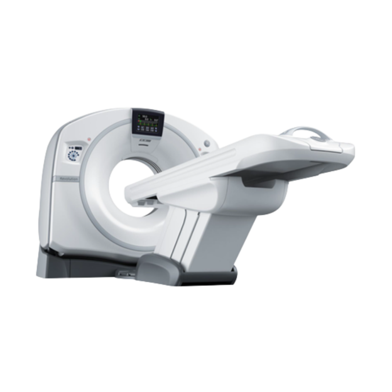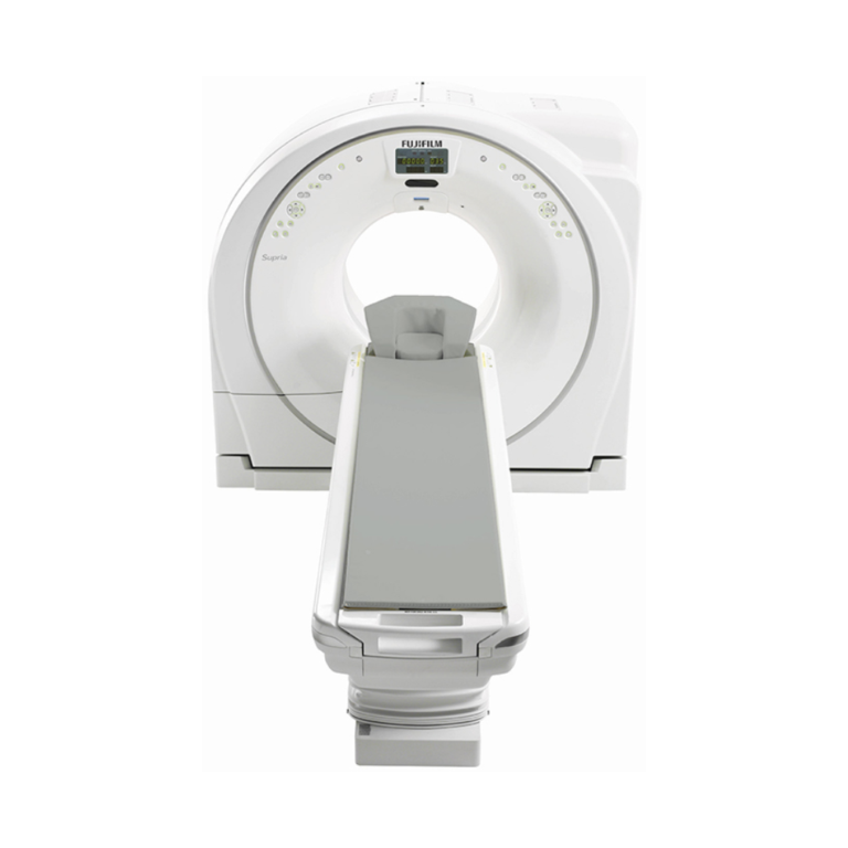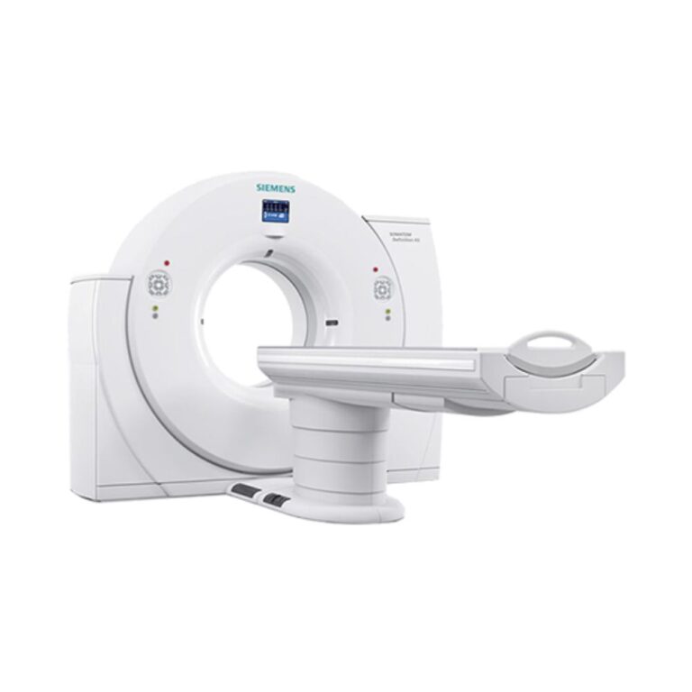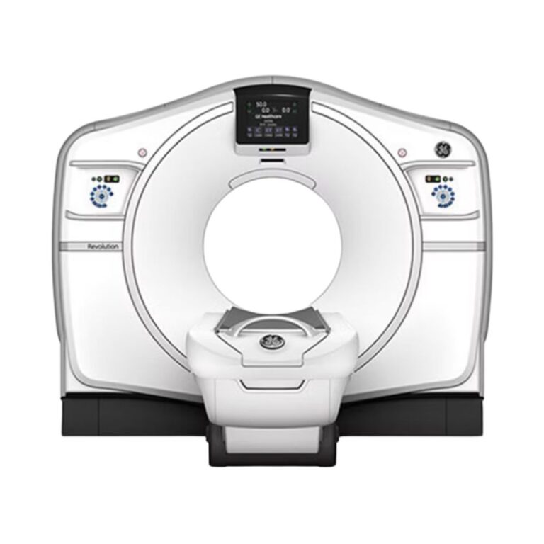
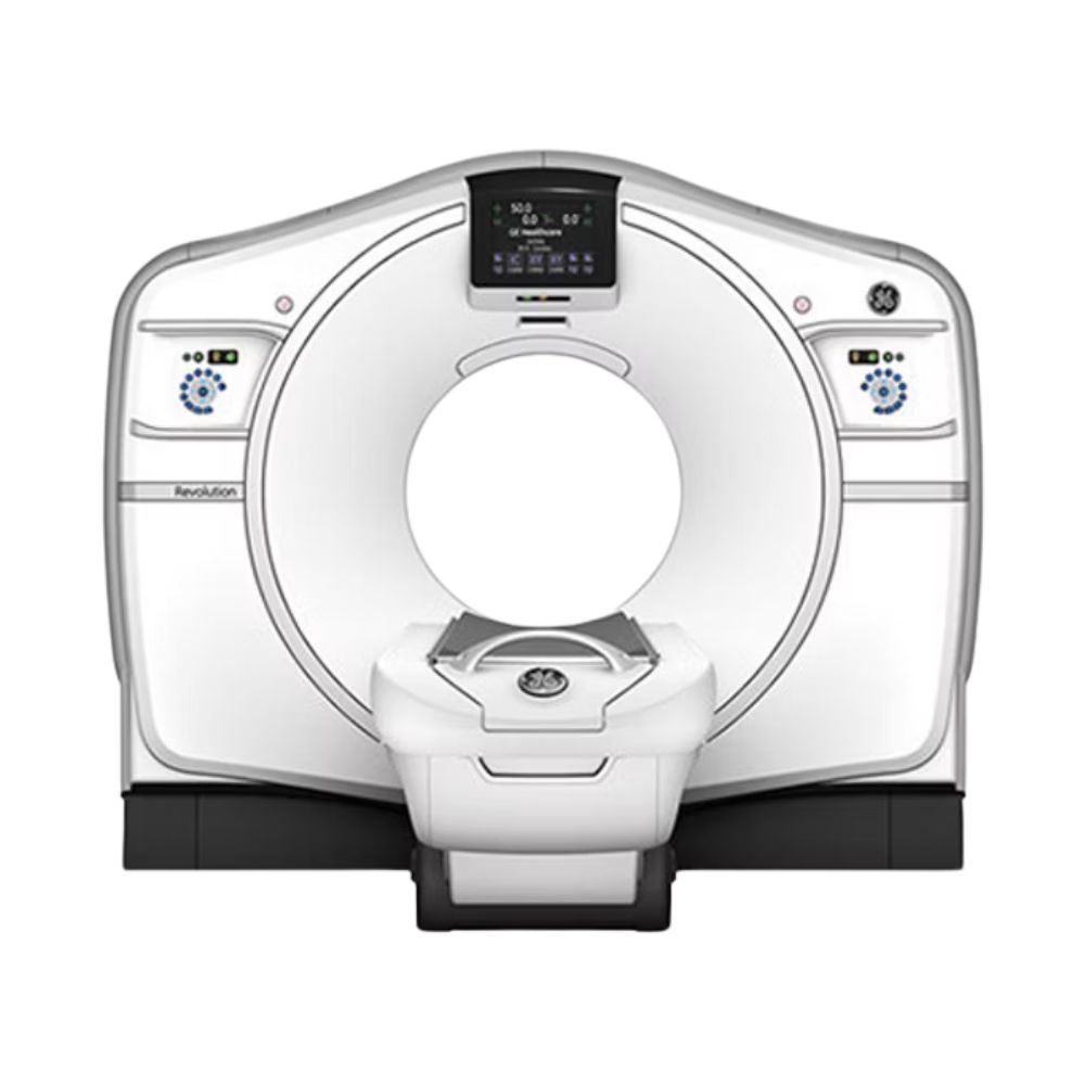
GE Revolution HD 64-Slice CT
- Category:
- CT Scanners
- Brand:
- GE
The GE Revolution HD 64 is a high-definition, 64-slice CT scanner well suited for hospitals, imaging centers, and specialty practices. Its Gemstone detector captures fine anatomical detail, while 0.35-second rotation supports cardiac, vascular, and routine imaging. Available reconstruction and spectral technologies extend its capabilities for advanced examinations.
Revolution HD 64 CT Highlights & Benefits
The GE Revolution HD 64 combines rapid CT acquisition with high-resolution detector technology and patient-specific dose controls. Its imaging capabilities support routine head-to-body examinations, while available advanced software extends its applications in cardiac, vascular, neurological, and other specialty imaging.
Key Features & Benefits
-
- 64-Slice CT Acquisition: Captures 64 slices per rotation with 40 mm (1.57 in) of detector coverage for detailed head-to-body imaging.
- Gemstone Detector: Delivers spatial resolution as fine as 0.23 mm (0.009 in), supporting evaluation of fine anatomical structures.
- 0.35-Second Gantry Rotation: Supports rapid scanning and helps limit motion-related image distortion.
- 100 kW X-Ray Generator: Provides high-output performance for routine and advanced examinations.
- ASiR Reconstruction: Reduces image noise while supporting patient-specific radiation-dose management.
- Optional Gemstone Spectral Imaging: Helps distinguish tissue and material composition beyond conventional CT images.
- Advanced Cardiac Imaging: Available SnapShot Freeze technology helps reduce coronary artery motion blur.
Revolution HD 64 Acquisition & High-Definition Imaging
The Revolution HD 64 uses a 64-channel acquisition system, GE’s Gemstone detector, and rapid gantry rotation to capture detailed CT images. These technologies support routine diagnostic studies and specialized examinations requiring fine anatomical detail or precise acquisition timing.
64-Slice Acquisition for Detailed Head-to-Body CT
The Revolution HD 64 acquires 64 slices per rotation across 40 mm (1.57 in) of anatomy. With acquired slice thicknesses as thin as 0.625 mm (0.025 in), it supports detailed head, chest, abdominal, pelvic, spine, and musculoskeletal examinations.
During helical scanning, continuous patient-table movement allows coverage of larger anatomical regions. The platform also supports overlapping reconstruction of up to 128 image sections from its 64-channel acquisition data, providing additional closely spaced views without requiring 128 separate physical detector rows.
Gemstone Detector for Fine Anatomical Detail
GE’s Gemstone detector uses fast-response scintillator material to capture X-ray measurements with minimal residual signal between successive readings. Its spatial resolution reaches as fine as 0.23 mm (0.009 in), supporting evaluation of coronary vessels, fine bone anatomy, and other small anatomical structures.
With 40 mm (1.57 in) of detector coverage, the system supports routine head-to-body examinations and compatible advanced acquisition protocols. The detector’s rapid response also enables the fast energy switching used by optional Gemstone Spectral Imaging.
0.35-Second Rotation for Fast CT Acquisition
The Revolution HD completes a gantry rotation in as little as 0.35 seconds, helping limit motion-related image distortion. Fast acquisition is particularly valuable for cardiac imaging, contrast-enhanced vascular examinations, and time-sensitive diagnostic studies.
Volara Data Acquisition for High-Resolution Images
GE’s Volara data acquisition system (DAS) uses high-density sampling to capture measurements from the Gemstone detector. Its increased sampling helps preserve fine anatomical detail and reduce certain imaging artifacts, supporting detailed bone, soft-tissue, and vascular examinations.
Revolution HD 64 Image Reconstruction & Dose Management
The Revolution HD combines image reconstruction and exposure-control technologies to help facilities balance diagnostic detail with radiation-dose management. Reconstruction software reduces image noise, while automated exposure tools help tailor scanning parameters to the patient and examination.
Iterative Reconstruction for Image Clarity & Dose Management
Iterative reconstruction helps reduce image noise while preserving clinically useful anatomical detail. The Revolution HD platform supports several reconstruction approaches with different performance characteristics.
-
- ASiR: Adaptive Statistical Iterative Reconstruction reduces image noise and supports diagnostic image quality at appropriately selected radiation-dose settings. ASiR is a standard Revolution HD reconstruction capability.
- ASiR-V: Optional advanced iterative reconstruction that offers additional noise reduction and supports dose optimization during compatible examinations.
- Veo: Optional model-based iterative reconstruction that uses a detailed mathematical model of the CT imaging process to improve fine detail and the visibility of subtle tissue differences.
ASiR-V and Veo require compatible reconstruction hardware, software, and licensing. Actual image-quality and radiation-dose benefits depend on patient anatomy, the clinical task, and the selected imaging protocol.
Patient-Specific Radiation-Dose Controls
The Revolution HD provides automated technologies that help technologists select exposure settings and manage examination protocols.
-
- SmartmA: Adjusts X-ray tube current according to changes in patient anatomy and body thickness, helping maintain appropriate image quality throughout an examination.
- kV Assist: Helps select X-ray tube voltage based on patient characteristics and clinical requirements.
- Organ Dose Modulation: Adjusts radiation exposure during selected examinations to help manage dose to sensitive anatomical regions.
- CT 4Kids: Provides pediatric scanning protocols designed for the imaging needs of smaller patients.
- Dose Check: Provides configurable notifications and alerts when planned radiation-dose parameters reach established thresholds.
These technologies complement iterative reconstruction by managing exposure during image acquisition. Available functions and protocol settings depend on the system configuration.
Smart MAR for Clearer Imaging Around Metal
Metal implants can cause streaks and distortions that obscure surrounding anatomy on CT images.
-
- Smart Metal Artifact Reduction (Smart MAR): Optional reconstruction technology that reduces artifacts caused by metallic objects, helping improve visibility around joint replacements, surgical hardware, and other implants. This is particularly useful for orthopedic and postoperative examinations.
Smart MAR requires compatible installed software.
Revolution HD 64 Patient Access & Positioning
The GE Revolution HD 64 features a 70 cm gantry opening with motorized patient positioning and available higher-capacity table options. These features help facilities accommodate different patient populations and examination requirements.
70 cm Gantry for Patient Access
The scanner features a 70 cm (27.6 in) gantry opening and tilt adjustments of up to 30° in either direction. Front and rear gantry controls, patient-alignment features, and compatible in-room functions support examination setup and positioning.
Patient Table Capacity & Available Scanning Range
Documented Revolution HD patient-table configurations include:
-
- 500 lb (227 kg) Patient Capacity: Supports a broad range of routine and specialty examinations.
- 675 lb (306 kg) High-Capacity Option: Accommodates higher patient weights with a compatible table.
- 170 cm (66.9 in) Scannable Range: Supports routine head-to-body coverage on the corresponding configuration.
- 200 cm (78.7 in) Scannable Range: Provides extended anatomical coverage with the corresponding table configuration.
The installed table determines patient capacity, scan coverage, positioning capabilities, and compatible accessories. MXR Imaging helps facilities select a configuration suited to their patient population and clinical requirements.
Revolution HD 64 Exam Planning & Workflow Automation
The Revolution HD offers technologies designed to simplify patient positioning, examination setup, contrast timing, and image processing. These functions support workflow consistency across routine and time-sensitive examinations.
Automated Patient Positioning & Gantry Controls
Compatible systems provide workflow features that reduce repetitive preparation steps and make selected examination controls accessible at the scanner.
-
- One Step Patient Positioning: Moves the patient table toward a predefined reference position through a simplified control action, reducing manual positioning adjustments.
- Xtream Gantry Display: Presents patient and examination information at the scanner. Compatible educational videos can also help explain CT procedures and support patient communication.
- One Stop ED: Provides access to frequently used emergency examination protocols through compatible gantry controls, helping streamline preparation for time-sensitive scans.
The available gantry controls and automated functions depend on the installed hardware and software.
Contrast Timing & Image Processing Automation
Compatible Revolution HD configurations offer additional workflow technologies for contrast-enhanced examinations, reconstruction, and clinical image review.
-
- Dynamic Transition: Helps coordinate acquisition with contrast arrival in the selected anatomy, supporting consistent timing during vascular and other contrast-enhanced examinations.
- Image Check: Supports rapid image reconstruction and review, with performance reaching up to 55 images per second on compatible configurations.
- DICOM Connectivity: Enables standardized image exchange with compatible picture archiving and communication systems (PACS), allowing completed studies to be stored and accessed through a facility’s clinical imaging network.
Reconstruction performance, available automation, and connectivity functions depend on the installed console, software, and network configuration.
Revolution HD 64 Advanced Clinical Imaging
Available clinical applications extend the Revolution HD beyond routine CT. Compatible configurations provide additional information about tissue composition, coronary anatomy, cerebral blood flow, and complex vascular structures.
Gemstone Spectral Imaging for Tissue Characterization
Optional Gemstone Spectral Imaging (GSI) rapidly switches between two X-ray energy settings during acquisition. Because different materials absorb these energies differently, GSI can help distinguish tissue and material characteristics that may appear similar on conventional CT images.
Compatible clinical applications include:
Kidney Stone Characterization: Helps distinguish certain stone compositions during urinary tract evaluation.
-
- Material Separation: Provides additional information about substances such as iodine and water.
- Virtual Noncontrast Imaging: Generates images that estimate the appearance of anatomy without contrast material using spectral acquisition data.
- Lesion Evaluation: Supports selected abdominal and oncology applications by providing additional tissue-characterization information.
GSI can also support specialized vascular imaging and advanced image analysis. It requires compatible acquisition hardware, reconstruction software, and active licenses.
SnapShot Freeze for Cardiac Motion Correction
Coronary artery movement can cause image blurring during cardiac CT, even with rapid gantry rotation.
-
- SnapShot Freeze: Optional motion-correction technology that analyzes coronary artery movement during reconstruction to reduce motion-related distortion and improve visibility during compatible cardiac CT angiography examinations.
Under specified cardiac phantom testing conditions, SnapShot Freeze has demonstrated an effective temporal resolution of 29 milliseconds. This describes motion-correction performance rather than the scanner’s physical gantry rotation time.
Volume Shuttle for Brain Perfusion Imaging
Selected neurological examinations require repeated acquisitions to evaluate changes in blood flow through brain tissue.
-
- Volume Shuttle: Moves the patient table between acquisition positions during repeated scans, extending anatomical coverage beyond the detector’s 40 mm width. Combined with compatible perfusion software, it supports assessment of blood flow across a larger region of brain tissue.
Available coverage, perfusion measurements, and clinical applications depend on the installed acquisition and reconstruction options.
Advanced Vascular & 3D Image Review
Compatible post-processing applications provide additional ways to analyze CT data and visualize complex anatomical structures.
-
- Advanced Vessel Analysis: Creates specialized views and measurements to support evaluation of vascular anatomy.
- 3D Volume Visualization: Generates rotatable anatomical images for examining complex fractures, blood vessels, and surgical anatomy.
- Specialty Clinical Applications: Provide additional analysis tools for selected cardiac, neurological, oncology, and surgical-planning examinations.
Application availability depends on the installed workstation, software version, and licenses.
Clinical Environments & Applications
The GE Revolution HD 64-slice CT scanner supports routine diagnostic imaging and specialized examinations requiring rapid acquisition, detailed anatomy, and advanced reconstruction. Its configurable capabilities are well suited to healthcare organizations with varied imaging requirements.
Clinical Environments
-
- Hospitals and health systems
- Diagnostic imaging and outpatient CT centers
- Emergency and trauma departments
- Cardiac and vascular imaging centers
- Neurology and neurosurgery departments
- Oncology and cancer care facilities
- Multispecialty medical practices
- Specialty surgical and diagnostic centers
- Academic medical centers and teaching hospitals
Clinical Specialties
-
- Radiology: Routine head, chest, abdominal, pelvic, and spine examinations.
- Cardiology: Coronary CT angiography and selected cardiac assessments on appropriately equipped systems.
- Vascular Medicine: CT angiography and evaluation of blood vessel anatomy.
- Neurology & Neurosurgery: Brain and spine imaging, vascular assessment, and selected perfusion examinations.
- Orthopedics: Complex fractures, bone abnormalities, and evaluation around metal implants.
- Oncology: Tumor assessment, disease staging, and treatment follow-up.
- Pulmonology: High-resolution chest imaging and lung evaluation.
- Urology: Renal stone and urinary tract assessment, including optional spectral characterization.
- Emergency Medicine: Acute diagnostic and selected trauma imaging.
Common CT Applications
-
- Routine head, chest, abdominal, and pelvic CT
- Noncontrast and contrast-enhanced examinations
- High-resolution lung and chest imaging
- CT angiography and vascular evaluation
- Coronary CT angiography on equipped systems
- Renal stone assessment
- Complex fracture and musculoskeletal imaging
- Oncology staging and treatment follow-up
- Metal implant evaluation with optional Smart MAR
- Selected brain perfusion examinations
- Emergency and trauma imaging
Revolution HD 64 Configurations & Installed Options
Pre-owned GE Revolution HD 64 scanners can vary by production year, X-ray tube condition, patient table, reconstruction hardware, workstation, and software licensing. These differences affect clinical applications, image-processing performance, and compatibility with a facility’s imaging requirements.
Gemstone Detector & X-Ray Hardware
The Revolution HD combines its Gemstone detector with a 100 kW X-ray generator. A documented configuration includes a Performix HD X-ray tube with 8 million heat units (MHU) of heat-storage capacity, supporting repeated scanning through appropriate heat management.
Installed or replacement X-ray tubes, usage history, cooling characteristics, and maintenance condition can vary across pre-owned systems.
Reconstruction Software & Installed Clinical Options
Depending on configuration, available technologies may include:
-
- ASiR, ASiR-V, and Veo reconstruction
- Gemstone Spectral Imaging
- SnapShot Freeze cardiac motion correction
- Smart MAR metal artifact reduction
- SmartmA, kV Assist, and Dose Check
- Volume Shuttle and perfusion applications
- Advanced vessel analysis and 3D visualization
- Specialized cardiac, neurological, and oncology software
These represent available platform capabilities rather than features guaranteed on every scanner.
Workstation & Connectivity Configurations
The installed operator console, reconstruction hardware, and clinical workstation affect image-processing speed, storage capacity, and available software applications. Software licensing and network configuration also influence integration with existing clinical imaging systems.
MXR Imaging helps facilities identify a configuration aligned with examination volume, advanced imaging requirements, and existing infrastructure.
Revolution HD 64 vs. Revolution HD 128
The GE Revolution HD 64 and Revolution HD 128 are separate slice-count product listings associated with the same high-definition CT platform. Both use 64-channel acquisition and Gemstone detector technology, with their designations emphasizing different acquisition and reconstruction characteristics.
- GE Revolution HD 64-Slice CT: Emphasizes the platform’s 64-slice acquisition architecture for routine and specialized diagnostic imaging.
- GE Revolution HD 128-Slice CT: Emphasizes the capability to reconstruct up to 128 overlapping image sections from 64-channel acquisition data.
The 128-slice designation does not indicate twice the physical detector coverage or necessarily a different acquisition system. Both listings share important platform technologies, while installed reconstruction software, hardware, and clinical applications determine the capabilities of individual systems.
MXR Imaging helps facilities compare available configurations according to clinical requirements, examination volume, and advanced imaging needs.
Revolution HD 64 Installation & Site Requirements
Installing a GE Revolution HD CT scanner requires an appropriately prepared imaging suite with electrical service, cooling, ventilation, radiation shielding, and structural support.
Site planning accounts for the gantry, patient table, operator console, supporting equipment, patient access, and maintenance clearances. Final room dimensions and infrastructure requirements depend on the installed configuration and facility layout.
MXR Imaging’s turnkey imaging project management services help coordinate equipment selection, site planning, room preparation, installation, and related project requirements.
Revolution HD 64 CT Price & Availability
MXR Imaging offers pre-owned GE Revolution HD 64-slice CT scanners for hospitals, imaging centers, and specialty practices seeking high-definition CT capabilities with configurable clinical applications.
Pricing depends on system condition, X-ray tube history, patient table, reconstruction hardware, licensed software, accessories, installation requirements, and service coverage. These factors also influence long-term operating costs and the suitability of a system for a facility’s examination volume.
Eligible configurations may be available through MXR Imaging’s Platinum Certified program, which provides a comprehensive equipment inspection and reconditioning pathway. CT maintenance and repair services are also available to support ongoing system performance.
Request a quote for the GE Revolution HD 64 to discuss available configurations, technical capabilities, installation support, and service options.
GE Revolution HD 64-Slice CT Specifications
These specifications reflect GE Revolution HD platform capabilities. Actual hardware, patient tables, reconstruction software, and clinical options may vary by installed system.
CT Acquisition & Detector
-
- Manufacturer: GE Healthcare
- Model: Revolution HD
- Slice designation: 64-slice CT
- Acquisition architecture: 64 channels, 64 acquired slices per rotation
- Detector technology: GE Gemstone
- Detector configuration: 64 × 0.625 mm
- Detector coverage: 40 mm (1.57 in)
- Minimum acquired slice thickness: 0.625 mm (0.025 in)
- Spatial resolution: As fine as 0.23 mm (0.009 in)
- Minimum gantry rotation: 0.35 seconds
- Scanning modes: Axial, helical, and compatible advanced protocols
- Overlapping single-energy reconstruction: Up to 128 image sections
Gantry & Patient Table
-
- Gantry opening: 70 cm (27.6 in)
- Gantry tilt: ±30°
- Standard patient capacity: Up to 227 kg (500 lb)
- Optional higher-capacity table: Up to 306 kg (675 lb)
- Documented scannable-range options: 170 cm (66.9 in) or 200 cm (78.7 in)
- Patient positioning: Motorized table
- Gantry controls: Front and rear controls with compatible in-room functions
X-Ray Generator & Tube
-
- Generator output: 100 kW
- X-ray tube: Performix HD in documented configurations; replacement tube models may vary
- Tube heat-storage capacity: 8 MHU with documented Performix HD tube
- Tube voltage: 80, 100, 120, and 140 kVp
- Tube current: 10–835 mA on documented configurations
- Tube cooling: Dependent on installed tube and cooling hardware
Reconstruction & Imaging Performance
-
- Data acquisition: GE Volara, 64-channel
- Acquired slices: 64 per rotation
- Overlapping single-energy reconstruction: Up to 128 image sections
- Spectral reconstruction: Up to 256 image sections with compatible GSI technology
- Reconstruction speed: Up to 55 images per second with Image Check
- Standard iterative reconstruction: ASiR
- Optional iterative reconstruction: ASiR-V and Veo
- Cardiac motion correction: Optional SnapShot Freeze
- Effective temporal resolution: As low as 29 milliseconds under specified cardiac phantom testing conditions
- Metal artifact reduction: Smart MAR, optional
- Spectral imaging: Gemstone Spectral Imaging, optional
- Perfusion imaging: Configuration-dependent
- 3D visualization: Available with compatible software
System & Site Requirements
-
- Image connectivity: DICOM
- PACS integration: Supported with compatible network configuration
- Electrical service: Site- and configuration-dependent
- Radiation shielding: According to facility and regulatory requirements
- Cooling and ventilation: Based on installed equipment
- Structural support: Required for gantry and patient table
- Room dimensions: Installation-dependent
- Service clearances: Configuration-dependent