20/20 Imaging H-DR 102 Hand X-Ray System
- Category:
- X-Ray Systems
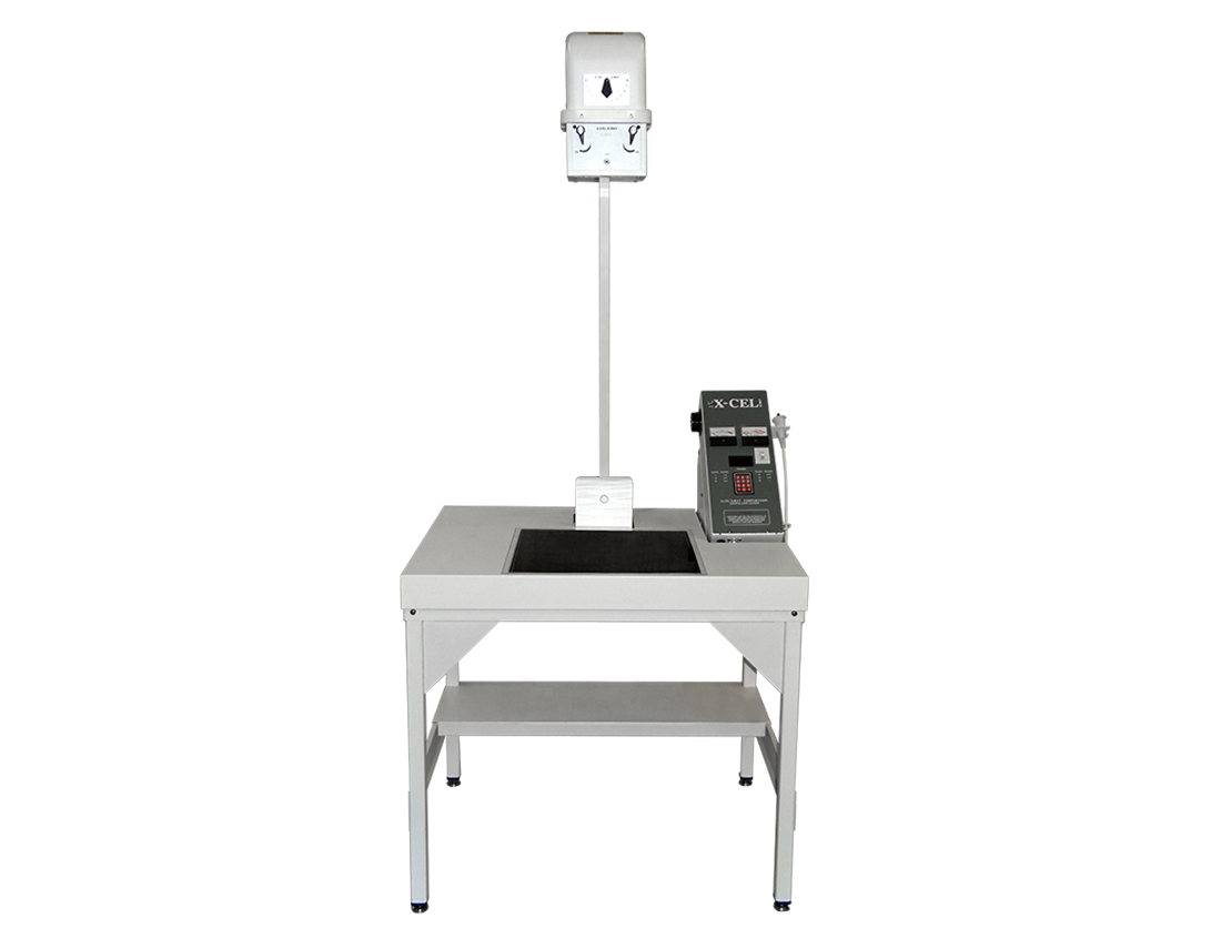
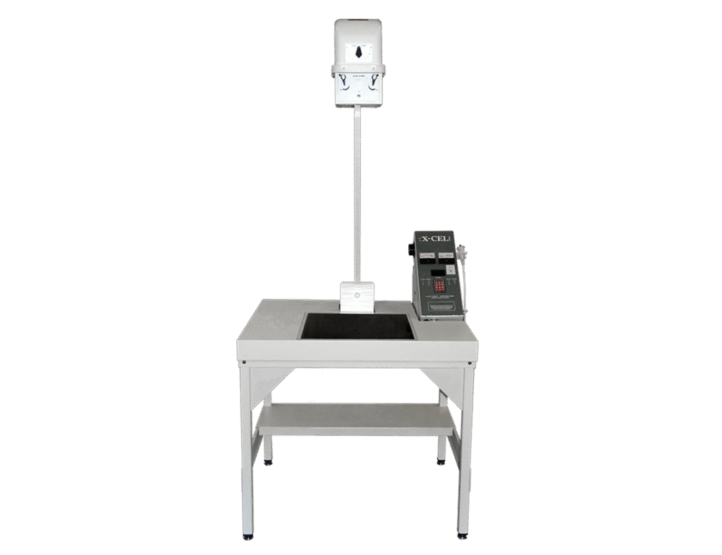
The 20/20 Imaging H-DR 102 is a dedicated orthopedic X-ray system for hand-focused practices and imaging. This complete package combines an X-Cel orthopedic/hand X-ray unit, a fixed 17 × 17-inch 20/20 Imaging DR detector, Symmetry-Sync software, and 45° carpal tunnel positioning.
H-DR 102 Digital Hand X-Ray Package
The 20/20 Imaging H-DR 102 is a complete digital orthopedic X-ray configuration built specifically for hand and wrist imaging. It pairs the X-Cel orthopedic/hand X-ray unit with a 20/20 Imaging C-FP fixed flat-panel detector and Symmetry-Sync digital imaging software. The H-DR 102 name refers to the complete digital system rather than only the underlying X-ray unit.
Key Benefits
-
- Dedicated orthopedic hand and wrist X-ray configuration
- 40 in. (101.6 cm) source-to-image distance
- 45° rotation for carpal tunnel views
- Adjustable X-ray collimation
- Flush tabletop for hand and wrist positioning
- 17 × 17 in. (43.2 × 43.2 cm) fixed DR detector
- Symmetry-Sync image acquisition, viewing, and storage
- Professional installation and system training
The 20/20 Imaging H-DR 102 is purpose-built for focused hand and wrist radiography rather than the broader positioning requirements of a general-purpose X-ray room.
X-Cel Positioning for Hand, Wrist & Carpal Tunnel X-Ray
40-Inch Source-to-Image Distance
The X-Cel radial-arm unit maintains a 40 in. (101.6 cm) source-to-image distance between the X-ray tube and detector. This fixed geometry provides a consistent setup for dedicated hand and wrist X-ray examinations.
45° Rotation for Carpal Tunnel Views
The radial arm rotates 45° to support carpal tunnel views. This built-in positioning capability allows the tube to reach the required angle within the dedicated hand X-ray system.
Flush Tabletop for Hand & Wrist Positioning
The flush tabletop provides a continuous surface for positioning the hand and wrist while maintaining alignment with the fixed detector and X-ray tube.
Adjustable Collimator for Focused X-Ray Fields
The adjustable collimator allows the technologist to control the size of the X-ray field around the anatomy being examined. This supports appropriate field sizing across different hand and wrist projections.
Orthopedic Hand & Wrist X-Ray Applications
The 20/20 Imaging H-DR 102 is designed for practices that regularly perform focused orthopedic X-ray imaging of the hand, wrist, and carpal anatomy.
Orthopedic & Hand Specialty Practices
The dedicated hand X-ray system is suited to environments such as:
-
- Orthopedic practices
- Hand and upper-extremity specialty practices
- Outpatient imaging centers performing hand and wrist X-ray
- Physician practices with in-office orthopedic X-ray services
Hand, Wrist & Carpal Tunnel Views
The tabletop and radial-arm configuration support radiography of:
-
- Hands
- Fingers
- Thumbs
- Wrists
- Carpal anatomy
- Carpal tunnel views using the 45° radial-arm rotation
The specialized configuration keeps the imaging workflow centered on hand and wrist studies rather than trying to function as a general radiography system.
17 × 17-Inch Fixed DR Detector in the H-DR 102
The complete 20/20 Imaging H-DR 102 configuration pairs the X-Cel X-ray unit with a fixed 17 × 17 in. (43.2 × 43.2 cm) C-FP flat-panel detector.
Full-Size Hand & Wrist Imaging Area
The detector provides a 17 × 17 in. (43.2 × 43.2 cm) imaging area for hand and wrist radiography. The H-DR 102 configuration specifies 3.5 lp/mm spatial resolution and 14-bit image conversion, with digital images available within seconds of exposure.
Fixed Detector Alignment
The C-FP uses a fixed interface within the dedicated hand imaging configuration. The detector does not need to be repositioned between routine studies, helping maintain a consistent relationship between the tabletop, detector, and X-ray tube.
17 × 17-Inch Fixed DR Detector in the H-DR 102
The complete 20/20 Imaging H-DR 102 configuration pairs the X-Cel X-ray unit with a fixed 17 × 17 in. (43.2 × 43.2 cm) C-FP flat-panel detector.
Full-Size Hand & Wrist Imaging Area
The detector provides a 17 × 17 in. (43.2 × 43.2 cm) imaging area for hand and wrist radiography. The H-DR 102 configuration specifies 3.5 lp/mm spatial resolution and 14-bit image conversion, with digital images available within seconds of exposure.
Fixed Detector Alignment
The C-FP uses a fixed interface within the dedicated hand imaging configuration. The detector does not need to be repositioned between routine studies, helping maintain a consistent relationship between the tabletop, detector, and X-ray tube.
Symmetry-Sync Digital X-Ray Workflow
The H-DR 102 integrates 20/20 Imaging’s Symmetry-Sync platform for digital X-ray acquisition, image processing, study management, viewing, and PACS image storage.
Automatic Image Processing After Exposure
The acquisition interface includes:
-
- Quick view selection
- Automatic contrast processing
- Automatic image shuttering and cropping
- Customizable image sharpness
- Grid-suppression configuration
- Automatic image saving during acquisition
These functions reduce manual processing steps between X-ray exposure and image review while keeping image acquisition within one digital workflow.
PACS & DICOM Study Management
Symmetry-Sync includes PACS functionality for storing and managing imaging studies along with a DICOM image viewer for clinical review. The platform also provides customizable workflow settings, study lists, viewing tools, and cybersecurity features.
Optional Browser-Based DICOM Viewer
An optional browser-based viewer allows authorized users to access DICOM studies without installing dedicated viewing software on each compatible device. Available functions include study-list access, image viewing, search, navigation, and customizable layouts.
20/20 Imaging H-DR 102 Specifications
X-Cel X-Ray Unit
-
- Configuration: Dedicated orthopedic/hand X-ray unit
- Tube support: Radial-arm design
- Source-to-image distance: 40 in. (101.6 cm)
- Radial-arm rotation: 45° for carpal tunnel views
- Collimation: Adjustable
- Tabletop: Flush design
- X-ray tube: SR Series proprietary tube
20/20 Imaging C-FP Detector
-
- Detector type: Fixed flat panel
- Imaging area: 17 × 17 in. (43.2 × 43.2 cm)
- Interface: Fixed
- Spatial resolution: 3.5 lp/mm
- Analog-to-digital conversion: 14-bit
- Detector warranty: 5 years
Symmetry-Sync Software
-
- Digital X-ray acquisition
- Multi-functional study list
- PACS functionality
- DICOM image viewer
- Customizable workflow and interface
- Automatic contrast processing
- Automatic shuttering and cropping
- Customizable image sharpness
- Automatic image saving during acquisition
- Optional browser-based DICOM viewer
Workstation Configuration
Current complete-system configurations can include:
-
- Small-form-factor computer
- Intel processor
- Memory: 32 GB RAM
- Storage: Dual 2 TB NVMe mirrored storage
- Network connectivity: Wi-Fi 6E and dual Ethernet
- Display: 24 in. LED IPS 2 MP touchscreen
- Touch interface: 10-point touchscreen
- Mounting: VESA-compatible
Computer and display brands, models, and component specifications may vary with current system configuration and availability.
Installation & Training
-
- Professional on-site system installation
- System setup by a 20/20 Imaging certified installer
- Training for physicians and X-ray technologists
Warranty
-
- X-Cel X-ray unit mechanical components: 5 years
- X-Cel X-ray unit electrical components: 2 years
- C-FP DR detector: 5 years
- PC components: 3-year on-site warranty, including parts