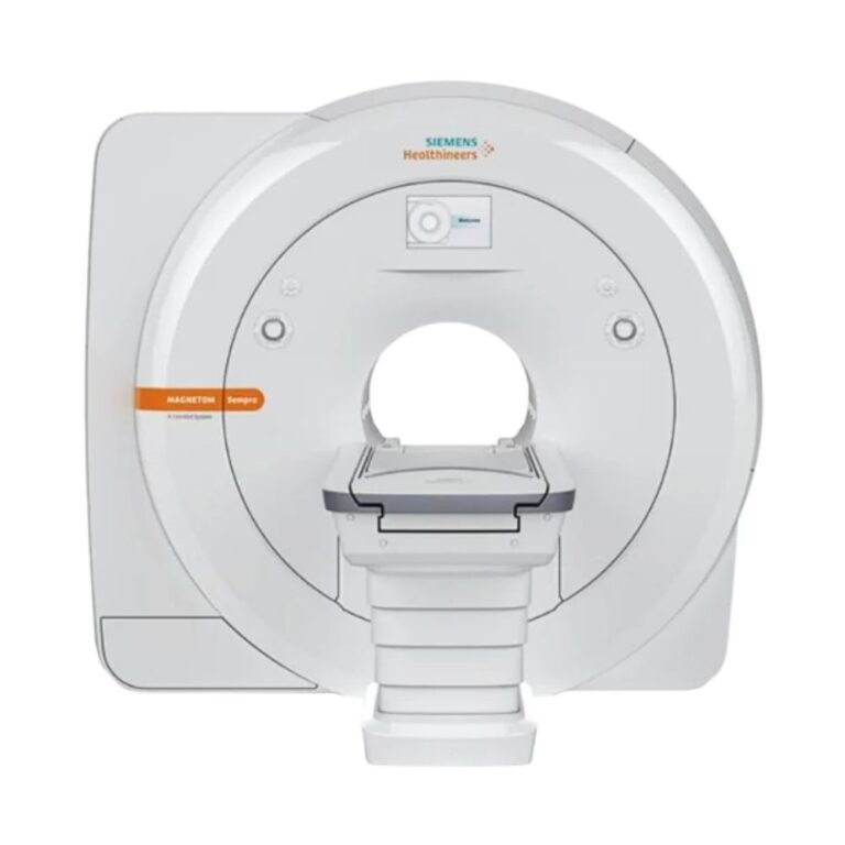

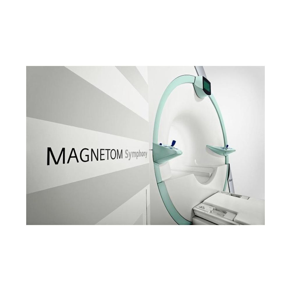
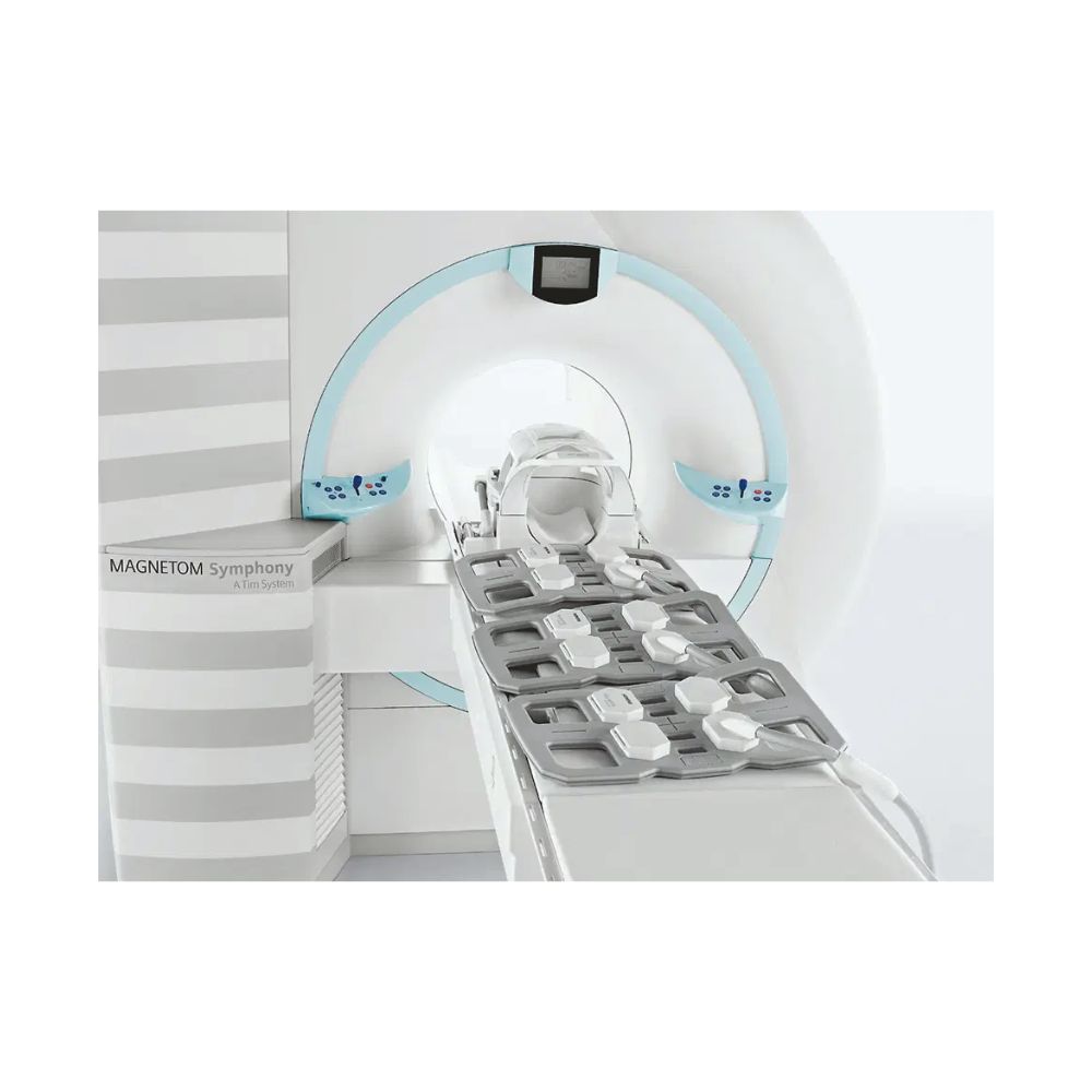
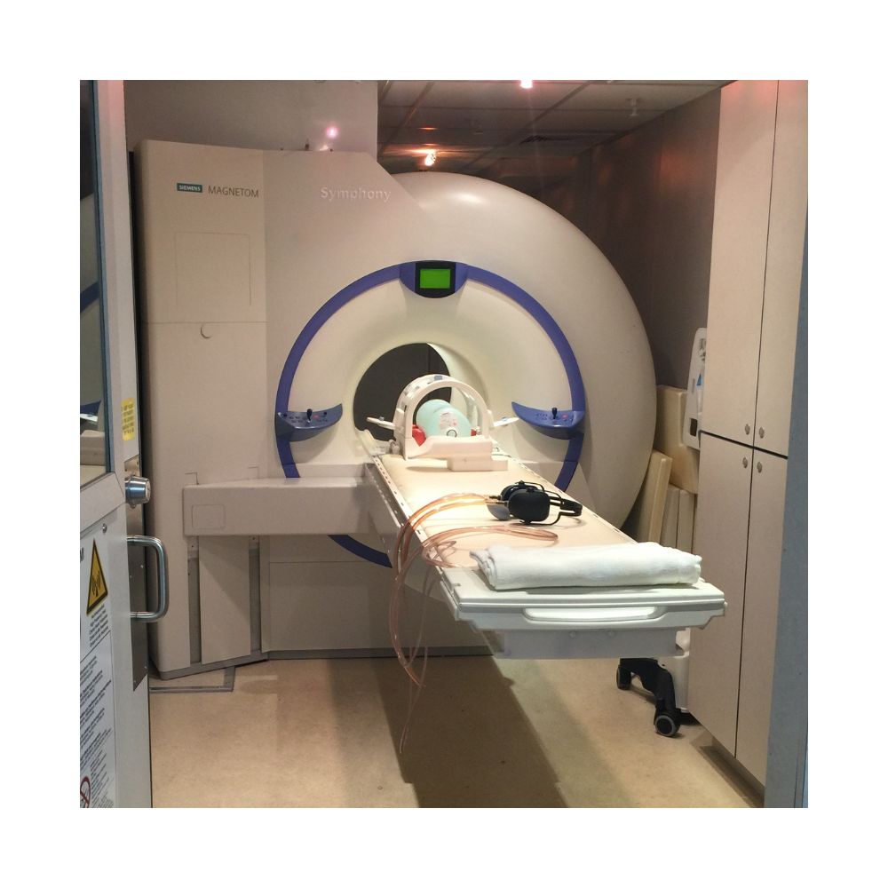
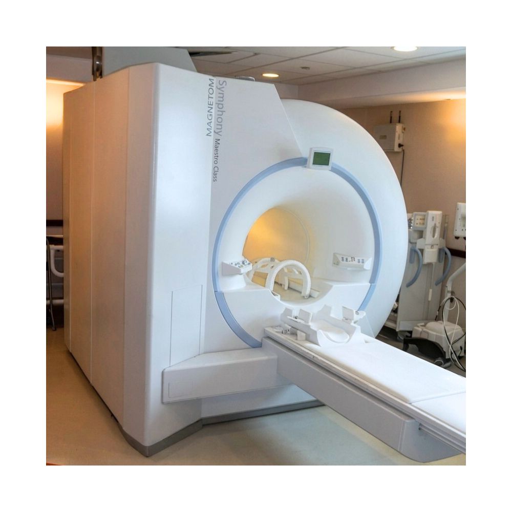
Siemens MAGNETOM Symphony 1.5T MRI
- Category:
- MRI Systems
- Brand:
- Siemens
The Siemens MAGNETOM Symphony is a 1.5T MRI scanner for hospitals, imaging centers, and specialty practices seeking flexible pre-owned equipment. Its conventional 60 cm bore supports routine and specialized exams, while Maestro Class enhancements and Siemens’ historical Tim upgrade path expanded workflow, coil coverage, and scanning capabilities.
MAGNETOM Symphony MRI Highlights & Benefits
The MAGNETOM Symphony combines 1.5T imaging with a compact magnet and configurable hardware. Original systems support routine and specialty examinations, while Maestro Class introduced enhanced imaging workflows and compatible accelerated scanning. Siemens later offered a Tim upgrade that added receiver channels, Matrix coils, and extended imaging capabilities to existing Symphony systems.
Key Features & Benefits
-
- 1.5T High-Field Imaging: Supports detailed brain, spine, orthopedic, abdominal, and specialty examinations with appropriate coils and software.
- 60 cm Bore: Provides a conventional 60 cm (23.6 in) opening for whole-body MRI.
- Compact Installation: Representative installations require approximately 30 m² (325 sq ft) of total space, which can help limit facility modifications in suitable locations.
- Original, Maestro Class & Tim Configurations: Original systems offer conventional receiving hardware, Maestro Class adds workflow and imaging enhancements, and Tim-equipped versions support 18 or 32 independent receiver channels.
- Historical Siemens Tim Upgrade: Siemens offered a hardware modernization pathway for existing Symphony scanners, adding advanced receiving hardware, Matrix coils, faster reconstruction, and expanded imaging capabilities.
- Up to 200 cm Imaging Coverage: Tim-equipped configurations support extended spine, vascular, and body examinations with fewer coil changes.
- iPAT Parallel Imaging: Compatible hardware and software shorten supported scans by reconstructing images from fewer measurements, including supported Maestro Class and Tim configurations.
- syngo MRI Workflow: Available exam-planning, protocol-management, and reconstruction tools reduce repetitive scanning tasks.
- Specialized Clinical Imaging: Compatible applications support neurological, orthopedic, cardiac, vascular, breast, and body examinations.
- Pre-Owned Equipment Options: Receiver, gradient, software, and coil configurations can be matched to facility requirements.
Symphony 1.5T Imaging Performance
The Symphony uses a superconducting 1.5T magnet and configurable gradient hardware to support routine MRI and advanced clinical examinations. Its imaging capabilities vary with the installed gradient system, receiving hardware, coils, and software.
1.5T Imaging for Routine and Specialty Exams
The Symphony’s 1.5 Tesla magnetic field provides the signal strength needed for detailed imaging of the brain, spine, joints, abdomen, pelvis, and other anatomical regions. With compatible coils and software, the scanner also supports specialized neurological, cardiac, breast, and vascular examinations.
Its configurable platform accommodates routine clinical workloads and selected advanced or research-oriented imaging applications when appropriately equipped. This flexibility allows facilities to select a Symphony configuration suited to their examination requirements.
Turbo Through Quantum Gradients: Imaging Speed Differences
The Symphony was produced with several gradient configurations, including Turbo, Ultra, Sprint, and Quantum hardware. MRI gradients create additional magnetic fields that identify where imaging signals originate within the patient’s anatomy, while their strength and switching speed influence which acquisition techniques a scanner can support.
Documented options include:
-
- Turbo, Ultra & Sprint Gradients: Earlier configurations with different gradient strengths and switching speeds. These determine which acquisition settings and imaging sequences a system can support.
- Quantum Gradients: Provide 30 mT/m maximum gradient amplitude and 125 T/m/s slew rate, supporting routine examinations and compatible accelerated imaging.
Tim-Equipped Symphony Gradients: Support approximately 30 mT/m gradient amplitude and 125 T/m/s slew rate. Siemens’ historical Tim upgrade included Quantum gradient hardware when required by the original configuration.
Gradient amplitude describes the strength of the additional magnetic fields used to locate anatomical information, while slew rate describes how quickly those fields change. Faster switching can benefit compatible imaging sequences, although performance also depends on receiving hardware, coils, and software.
MXR Imaging helps customers identify the installed gradient configuration and determine whether its capabilities match their planned examinations.
Symphony Patient Access with a 60 cm Bore
The Symphony uses a conventional cylindrical magnet with dedicated imaging coils and compatible patient-positioning equipment. Available tabletop and transfer arrangements vary by system, making the installed configuration an important consideration for facilities with specific patient-handling requirements.
60 cm Bore for Conventional Whole-Body MRI
The Symphony features a 60 cm (23.6 in) bore and an approximately 160 cm (63 in) magnet length. Its whole-body design supports neurological, musculoskeletal, abdominal, and other examinations with appropriate coils and positioning.
Compatible configurations allow head-first or feet-first scanning for selected examinations, depending on the anatomy and patient table. Facilities serving patients who need additional room around their bodies may benefit from considering a wide-bore MRI system.
Exchangeable Tabletop for Patient Transfers
Compatible Symphony Tim systems support an exchangeable tabletop designed to simplify patient preparation and transfers. This arrangement allows patients to be positioned on a compatible tabletop and transport trolley before entering the scanner, reducing the need to perform every transfer directly onto the scanning table.
Key capabilities include:
-
- Exchangeable Tabletop: Supports patient preparation and transfers using compatible transport equipment.
- Powered Positioning: Moves the patient into the magnet during examination setup.
- 200 kg Patient Capacity: Documented compatible exchangeable-tabletop configurations support up to 200 kg (441 lb).
The exchangeable tabletop is an available configuration rather than standard equipment on every Symphony. MXR Imaging confirms the installed table, patient capacity, and transfer accessories when helping customers select a system.
Symphony Maestro Class & Tim Upgrade Configurations
The MAGNETOM Symphony was originally available with conventional receiving hardware. Siemens subsequently introduced Maestro Class enhancements and offered Tim (Total imaging matrix) as a more extensive hardware upgrade for existing Symphony scanners.
These developments expanded workflow and imaging capabilities while retaining the underlying Symphony 1.5T platform. As a result, pre-owned systems can differ substantially in installed hardware, software, and upgrade history.
Original Symphony: 4- and 8-Channel Systems
Original Symphony systems use conventional receiving hardware and compatible Siemens Integrated Panoramic Array (IPA) coils. IPA allows compatible coil arrays to collect MRI signals across different anatomical regions, providing flexible coverage for routine examinations and supported specialty applications.
Available configurations include:
-
- Four-Channel Receiving: Processes independent signals from compatible coils for routine MRI and supported specialty applications.
- Eight-Channel Receiving: Provides additional signal-processing capacity for compatible coil arrangements and imaging techniques.
- Integrated Panoramic Array: Combines compatible coil coverage to support larger anatomical regions with fewer changes between selected coil arrangements.
Receiver capacity determines how many independent coil signals the scanner can process during an acquisition. Original Symphony configurations therefore differ from Tim-equipped versions in receiving hardware, coil compatibility, and supported advanced applications.
Maestro Class Workflow & Imaging Enhancements
Siemens introduced Maestro Class enhancements for the MAGNETOM Symphony to improve examination setup, image processing, and scanning efficiency. These improvements expanded the capabilities of the established 1.5T platform without requiring the extensive receiver and coil changes associated with the later Tim upgrade.
Compatible Maestro Class capabilities include:
-
- Updated syngo MR Workflows: Improved examination management and access to imaging protocols, helping technologists organize and perform routine studies.
- Phoenix Protocol Management: Allows saved examination protocols to be retrieved and reused, reducing repetitive setup and supporting consistent scanning procedures.
- Inline Image Processing: Automates selected reconstruction tasks, reducing manual image preparation after scanning.
- iPAT Parallel Imaging: Supports faster compatible acquisitions by combining multicoil receiving information with accelerated reconstruction techniques, depending on the installed hardware and software.
Maestro Class retained the Symphony’s underlying magnet platform and conventional coil architecture. It also provided a foundation from which eligible existing systems could receive further modernization through Siemens’ Tim upgrade program.
A Symphony labeled Maestro Class is not necessarily Tim-equipped. MXR Imaging helps customers identify the installed receiver channels, gradient hardware, coils, and software when evaluating pre-owned systems.
Siemens Tim Upgrade for Existing Symphony Systems
The Siemens Tim upgrade provided a further modernization pathway for facilities operating an original or Maestro Class MAGNETOM Symphony. Rather than replacing the complete MRI system, the upgrade introduced new imaging hardware, coils, reconstruction equipment, and compatible software while retaining the existing scanner platform.
Depending on the starting configuration, the historical upgrade included:
-
- 18- or 32-Channel RF System: Replaced the original receiving hardware, allowing more independent signals to be collected from compatible Matrix coils.
- Quantum Gradient Hardware: Added the 30 mT/m, 125 T/m/s gradient configuration when the necessary hardware was not already installed.
- Tim Matrix Coils: Introduced integrated head, neck, and spine coils, along with additional compatible body and specialty coil options.
- Updated Patient Table: Added patient-handling features and connections for compatible coil arrangements.
- New Reconstruction Computers: Improved image-processing capacity to support the upgraded receiving system and clinical applications.
- Patient Comfort Improvements: Included updated system covers and audio-comfort features in the documented upgrade package.
The upgrade expanded the Symphony’s ability to perform accelerated scans and extended examinations while preserving its original magnet platform. It was a genuine hardware modernization, not simply a software update or a separate MRI model.
Siemens offered this upgrade program historically, and current retrofit availability should not be assumed. For pre-owned equipment, MXR Imaging helps customers identify systems that have already received Tim modernization and evaluate their installed capabilities.
Tim-Equipped Symphony: 18- and 32-Channel Systems
A Symphony equipped with Tim technology supports higher-channel receiving configurations and compatible Matrix coils. These additions allow the scanner to collect more independent signal information during supported acquisitions and use coil combinations suited to extended anatomical coverage.
Two documented configurations include:
-
- Tim [76 × 18]: Connects up to 76 coil elements with 18 independent receiver channels, supporting compatible accelerated and extended examinations.
- Tim [76 × 32]: Connects up to 76 coil elements with 32 independent receiver channels, providing additional receiving capacity for compatible multichannel imaging.
The first number identifies the maximum connected coil elements, while the second identifies the independent receiver channels available during acquisition. Higher-channel configurations support more demanding coil combinations and compatible parallel imaging techniques.
The designation MAGNETOM Symphony, A Tim System identifies a Symphony equipped with Tim technology, including systems modernized through Siemens’ upgrade program. It does not require a separate product page.
Tim-Equipped Symphony for Extended MRI Exams
Tim-equipped Symphony systems combine Matrix coils with compatible accelerated imaging software to support examinations covering multiple anatomical regions. By allowing different coils to remain connected throughout appropriate studies, Tim technology reduces interruptions for patient repositioning and coil changes.
Matrix Coils Reduce Changes Between Body Regions
Tim Matrix technology combines signals from multiple imaging coils positioned around different anatomical regions. Compatible coils can remain connected while the scanner selects the receiving elements needed for each acquisition, allowing technologists to prepare several areas before scanning begins.
Practical benefits include:
-
- Fewer Coil Changes: Compatible coils remain connected during selected multiregion examinations.
- Less Patient Repositioning: Technologists can examine different anatomical regions without repeatedly moving patients to replace coils.
- Flexible Anatomical Coverage: Matrix coil arrangements support head, spine, body, and vascular imaging.
- Accelerated Scanning: Multiple receiving elements provide information used by compatible software to shorten acquisitions.
These advantages are especially useful during extended examinations where repeated patient movement can interrupt workflow. Available coil combinations depend on the installed Tim configuration and accessories.
Up to 200 cm Coverage for Spine & Vascular Imaging
Compatible Tim-equipped Symphony systems support whole-body imaging coverage of up to approximately 200 cm (78.7 in). The scanner acquires information across multiple anatomical regions while compatible Matrix coils remain connected, reducing preparation between imaging stations.
Applications include:
-
- Whole-Spine MRI: Extended cervical, thoracic, and lumbar examinations.
- Peripheral Angiography: Blood vessel imaging across larger anatomical regions.
- Multiregion Body MRI: Examination of several body areas with fewer coil changes.
- Oncology Imaging: Selected extended-coverage tumor assessments and follow-up studies.
This extended coverage helps facilities perform longer examinations more efficiently, particularly when several anatomical regions must be evaluated during the same appointment.
iPAT Parallel Imaging for Shorter Scans
Siemens iPAT (integrated Parallel Acquisition Technique) uses signals from multiple receiving coils to reconstruct images from fewer measurements. Instead of collecting every measurement directly, compatible reconstruction methods use the coil information to complete the image, reducing acquisition time during supported examinations.
Available techniques include:
-
- GRAPPA: Uses signals from multiple coil elements to reconstruct information that would otherwise require additional measurements, shortening compatible scans.
- mSENSE: Uses differences in individual coil sensitivity to reconstruct images from reduced acquisition data, supporting faster compatible examinations.
iPAT is not exclusive to Tim-equipped Symphony systems. Compatible Maestro Class configurations also supported parallel imaging, while Tim expanded the available receiver channels and coil combinations. Actual acceleration depends on the installed hardware, software, coil arrangement, and examination protocol.
Symphony syngo MR Workflow for Consistent Exams
The Symphony supports Siemens syngo MR software for patient registration, examination management, protocol selection, and image reconstruction. Maestro Class introduced compatible workflow and processing enhancements, while later system configurations supported additional clinical applications.
AutoAlign & Phoenix for Repeatable Scan Setup
Compatible Symphony software supports automated positioning, saved examination protocols, and remote technical assistance. These functions help technologists reproduce established scanning procedures while maintaining the flexibility needed for individual patients.
-
- AutoAlign: Uses anatomical reference information to position imaging slices during compatible examinations. This reduces manual planning and supports consistent coverage, particularly during follow-up brain studies.
- Phoenix: Stores and retrieves MRI protocols so technologists can reuse established acquisition settings instead of entering each parameter manually.
- syngo Expert-i: Provides compatible remote access to examination information and images through a hospital network, allowing experienced personnel to assist with complex studies.
Together, these tools support more consistent examinations across operators and reduce manual work during repeat studies. Their availability depends on the installed software and clinical options.
Inline Processing for Diffusion & Extended Images
Siemens’ Inline technologies automate selected reconstruction and processing steps as imaging information becomes available. This reduces the need to manually prepare every result after scanning and makes compatible examinations easier to review.
-
- syngo Inline Processing: Automatically performs selected reconstruction and processing tasks, reducing manual work after scanning.
- Inline Diffusion: Calculates diffusion-related images and apparent diffusion coefficient (ADC) maps. These maps measure water movement within tissue, supporting compatible neurological and body examinations.
- Image Composing: Combines images acquired at different anatomical positions into an extended view, helping radiologists evaluate regions such as the whole spine or peripheral blood vessels.
These functions are particularly useful when examinations produce multiple image sets or require specialized reconstruction. Available capabilities depend on the system’s software and processing hardware.
DICOM Transfer to Hospital Imaging Systems
The Symphony supports DICOM image transfer for storing and distributing examinations through compatible picture archiving and communication systems (PACS). DICOM is the standard format used by medical imaging systems to exchange images and examination information, allowing completed studies to move from the scanner to hospital systems for storage, interpretation, and clinical review.
Available connectivity and processing functions depend on the installed workstation and software.
Symphony Imaging for Motion, 3D Detail and Contrast
Compatible Symphony software helps reduce motion artifacts, provide three-dimensional anatomical views, and capture contrast enhancement during selected examinations. These technologies support different clinical needs, from improving images affected by patient movement to evaluating anatomy and blood vessels from multiple perspectives.
BLADE Reduces Artifacts from Patient Movement
syngo BLADE collects overlapping imaging information in rotating segments, allowing certain inconsistencies caused by patient movement to be corrected during reconstruction. This reduces blurring and repeated image patterns that can obscure anatomy during compatible brain, spine, joint, and body examinations.
BLADE is particularly useful when patients have difficulty remaining still, helping improve image clarity and potentially reducing the need to repeat motion-affected acquisitions.
SPACE Provides Detailed 3D Anatomical Views
SPACE collects three-dimensional MRI information that can be reconstructed into different viewing planes. Rather than acquiring separate scans for every orientation, radiologists can review the same dataset from multiple directions, supporting a more detailed evaluation of complex anatomy.
This capability is useful for compatible neurological, musculoskeletal, and selected body examinations where the relationship between structures is easier to assess across multiple views.
VIBE and TWIST for Contrast-Enhanced Imaging
VIBE and TWIST support different aspects of contrast-enhanced MRI. VIBE provides detailed three-dimensional imaging during selected body examinations, while TWIST captures repeated images as contrast moves through the blood vessels.
-
- VIBE: Acquires three-dimensional images during compatible abdominal and breast examinations. It supports detailed anatomical assessment and selected phases of contrast enhancement.
- syngo TWIST: Repeatedly acquires accelerated three-dimensional images as contrast travels through blood vessels. This supports time-resolved angiography, helping clinicians evaluate vascular filling patterns.
These applications require compatible software, coils, and examination protocols. TWIST is a documented option for appropriately equipped Symphony Tim systems.
Symphony Specialized MRI for Brain, Heart and Body
Appropriately equipped Symphony systems support neurological, cardiovascular, and body imaging applications that provide information beyond conventional anatomical MRI. These techniques extend the scanner’s clinical capabilities by evaluating tissue characteristics, brain activity, cardiac function, and other specialized findings.
Brain Pathways, Small Hemorrhages and Functional MRI
Compatible neurological applications provide information about nerve pathways, blood-related changes, and brain activity. Each technique measures a different tissue characteristic, allowing specialized examinations to address clinical questions that may not be answered by conventional anatomical images alone.
-
- Diffusion Tensor Imaging (DTI): Measures the direction of water movement within tissue to provide information about nerve pathways, supporting specialized neurological and selected presurgical examinations.
- Susceptibility-Weighted Imaging (SWI): Uses magnetic-susceptibility differences to improve visualization of small hemorrhages, venous structures, and other tissue characteristics.
- Functional MRI: Detects changes associated with blood oxygenation to help identify activity in selected brain regions during specialized neurological examinations.
These applications require compatible acquisition sequences and processing software, with availability determined by the installed configuration.
BEAT Cardiac Imaging for Heart Motion and Blood Flow
Siemens BEAT supports cardiac examinations synchronized with the patient’s heart cycle, helping the scanner collect imaging information at appropriate points during heart movement. Compatible cardiac software also supports dynamic views of chamber function and selected blood-flow measurements.
Available techniques include:
-
- BEAT Cardiac Imaging: Coordinates acquisitions with cardiac motion, helping collect images at appropriate points in the heart cycle.
- Cine MRI: Produces image sequences showing heart movement for evaluating chamber function and wall motion.
- Cardiac Flow Imaging: Measures selected blood-flow characteristics during compatible cardiovascular examinations.
Together, these capabilities support more detailed evaluation of cardiac anatomy and function. Cardiac MRI requires appropriate software, coils, physiological monitoring equipment, and clinical protocols.
Body Diffusion for Tumor Studies and MR Spectroscopy
Compatible Symphony software also supports specialized techniques that assess tissue characteristics beyond conventional image appearance. Diffusion imaging evaluates water movement within tissue, while spectroscopy measures selected chemical signals that provide additional information about tissue composition.
-
- syngo REVEAL: Uses diffusion-weighted MRI to measure differences in water movement within body tissues, providing additional information during selected tumor assessments and follow-up examinations.
- MR Spectroscopy: Measures chemical signals within selected tissue regions to provide information about tissue composition beyond conventional anatomical images.
Both techniques require compatible software and imaging equipment. Their clinical applications depend on the anatomy, examination protocol, and installed system capabilities.
Clinical Environments & Applications
The Siemens MAGNETOM Symphony supports routine and specialized 1.5T MRI in hospitals, diagnostic imaging centers, and specialty facilities. Its conventional 60 cm bore and configurable imaging hardware accommodate a range of examinations, while Tim-equipped systems provide additional receiver channels, extended anatomical coverage, and compatible accelerated imaging.
Clinical Environments
-
- Hospitals & Radiology Departments
- Outpatient Diagnostic Imaging Centers
- Orthopedic & Sports Medicine Imaging Centers
- Neurology & Neuroscience Imaging Departments
- Cardiac & Vascular Imaging Departments
- Breast Imaging Centers
- Oncology Imaging Departments
- Pediatric Hospitals & Imaging Departments
- Academic Medical Centers & Research Facilities
- Veterinary Teaching Hospitals & Specialty Referral Centers
Clinical Specialties & Applications
-
- Neurology & Neurosurgery: Brain MRI, diffusion studies, nerve pathway evaluation, and compatible functional imaging examinations.
- Orthopedics & Sports Medicine: Shoulder, knee, hip, wrist, and other joint examinations, including ligament, tendon, cartilage, and soft-tissue assessment.
- Spine Care & Chiropractic Referrals: Cervical, thoracic, lumbar, and compatible whole-spine MRI, including evaluation of disc herniations, spinal stenosis, radiculopathy, and suspected nerve-root compression.
- Podiatry & Foot/Ankle Care: Foot and ankle imaging for tendon, ligament, joint, cartilage, and selected bone or soft-tissue abnormalities using compatible extremity coils.
- Pediatric Radiology: Brain, spine, and musculoskeletal imaging, including selected neurological and developmental abnormalities, using age-appropriate protocols and patient-monitoring equipment.
- Abdominal Imaging: Liver, kidney, pancreatic, abdominal, and pelvic examinations using appropriate coils and software.
- Urology: Prostate and pelvic MRI using compatible coils, including selected diffusion and contrast-enhanced examinations.
- Cardiology: Cardiac anatomy, ventricular function, wall motion, and selected blood-flow studies using compatible cardiac applications.
- Vascular Medicine: MR angiography and extended peripheral vascular imaging using compatible coils and acquisition techniques.
- Breast Radiology: Diagnostic breast MRI, including contrast-enhanced examinations using appropriate coils and clinical software.
- Oncology: Tumor assessment, treatment follow-up, and selected extended-coverage examinations using compatible anatomical and diffusion imaging.
- Equine Veterinary Medicine: Imaging of the feet, distal limbs, joints, tendons, ligaments, and related soft tissues in appropriately equipped equine MRI facilities.
- Small-Animal Veterinary Medicine: Selected neurological, spinal, orthopedic, and soft-tissue examinations in dogs and cats using suitable coils, anesthesia, and veterinary imaging protocols.
Clinical applications vary between original Symphony and Tim-equipped configurations. Specialized examinations require compatible hardware, software, imaging coils, and appropriate clinical procedures.
Symphony Configurations, Coils & Available Options
Individual Symphony systems can differ in receiver hardware, gradient performance, patient tables, imaging coils, software, and accessories. These variations affect the examinations each system can perform and help determine which pre-owned configuration best matches a facility’s clinical needs.
Hardware and Patient Table Differences Between Systems
Symphony configurations vary in the hardware used for image acquisition, reconstruction, and patient positioning. Some systems retain their original equipment, others include Maestro Class enhancements, and still others were modernized through Siemens’ Tim upgrade program.
Important variations include:
-
- Receiver Hardware: Original four- or eight-channel equipment or Tim-equipped 18- and 32-channel systems.
- Maestro Class Enhancements: Installed workflow, image-processing, and compatible accelerated imaging capabilities.
- Gradient Configuration: Turbo, Ultra, Sprint, Quantum, or compatible upgraded hardware.
- Tim Upgrade Components: Installed RF hardware, Matrix coil connections, reconstruction computers, and associated equipment.
- Patient Table: Installed tabletop, powered positioning, exchangeable-tabletop equipment, and rated capacity.
- Image Reconstruction Hardware: Processing equipment compatible with the software generation and clinical applications.
MXR Imaging helps customers compare these configurations according to their examination requirements, including the hardware and accessories installed on a particular system.
Original IPA Coils vs Tim Matrix Coils
Original and Maestro Class Symphony systems use compatible conventional and Integrated Panoramic Array coils, while Tim-equipped configurations support Siemens Matrix coils. Both technologies collect signals close to the anatomy being examined, but Tim provides additional receiving elements and compatible coil combinations for extended and accelerated imaging.
Available options include:
-
- Head & Neck Coils: Support neurological, head, neck, and cervical spine examinations.
- Spine Coils: Provide regional and extended spinal coverage.
- Body Coils: Support abdominal, pelvic, and selected multiregion examinations.
- Orthopedic Coils: Support dedicated knee, shoulder, wrist, ankle, and other joint imaging.
- Breast Coils: Provide coverage for diagnostic breast MRI.
- Peripheral Angiography Coils: Support selected extended vascular studies.
- Tim Matrix Coils: Provide additional receiving elements for compatible extended and accelerated imaging.
Original IPA coils and Tim Matrix coils require the appropriate receiving hardware and are not universally interchangeable. Some legacy coils may remain compatible with upgraded equipment, depending on the exact coil and system configuration.
MXR Imaging helps customers identify compatible coil packages and provides MRI coil repair services for supported equipment.
Installed Software and Specialty Imaging Options
Available software depends on the Symphony’s hardware generation, Maestro Class enhancements, syngo MR release, and installed clinical licenses. These options determine which specialized examinations and automated workflows a particular system can support.
Compatible applications include:
-
- Examination Planning: AutoAlign, Phoenix, and compatible remote assistance.
- Accelerated Imaging: iPAT, GRAPPA, and mSENSE.
- Image Processing: syngo Inline, diffusion reconstruction, and image composing.
- Motion Reduction: syngo BLADE.
- 3D Imaging: SPACE and VIBE.
- Neurological Imaging: DTI, SWI, functional MRI, and spectroscopy.
- Cardiac Imaging: BEAT, cine MRI, and compatible blood-flow applications.
- Vascular Imaging: MR angiography and TWIST.
- Body Imaging: syngo REVEAL and supported diffusion techniques.
These are compatible options rather than a fixed software package included with every Symphony. MXR Imaging helps customers identify systems equipped with the appropriate applications for their intended examinations.
Symphony Magnet, Helium & Site Requirements
The Symphony’s conventional 1.5T magnet and configurable supporting equipment accommodate different MRI installation requirements. Final specifications depend on the installed hardware, facility layout, and approved site plan.
Liquid Helium Replenishment and Refrigeration Maintenance
The Symphony uses liquid helium to keep its superconducting magnet at the required operating temperature. Its refrigeration system helps manage helium consumption, but the Symphony should not be assumed to have zero helium boil-off.
Documented Tim-equipped configurations experience gradual helium loss during normal operation and may require periodic replenishment. Magnet operation also requires refrigeration maintenance, helium monitoring, and emergency venting.
MXR Imaging provides MRI helium and refrigeration services for supported scanners.
Representative 325 sq ft MRI Installation Footprint
The Symphony supports representative installations requiring approximately 30 m² (325 sq ft) of total space under specified planning conditions. This includes the magnet, control, and supporting equipment areas, rather than the examination-room area alone.
Installation planning must address:
-
- Equipment Delivery: Access for transporting the magnet, patient table, and supporting equipment.
- Structural Support: Floor capacity for the magnet assembly and concentrated loads.
- RF Shielding: Protection against external radiofrequency interference that can reduce MRI image quality.
- Magnetic Field Boundaries: Appropriate safety zones and access restrictions.
- Quench Venting: Emergency infrastructure for directing released helium outside the building.
- Service Clearances: Access for maintenance, inspections, and repairs.
MXR Imaging provides custom MRI shielding solutions for new and replacement installations. For projects requiring equipment coordination, construction planning, and installation support, MXR Imaging also offers turnkey imaging project management.
Electrical Service & Equipment Cooling
The Symphony requires dedicated electrical service and cooling appropriate to its gradient, refrigeration, and control equipment. Electrical loads and cooling requirements depend on the installed configuration and facility design, so final utility specifications must be established for the selected system.
MXR Imaging helps coordinate site planning and installation requirements as part of the equipment selection process.
Pre-Owned Symphony Systems: Hardware, Software, & Condition
MXR Imaging helps healthcare facilities select pre-owned Siemens MAGNETOM Symphony systems based on clinical requirements, equipment condition, and installation needs. Because original, Maestro Class, and Tim-equipped systems can differ significantly, evaluating installed hardware and upgrade history is an important part of selecting the appropriate scanner.
Available units can differ in:
- Original, Maestro Class or Tim Configuration: Conventional receiving equipment, Maestro Class enhancements, or a Symphony with an installed Tim upgrade.
- Receiver Channels: Four- or eight-channel original configurations, or 18- and 32-channel Tim systems.
- Gradient Hardware: Installed gradient generation and performance.
- Upgrade History: Previously installed workflow, receiving, coil, reconstruction, and patient-table upgrades.
- Software & Licenses: syngo MR generation and available clinical applications.
- Imaging Coils: Included neurological, orthopedic, body, breast, and vascular coils.
- Patient Table: Installed tabletop, positioning equipment, and rated capacity.
- Magnet & Refrigeration: Helium condition, cooling performance, and maintenance history.
- Equipment History: Previous repairs, inspections, and replacement components.
MXR Imaging works with customers to match each system’s capabilities to their planned examinations, evaluate installed options, and coordinate installation requirements. This helps facilities distinguish original Symphony hardware, Maestro Class enhancements, and scanners that have received Tim modernization.
MAGNETOM Symphony MRI Price & Availability
MXR Imaging helps facilities purchase pre-owned and reconditioned Siemens MAGNETOM Symphony 1.5T MRI systems suited to their clinical and installation requirements. Pricing depends on receiver channels, gradient hardware, Maestro Class enhancements, installed Tim upgrades, software, coils, equipment condition, service history, and installation needs.
Qualifying equipment may be available through the MXR Imaging Platinum Certified program, subject to availability.
Platinum Certified Refurbishment and Warranty
Qualifying Platinum Certified systems undergo a 100-point refurbishment process under MXR Imaging’s ISO 13485:2016-certified quality management system. The program includes a full one-year warranty, along with applicable site planning, installation, and clinical applications training.
For complete program details, visit Platinum Certified Imaging Equipment. MXR Imaging also provides MRI maintenance and repair and nationwide technical support for qualifying equipment.
Family-owned since 1964, MXR Imaging supports healthcare facilities with imaging equipment, replacement parts, and service throughout the equipment lifecycle.
Contact MXR Imaging to discuss available Symphony configurations and request a quote.
Siemens MAGNETOM Symphony 1.5T Specifications
Magnet System
-
- Field Strength: 1.5T
- Magnet Type: Superconducting, actively shielded
- Magnet Design: Siemens OR70
- Bore Diameter: 60 cm (23.6 in)
- Representative Magnet Length: Approximately 160 cm (63 in)
- Cryogenic Cooling: Liquid helium with mechanical refrigeration
- Helium Consumption: Configuration-dependent; periodic replenishment may be required
Gradient System
-
- Original Gradient Options: Turbo, Ultra, Sprint, and Quantum
- Gradient Amplitude: Configuration-dependent, with documented systems ranging approximately 20–30 mT/m
- Slew Rate: Configuration-dependent, with documented systems ranging approximately 25–125 T/m/s
- Quantum Gradient Amplitude: 30 mT/m per axis
- Quantum Slew Rate: 125 T/m/s per axis
- Symphony Tim Gradient Performance: Approximately 30 mT/m and 125 T/m/s
- Historical Tim Upgrade: Included Quantum gradient hardware when required by the original configuration
- Installed Gradient Hardware: Determined by the selected system
RF System
-
- Original RF Technology: Siemens conventional receiving hardware with compatible IPA coils
- Original Receiver Channels: Four or eight, depending on configuration
- Maestro Class: Workflow and imaging enhancements; actual RF hardware depends on the installed configuration
- Tim Technology: Siemens Total imaging matrix, available through a historical Symphony hardware upgrade
- Tim Configurations: Tim [76 × 18] and Tim [76 × 32]
- Maximum Connected Tim Coil Elements: Up to 76
- Independent Tim Receiver Channels: 18 or 32
- Parallel Imaging: Compatible iPAT, GRAPPA, and mSENSE
- Extended Tim Imaging Coverage: Up to approximately 200 cm (78.7 in)
Bore & Patient Table
-
- Bore Diameter: 60 cm (23.6 in)
- Patient Table: Configuration-dependent powered positioning
- Exchangeable Tabletop: Available with compatible Symphony Tim equipment
- Documented Exchangeable Tabletop Capacity: Up to 200 kg (441 lb)
- Patient Positioning: Head-first and feet-first for supported examinations
- Final Patient Capacity: Determined by the installed table
Coils & Accessories
-
- Original Coil Technology: Siemens Integrated Panoramic Array and compatible conventional coils
- Maestro Class Coil Architecture: Conventional Symphony coils, depending on installed hardware
- Tim Coil Technology: Siemens Matrix coils, introduced through the Tim hardware upgrade
- Head & Neck Coils: Compatible neurological coils
- Spine Coils: Compatible spine arrays
- Body Coils: Compatible body arrays
- Orthopedic Coils: Compatible shoulder, knee, wrist, and extremity coils
- Breast Coils: Compatible diagnostic breast coils
- Vascular Coils: Compatible peripheral angiography coils
- Included Equipment: Depends on the selected system package
Workflow & Imaging Software
-
- Software Platform: Siemens syngo MR
- Software Generation: Configuration-dependent, including original, Maestro Class, and Tim-compatible releases
- Exam Planning: Compatible AutoAlign
- Protocol Management: Phoenix
- Remote Assistance: Compatible syngo Expert-i
- Image Processing: Compatible syngo Inline, diffusion reconstruction, and image composing
- Accelerated Imaging: Compatible iPAT, GRAPPA, and mSENSE
- Motion Reduction: Compatible syngo BLADE
- 3D Imaging: Compatible SPACE and VIBE
- Advanced Applications: Configuration-dependent DTI, SWI, functional MRI, REVEAL, BEAT, TWIST, and spectroscopy
Siting & Utilities
-
- Representative Minimum Total Installation Area: Approximately 30 m² (325 sq ft)
- RF Shielding: Required
- Magnetic-Field Safety Planning: Required
- Emergency Helium Venting: Required
- Electrical Service: Configuration-specific dedicated service
- Equipment Cooling: Configuration-specific refrigeration and cooling infrastructure
- Final Site Requirements: Determined by the selected equipment and approved installation plan
System Dimensions & Weight
-
- Bore Diameter: 60 cm (23.6 in)
- Representative Magnet Length: Approximately 160 cm (63 in)
- Nominal Magnet-Only Weight: Approximately 4,050 kg (8,929 lb)
- Representative OR70 Magnet Transport Assembly: Approximately 5,500 kg (12,125 lb), including cryogens and gradient coil
- Patient Table Capacity: Configuration-dependent; 200 kg (441 lb) documented for the compatible exchangeable tabletop
- Installation Dimensions & Structural Loads: Determined by the installed assembly, equipment arrangement, shielding, and approved site plan