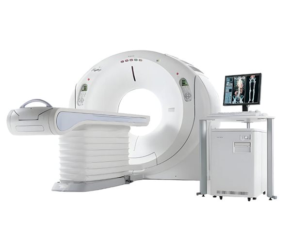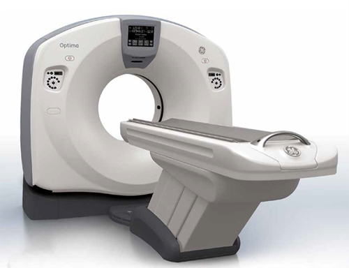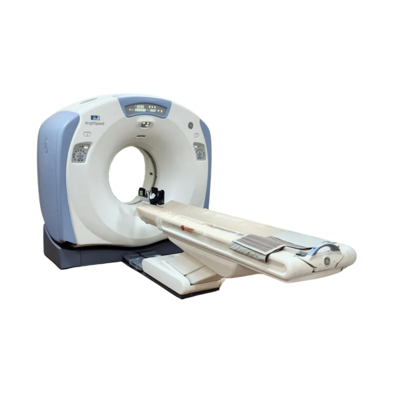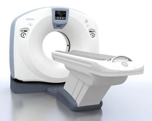
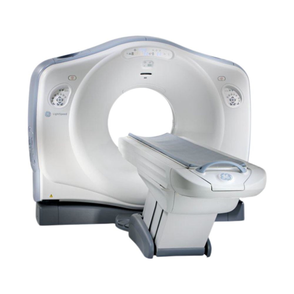
GE LightSpeed 16-Slice CT
- Category:
- CT Scanners
- Brand:
- GE
The GE LightSpeed 16 CT Scanner is a proven 16-slice system for routine head-to-body imaging in hospitals, outpatient imaging centers, emergency departments, and specialty practices. Submillimeter acquisition, 0.5-second rotation, SmartmA dose modulation, and a compact system design support efficient diagnostic CT across a broad range of applications.
GE LightSpeed 16 Highlights & Benefits
The GE LightSpeed 16 combines thin-slice acquisition, practical workflow tools, and a compact 70 cm gantry design for routine diagnostic and whole-body CT. As a pre-owned CT scanner platform, available hardware, software, accessories, and advanced applications can vary by system.
Key LightSpeed 16 Capabilities
-
- 16 × 0.625 mm submillimeter acquisition
- 0.5-second fastest rotation
- HiLight Matrix II detector
- Up to 35 mm per rotation helical coverage
- SmartmA automatic tube-current modulation
- 70 cm (27.6 in) gantry aperture
- Optional cardiac, vascular, perfusion, lung, colonography, and post-processing applications
This GE LightSpeed CT scanner can be well suited to hospitals, diagnostic imaging departments, and outpatient imaging centers seeking a 16-slice system for established head-to-body imaging workflows. Its relatively compact installation requirements can also benefit facilities working within an existing CT suite.
CT Acquisition & Image Detail
Detector & Thin-Slice Imaging
The GE LightSpeed 16 uses the HiLight Matrix II detector with 21,888 detector elements. Its 16 × 0.625 mm acquisition mode provides thin volumetric data for detailed anatomy, vascular imaging, bone imaging, and multiplanar reconstruction.
Additional detector configurations allow protocols to balance anatomical coverage and reconstructed image thickness according to the examination.
0.5-Second Head-to-Body Scanning
The LightSpeed 16 supports full 360° helical rotation times as fast as 0.5 seconds.
Helical coverage reaches up to 35 mm per rotation depending on detector configuration and pitch, supporting efficient head-to-body and whole-body CT while retaining thin-slice acquisition when greater anatomical detail is required.
Flexible Thin-Slice Reconstruction
The scanner reconstructs images at speeds up to six images per second.
Prospective Multiple Reconstruction can generate multiple reconstruction sets from one acquisition, while GE VariViewer allows thin acquired data to be displayed at different effective slice thicknesses.
This supports detailed thin-section review while also allowing thicker image sets to be created for routine interpretation.
Image Quality & Dose Management
SmartmA Dose Management
SmartmA automatically adjusts tube current according to the selected image-quality target and scan parameters.
Operators set a Noise Index and maximum mA, allowing the scanner to adapt tube current throughout the examination rather than applying the same technique across all anatomy.
The system also provides dose-length product information before acquisition for review during scan preparation.
Artifact Reduction for Challenging Exams
HyperPlane, CrossBeam, and Advanced Artifact Reduction technologies help manage helical, cone-beam, and streak artifacts.
These tools can be particularly useful when imaging dense anatomy or patients with orthopedic hardware.
Patient Access, Table & Workflow
Gantry Access & Patient Positioning
The GE LightSpeed 16 has a 70 cm (27.6 in) gantry aperture and a 50 cm (19.7 in) maximum scan field of view.
Gantry tilt extends to ±30°, providing additional flexibility for head positioning and scan-plane selection. Gantry-mounted controls and alignment lights support patient setup from inside the scan room.
170 cm Table Travel & 450 lb Capacity
The cantilever patient table provides 170 cm (66.9 in) of horizontal travel for head-to-body imaging and supports patient loads up to 450 lb (205 kg).
An adjustable table height, bilateral foot controls, and an integrated IV pole support positioning and contrast-enhanced CT workflow.
Programmable Protocols & Contrast Timing
The LightSpeed 16 uses GE Xtream workflow and Exam Rx protocol tools to streamline examination setup.
Programmable adult and pediatric protocols can help standardize common studies while remaining adjustable for patient-specific needs. SmartPrep supports real-time monitoring of contrast enhancement to help time contrast-enhanced acquisitions.
In-room controls further support efficient positioning and scan preparation.
Clinical Specialties & Applications
The GE LightSpeed 16 CT Scanner supports diagnostic imaging across a range of clinical specialties, with advanced applications depending on the installed system configuration and software.
- Radiology & Diagnostic Imaging
- Neurology
- Orthopedics & Musculoskeletal Imaging
- Cardiology & Vascular Imaging
- Oncology
- Emergency & Trauma Care
- Pediatrics
- Veterinary Medicine
Advanced CT Applications & Software
Advanced applications vary between pre-owned LightSpeed 16 systems according to installed software, hardware, and workstation configuration.
Cardiac, Vascular & Perfusion Imaging
Available configurations may support ECG-gated cardiac acquisition, coronary calcium scoring with SmartScore Pro, SnapShot cardiac imaging, CT perfusion, and Advanced Vessel Analysis.
These capabilities depend on the hardware and software installed on the individual scanner.
Lung, Colonography & Advanced Review
Optional applications included Advanced Lung Analysis, CT Colonography, Direct3D, and additional advanced visualization tools for reviewing volumetric CT data.
Direct3D supports three-dimensional image visualization from reconstructed CT data when included in the installed configuration.
CT Simulation for Radiation Therapy Planning
Advantage Sim was available for CT simulation and radiation therapy planning workflows when included in the installed software configuration.
PACS, DICOM & Image Transfer
The GE LightSpeed 16 supports DICOM 3.0 connectivity for integration with compatible PACS and medical imaging networks.
Available workflow functions can include image transfer, Query/Retrieve, Storage Commitment, DICOM Print, Modality Worklist, and Modality Performed Procedure Step, depending on system configuration.
Network and storage configurations may vary by installed system and workstation setup.
GE LightSpeed 16 Configurations & Options
Pre-owned GE LightSpeed 16 CT scanners can include different combinations of clinical software, workstations, accessories, and advanced applications.
Clinical Software & Imaging Options
Available configurations may support:
-
- Cardiac imaging and calcium scoring
- SnapShot cardiac imaging
- CT perfusion
- Advanced Vessel Analysis
- CT Colonography
- Advanced Lung Analysis
- Bone Mineral Densitometry (BMD)
- Direct3D visualization
- Advanced visualization and post-processing
- Radiation therapy planning
- Additional workflow and connectivity options
Hardware, Workstations & Accessories
Configurations may also vary by operator console, workstation setup, patient table accessories, X-ray tube history, and positioning accessories.
The available configuration can be matched to the facility’s clinical workload and workflow needs.
Installation & Site Planning
The GE LightSpeed 16 was designed with a relatively compact installation footprint for a multislice CT scanner.
Original GE planning specifications identify a minimum suite of approximately 302 sq ft (28 m²). Actual room size, shielding, electrical service, HVAC, service clearances, and installation requirements should be confirmed for the specific scanner and facility.
GE LightSpeed 16 Price & Availability
MXR offers pre-owned GE LightSpeed 16 CT Scanner configurations for a range of diagnostic imaging needs.
Pricing and availability depend on factors such as system configuration, installed software, X-ray tube history, accessories, installation requirements, and service needs.
GE LightSpeed 16 CT Scanner Specifications
The GE LightSpeed 16 specifications below cover CT acquisition, detector configuration, gantry and patient table dimensions, X-ray generator and tube performance, image reconstruction, connectivity, and site requirements.
Acquisition & Detector Specifications
-
- Slice count: 16 slices
- Detector: HiLight Matrix II
- Detector elements: 21,888
- Detector configuration: 16 rows × 0.625 mm plus 8 rows × 1.25 mm
- Acquisition modes: 16 × 0.625 mm, 16 × 1.25 mm, 8 × 1.25 mm, 8 × 2.5 mm
- Scan modes: Helical, axial, and Scout
- Fastest helical rotation: 0.5 seconds
- Helical rotation settings: 0.5, 0.6, 0.7, 0.8, 0.9, and 1.0 seconds
- Maximum helical coverage: Up to 35 mm per rotation, depending on protocol
- Maximum scan field of view: 50 cm (19.7 in)
Gantry & Patient Table Specifications
-
- Gantry aperture: 70 cm (27.6 in)
- Gantry tilt: ±30°
- Table vertical range: 51.6 to 99.1 cm (20.3 to 39 in)
- Horizontal table range: 170 cm (66.9 in)
- Table capacity at ±0.25 mm positioning accuracy: 180 kg (400 lb)
- Maximum table load during normal operation: 205 kg (450 lb) at ±1 mm positioning accuracy
X-Ray Generator & Tube Specifications
-
- X-ray tube: Performix ADV metal-ceramic tube
- Tube heat storage capacity: 6.3 MHU
- Maximum generator output: 53.2 kW
- Tube voltage: 80, 100, 120, and 140 kVp
- Tube current: 10 to 440 mA in 5 mA increments
- Small focal spot: 0.7 × 0.6 mm nominal
- Large focal spot: 0.9 × 0.9 mm nominal
Image Reconstruction & Processing
-
- Reconstruction matrix: 512 × 512
- Maximum reconstruction speed: Up to 6 images per second
- Prospective Multiple Reconstruction: Up to three programmed reconstruction sets
- Reconstruction algorithms: Soft Tissue, Standard, Detail, Bone, Bone Plus, Lung, Edge
- VariViewer: Variable displayed slice-thickness reconstruction from thin acquired data
- Standard high-contrast limiting resolution: Up to 8.5 lp/cm at 0% MTF
- High-resolution limiting resolution: Up to 15.4 lp/cm at 0% MTF
Connectivity & Storage Specifications
-
- Networking: 10/100Base-T Ethernet
- DICOM: DICOM 3.0 image send/receive, Query/Retrieve, Storage Commitment, and Print
- Additional workflow services: Configuration-dependent
- Original total disk capacity: Approximately 254 GB
- Original image capacity: Approximately 250,000 uncompressed 512 × 512 images
Room & Electrical Requirements
-
- Minimum documented suite: 15.5 × 19.5 × 9 ft (4.74 × 5.96 × 2.75 m)
- Recommended documented suite: 19.5 × 24.5 × 9 ft (5.96 × 7.49 × 2.75 m)
- Electrical input: 380–480 V nominal, three-phase, 50/60 Hz
- Service capacity: 90 kVA
- Average power: Approximately 20 kVA
- Operating temperature: 60°–75°F (15°–26°C)
- Relative humidity: 30%–60%, non-condensing