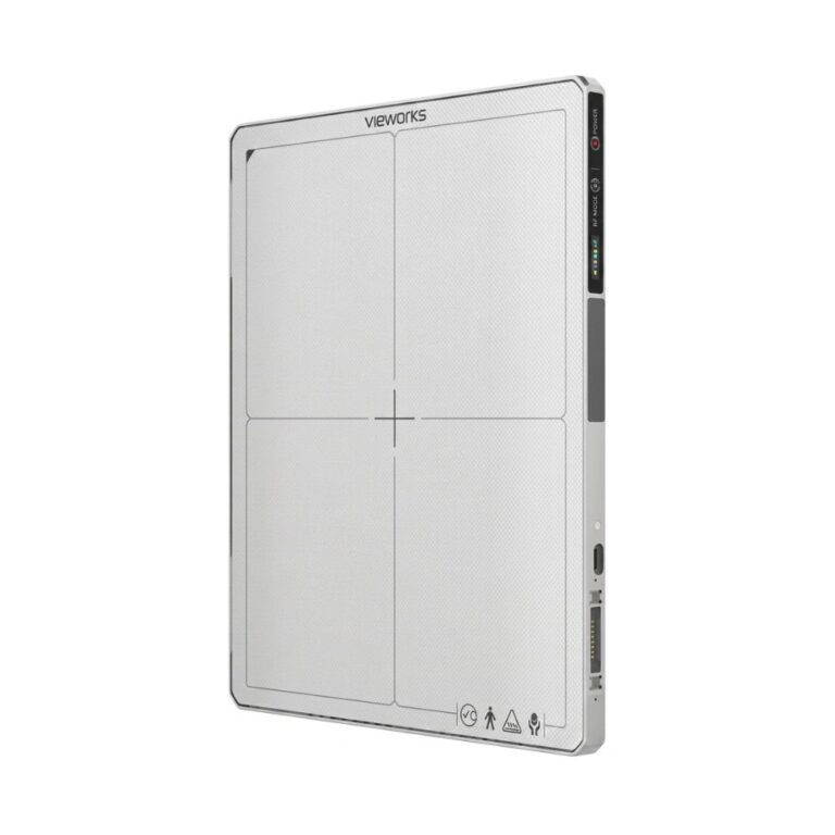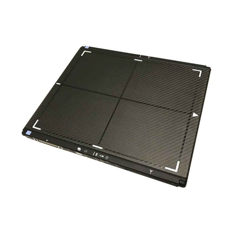


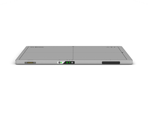
KA Imaging Reveal 35C Dual-Energy DR Detector
- Category:
- X-Ray Systems
- Brand:
- KA Imaging
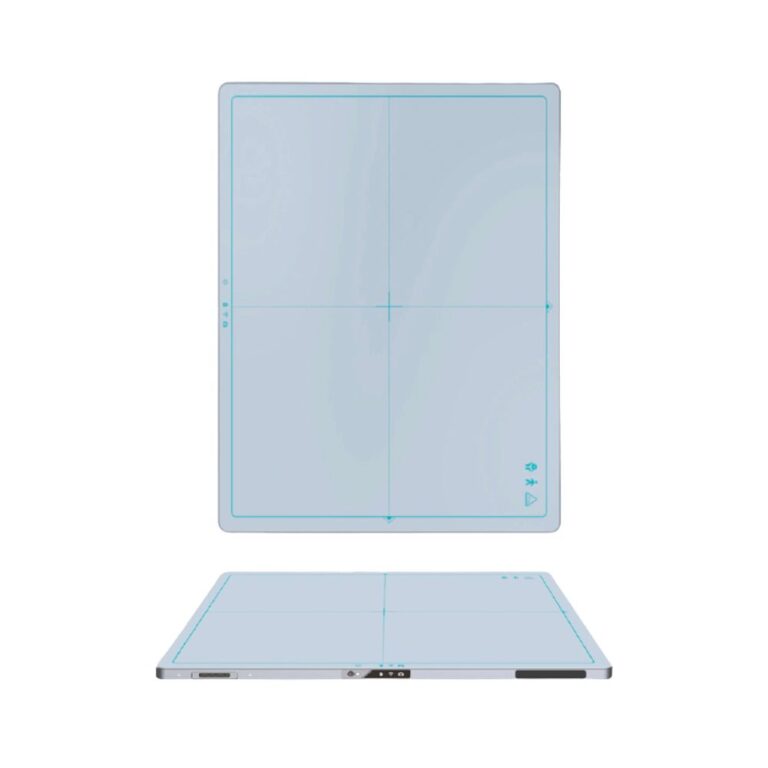
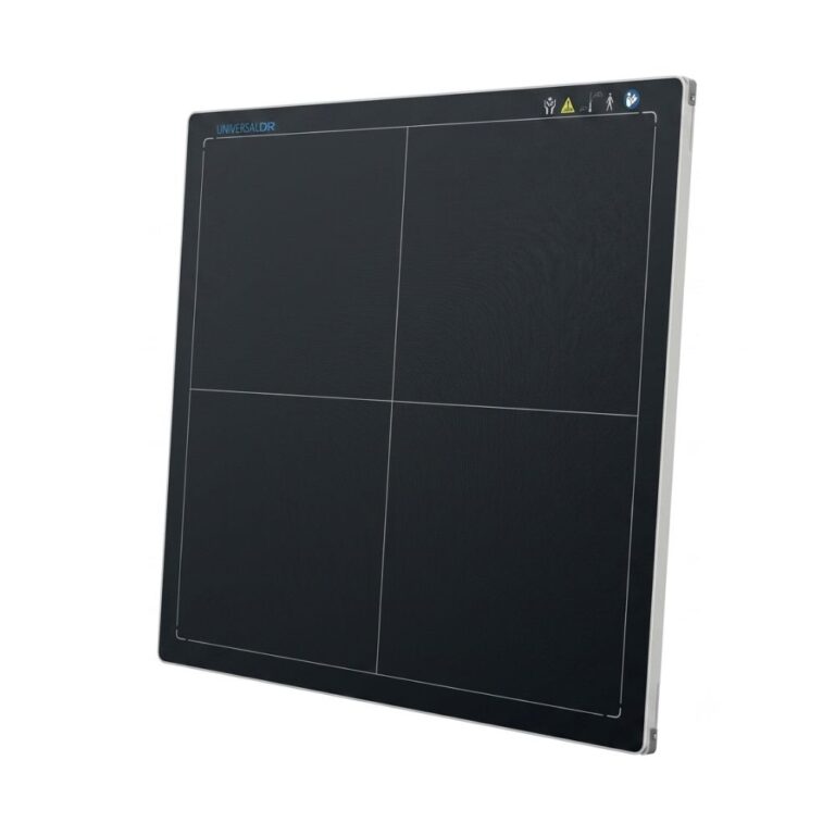
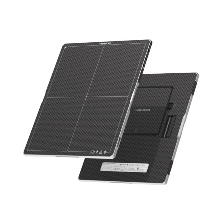
The KA Imaging Reveal 35C is a 14×17 dual-energy DR detector best suited for facilities adding dual-energy imaging to compatible fixed, mobile, or portable X-ray systems. Its SpectralDR technology produces conventional DR, bone, and soft-tissue images from a single exposure without requiring a second acquisition.
Reveal 35C Dual-Energy Imaging: 3 Images From 1 X-Ray Exposure
The KA Imaging Reveal 35C X-ray detector uses SpectralDR, KA Imaging’s single-exposure dual-energy technology, to produce a conventional digital radiograph plus separate bone and soft-tissue images simultaneously.
Dual-energy imaging separates information from different X-ray energy levels so clinicians can view bone and soft tissue independently in addition to the standard radiograph.
Key Benefits
-
- One exposure produces standard DR, bone, and soft-tissue images
- 14×17 in. / 35×43 cm ISO 4090 flat panel detector
- Compatible with fixed, mobile, and portable X-ray applications
- Lossless Automatic Exposure Detection
- Wired or wireless communication with hot-swappable battery or tethered power
Because all three images are generated from the same exposure, Reveal 35C provides additional spectral information without requiring a second X-ray acquisition solely to create the bone and soft-tissue images.
See How Reveal 35C Adds Dual-Energy Imaging
See how the Reveal 35C dual-energy X-ray detector adds standard DR, bone, and soft-tissue imaging to a single exposure while maintaining a familiar digital radiography workflow.
How SpectralDR Creates 3 Images From 1 Exposure
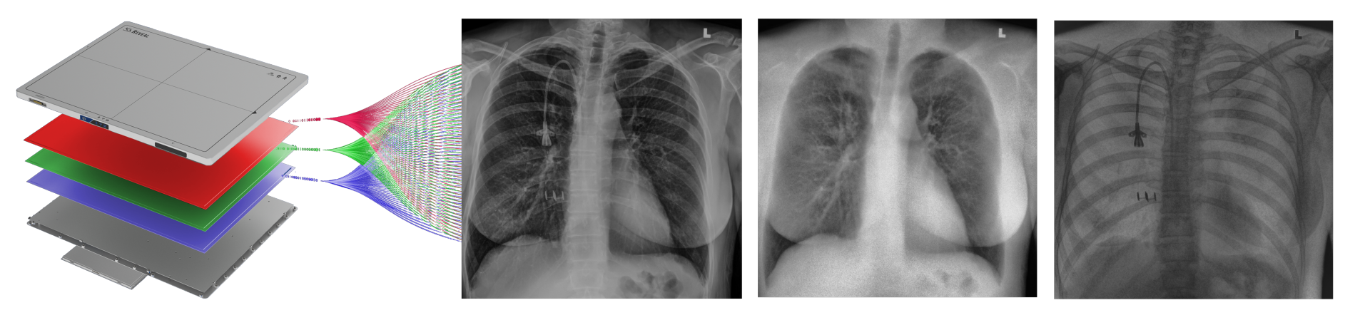
SpectralDR is KA Imaging’s detector-based spectral imaging technology. It captures information from different X-ray energy levels during one exposure and uses that information to create conventional DR, bone, and soft-tissue images.
Triple-Layer Detector Architecture Separates X-Ray Energy
Reveal 35C uses a multilayer detector architecture that separates X-ray energy information during image acquisition. In practical terms, different detector layers capture different portions of the X-ray spectrum, allowing the system to distinguish bone and soft tissue while also producing the conventional radiograph.
This detector-based approach brings spectral X-ray imaging into the same acquisition rather than requiring separate X-ray exposures at different energy levels.
Standard DR, Bone & Soft-Tissue Images From One Exposure
Each Reveal 35C exposure produces three complementary image types:
-
- Standard DR image: The conventional digital radiograph used for routine interpretation
- Soft-tissue image: Reduces the visibility of bone to help visualize soft-tissue structures that may otherwise overlap with skeletal anatomy
- Bone image: Reduces soft-tissue interference to improve visualization of bone and other higher-density structures
The supplemental bone and soft-tissue images provide additional anatomical information while preserving the conventional DR image used in routine radiography.
Single-Exposure Imaging Avoids Dual-Energy Image Misalignment
Sequential dual-energy techniques can be affected when the patient moves between separate acquisitions.
Reveal 35C captures the information required for all three images during the same X-ray exposure. This avoids the inter-exposure image misalignment associated with dual-energy techniques that depend on sequential acquisitions.
SpectralDR Supports Lateral & Oblique Views
SpectralDR can provide dual-energy information during lateral and oblique imaging in addition to frontal projections. These views can provide another way to evaluate anatomy that may be obscured by overlapping bone or soft tissue on a conventional radiograph.
Watch How SpectralDR Works
Learn how KA Imaging’s SpectralDR technology separates one X-ray exposure into conventional DR, bone, and soft-tissue images.
Add Reveal 35C Dual-Energy DR to Fixed, Mobile & Portable X-Ray Systems
Reveal 35C is designed to bring dual-energy digital radiography to compatible existing X-ray equipment as well as new imaging configurations. Its cassette-size format, detector-based SpectralDR technology, wired and wireless communication, and automatic exposure detection support deployment across fixed, mobile, and portable radiography workflows.
14×17 ISO 4090 Format for Compatible DR Retrofits
The Reveal 35C flat panel detector measures 14×17 in. / 35×43 cm and follows the ISO 4090 cassette format.
This standard detector footprint can support DR retrofit projects where facilities want to add direct digital radiography and SpectralDR imaging while retaining compatible X-ray equipment.
Lossless AED Automatically Detects the X-Ray Exposure
Automatic Exposure Detection (AED) allows the detector to recognize when an X-ray exposure occurs. KA Imaging’s Lossless AED supports exposure triggering without requiring a separate synchronization connection for compatible applications.
This can simplify integration when adding the detector to an existing X-ray system.
Gigabit Ethernet or Wi-Fi Connectivity
Reveal 35C supports Gigabit Ethernet for wired data transmission and IEEE 802.11ac/n Wi-Fi for wireless communication.
Facilities can select the communication method that fits the compatible X-ray system and imaging environment, whether the detector is being used in a fixed room or moved between imaging positions.
Hot-Swappable Batteries or Tethered Power
Reveal 35C can operate using hot-swappable batteries or tethered AC power.
Hot-swappable batteries support mobile and portable workflows where detector mobility is important, while tethered power provides another option for fixed or continuously powered applications.
Reveal 35C is also integrated into the KA Imaging Reveal Mobi Pro Mobile X-Ray System for facilities that need SpectralDR technology as part of a complete mobile X-ray platform.
Clinical Environments & Applications for Reveal 35C
The KA Imaging Reveal 35C is designed for general radiography environments where conventional DR plus supplemental bone and soft-tissue images can provide additional information from the same X-ray exposure.
FDA-Cleared for General Radiography
Reveal 35C is FDA-cleared for use with radiographic imaging systems to generate images of human anatomy for general radiography.
When its dual-energy subtraction function is enabled, the supplemental images are intended to assist physicians by reducing the visibility of overlying or underlying anatomical structures. The detector is not intended for mammography.
Clinical Environments
-
- Hospitals and health systems
- Emergency departments
- Intensive and critical care units
- Diagnostic imaging departments
- Outpatient imaging centers
- Fixed X-ray rooms
- Mobile and bedside radiography workflows
- Portable and point-of-care X-ray applications using compatible equipment
Applications Supported by SpectralDR Imaging
Reveal 35C supports routine general radiography while its spectral images can provide additional visualization for applications including:
-
- Chest radiography
- Lung nodules and lesions
- Pneumonia
- Pneumothorax
- Line and tube tips
- Retained surgical objects
- Fractures obscured by overlapping anatomy
- Calcifications and other high-density structures
The supplemental bone and soft-tissue images are designed to add information to the conventional radiograph by reducing the visibility of overlapping structures. They do not replace additional imaging when another modality or examination is clinically required.
Veterinary Imaging with Reveal 35C
The Reveal 35C can also be integrated into compatible veterinary X-ray systems. KA Imaging positions the detector for both large- and small-animal imaging, with SpectralDR providing standard DR, bone, and soft-tissue images from one exposure in portable and fixed veterinary applications.
Veterinary applications include:
-
- Small-animal respiratory imaging
- Small-animal orthopedic imaging
- Abdominal radiography
- Equine sports medicine
- Mobile veterinary and field imaging
- Wildlife and zoo medicine
Its 14×17 detector format and portable workflow are particularly useful where moving the animal may be difficult or undesirable. KA Imaging specifically identifies veterinary clinics, animal hospitals, mobile equine practices, and multi-species imaging among the environments supported by SpectralDR.
Clinical Evidence for Reveal 35C SpectralDR Imaging
KA Imaging cites clinical research evaluating how supplemental dual-energy images can add information to conventional radiography. The results below reflect the specific studies and patient populations evaluated and should not be interpreted as guaranteed results for every examination.
Greater Lung Lesion Visibility in Preliminary Results
In preliminary results from a lung-cancer imaging study cited by KA Imaging, adding SpectralDR images was associated with 43% greater lesion visibility.

Additional Pneumonia Findings in a Cited Study
In a separate study cited by KA Imaging, dual-energy imaging identified 33% more pneumonia cases than conventional digital radiography alone within the evaluated patient population.
Increased Confidence in Evaluated ICU Cases
KA Imaging also cites research reporting increased confidence in 57% of evaluated ICU cases when dual-energy images were available, with no additional reading time reported in the study.
These findings illustrate where bone and soft-tissue separation may provide additional information beyond the conventional DR image while keeping the standard radiograph available for interpretation.
Reveal 35C Detector Performance & Image Workflow
140 μm Pixel Pitch & CsI Detector Design
Reveal 35C combines an amorphous silicon sensor with a Cesium Iodide (CsI) scintillator and a 140 μm pixel pitch.
Pixel pitch describes the spacing between individual detector pixels. The CsI scintillator converts incoming X-rays into light that the detector captures to create the digital image.
16-Bit Acquisition for Grayscale Detail
Reveal 35C uses 16-bit analog-to-digital conversion, providing a broad range of grayscale information for digital image processing and display.
Up to 75% DQE for Efficient X-Ray Signal Capture
Detective Quantum Efficiency (DQE) measures how efficiently a detector uses the available X-ray signal to create useful image information.
KA Imaging specifies DQE as high as 75% at 0 lp/mm for Reveal 35C, with 67% specified at 1 lp/mm.
7-Second Preview & 20-Second Cycle Time
KA Imaging specifies a typical 7-second preview time and 20-second cycle time.
Preview time indicates how quickly an image becomes available for initial review, while cycle time describes the approximate interval before the detector is ready for another acquisition.
Conventional DR & Dual-Energy Imaging in One Detector
The KA Imaging Reveal 35C gives facilities a way to add single-exposure dual-energy imaging without abandoning the workflow of conventional digital radiography. Its 14×17 flat panel format, SpectralDR technology, retrofit capability, wired or wireless operation, and compatibility with fixed, mobile, and portable applications bring conventional DR and supplemental bone and soft-tissue imaging together in one detector.
KA Imaging Reveal 35C Specifications
Supported Deployment
-
- General-purpose digital radiography
- Compatible fixed X-ray systems
- Mobile X-ray systems
- Portable X-ray applications
- Compatible DR retrofit projects
- Compatible veterinary X-ray systems
- Fixed and portable veterinary imaging
- Integration into complete X-ray systems
Detector & Imaging Specifications
-
- Detector type: Single-exposure dual-energy flat panel DR detector
- Detector size: 14×17 in. / 35×43 cm
- Cassette standard: ISO 4090
- Pixel pitch: 140 μm
- Weight: 8 lb. / 3.6 kg including battery
- Sensor type: Amorphous silicon
- Scintillator: Cesium Iodide (CsI)
- A/D conversion: 16-bit
- Energy range: 40 to 150 keV
- Intended use: General radiography
- FDA status: 510(k) cleared
- Mammography: Not intended for mammography
- Connectivity, Trigger & Power
- Wired data transmission: Gigabit Ethernet, 1000BASE-T
- Wireless data transmission: IEEE 802.11ac/n
- Exposure trigger: Lossless Automatic Exposure Detection (AED)
- Power: Hot-swappable batteries and/or tethered power
- AC input: 100–240 VAC, 50–60 Hz
Image Acquisition Timing
-
- Typical preview time: 7 seconds
- Typical cycle time: 20 seconds
Detector Efficiency & Resolution
-
- DQE at 0 lp/mm: 75%
- DQE at 1 lp/mm: 67%
- DQE at 2 lp/mm: 53%
- DQE at 3 lp/mm: 34%
- DQE at Nyquist: 21%
- MTF at 1 lp/mm: 64%
- MTF at 2 lp/mm: 35%
- MTF at 3 lp/mm: 18%
- MTF at Nyquist: 13%
DQE measures how efficiently the detector uses the available X-ray signal to create useful image information.
MTF, or Modulation Transfer Function, measures how well the detector preserves image detail at different spatial frequencies.
Detector Dose Range
-
- Saturation dose: 140 μGy
- Maximum linear dose: 120 μGy
- Measurements performed under RQA5 test conditions
These values describe detector response under standardized RQA5 test conditions and are not patient dose recommendations.
Supported Deployment
-
- General-purpose digital radiography
- Compatible fixed X-ray systems
- Mobile X-ray systems
- Portable X-ray applications
- Compatible DR retrofit projects
- Integration into complete X-ray systems
For additional information and specifications, access the PDFs provided below.