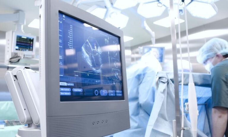
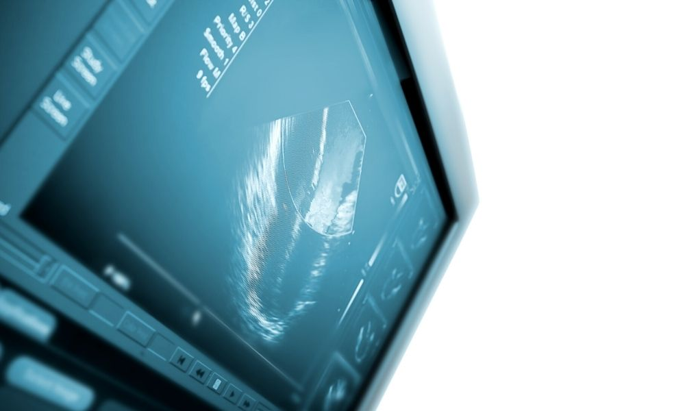
Imaging techniques like ultrasounds are critical diagnostic tools used for locating and evaluating abnormalities in bodily tissues and organs. While these techniques are normally effective, imaging artifacts—distortions or misrepresentations of tissue structures—can affect the detection and diagnosis of life-threatening diseases. To prevent these artifacts from tampering with the diagnostic process, read up on these common ultrasound imaging artifacts and how to recognize them.
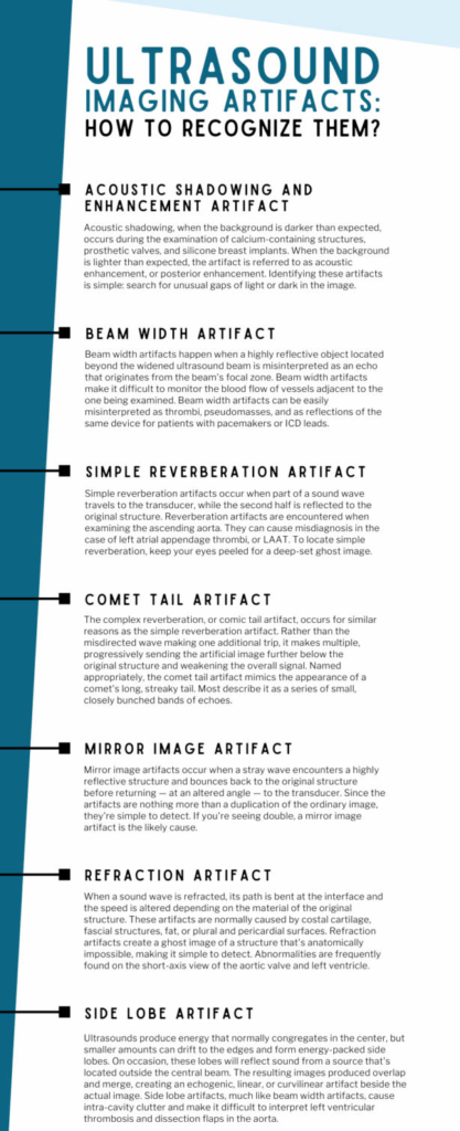
Acoustic Shadowing and Enhancement Artifact
Ultrasounds act on the assumption that all tissues attenuate uniformly. When they don’t, the image features either a dark, shadowy background or one that’s abnormally bright. Acoustic shadowing, when the background is darker than expected, occurs during the examination of calcium-containing structures, prosthetic valves, and silicone breast implants. When the background is lighter than expected, the artifact is referred to as acoustic enhancement, or posterior enhancement. It’s seen during the examination of fluid-filled organs, like the gallbladder and urinary bladder, or ones in the subxiphoid window, like the liver. Identifying these artifacts is simple: search for unusual gaps of light or dark in the image.
Beam Width Artifact
Beam width artifacts happen when a highly reflective object located beyond the widened ultrasound beam is misinterpreted as an echo that originates from the beam’s focal zone. For a better understanding of this artifact, consider the beam’s hourglass-like shape. It starts off wide, narrows as it approaches the focal zone, and then returns to its original width. This means that the beam isn’t uniform in its depth.
Beam width artifacts make it difficult to monitor the blood flow of vessels adjacent to the one being examined. They create artificial images of the reflective object, which form along the same plane as the authentic image and overlap the object of interest. This can lead to vast amounts of intra-cavity clutter. Beam width artifacts can be easily misinterpreted as thrombi, pseudomasses, and as reflections of the same device for patients with pacemakers or ICD leads.
Simple Reverberation Artifact
Simple reverberation artifacts occur when part of a sound wave travels to the transducer, while the second half is reflected to the original structure. The half that fails to make it back to the transducer carries on to make a second round trip. Because the second wave takes longer to arrive than the first, the transducer interprets it as coming from a deeper structure. It maps this as an artificial image that’s set lower than the original structure.
Reverberation artifacts are encountered when examining the ascending aorta. They can cause misdiagnosis in the case of left atrial appendage thrombi, or LAAT. To locate simple reverberation, keep your eyes peeled for a deep-set ghost image. Since its location is perceived as being deeper than it is, the artificial image is often smaller and located directly below the original structure.
Comet Tail Artifact
The complex reverberation, or comet tail artifact, occurs for similar reasons as the simple reverberation artifact. Rather than the misdirected wave making one additional trip, it makes multiple, progressively sending the artificial image further below the original structure and weakening the overall signal.
The comet tail artifact is commonly seen in patients with mechanical valves. Unlike other artifacts, the comet tail artifact is a useful clinical marker. It’s used in the diagnosis of pulmonary conditions like pneumothorax, alveolar-interstitial syndrome, interstitial pneumonia, and pulmonary fibrosis. Named appropriately, the comet tail artifact mimics the appearance of a comet’s long, streaky tail. Most describe it as a series of small, closely bunched bands of echoes.
Mirror Image Artifact
Mirror image artifacts occur when a stray wave encounters a highly reflective structure and bounces back to the original structure before returning—at an altered angle—to the transducer. Instead of recognizing that the signal took an indirect path, the transducer assumes the reflection took a straight path below the initial object. It maps accordingly, producing a reflected image of the original structure.
These artifacts are often caused by the pleura and can cause difficulties in evaluating mitral valve prostheses and major blood vessels, like the carotid arteries and abdominal aorta. Since the artifacts are nothing more than a duplication of the ordinary image, they’re simple to detect. If you’re seeing double, a mirror image artifact is the likely cause.
Refraction Artifact
When a sound wave is refracted, its path is bent at the interface and the speed is altered depending on the material of the original structure. This is referred to as a refraction artifact. These artifacts are normally caused by costal cartilage, fascial structures, fat, or plural and pericardial surfaces.
Refraction artifacts create a ghost image of a structure that’s anatomically impossible, making it simple to detect. Abnormalities are frequently found on the short-axis view of the aortic valve and left ventricle. The artificial images, or duplications they make, tend to resemble the mitral, aortic, or pulmonary valve.
Side Lobe Artifact
Ultrasounds produce energy that normally congregates in the center, but smaller amounts can drift to the edges and form energy-packed side lobes. On occasion, these lobes will reflect sound from a source that’s located outside the central beam. The resulting images produced overlap and merge, creating an echogenic, linear, or curvilinear artifact beside the actual image.
Side lobe artifacts, much like beam width artifacts, cause intra-cavity clutter and make it difficult to interpret left ventricular thrombosis and dissection flaps in the aorta. Normally, the artifact reflects the sinotubular junction and creates a curvilinear distortion within the aorta. An unexpected artifact can disrupt the diagnostic process, leading to misdiagnosis or, in rare circumstances, unnecessary medical intervention.
Being aware of the common ultrasound imaging artifacts and how to recognize them can help with early detection and ultrasound repair service, thereby reducing the likelihood of an undetected artifact impacting a patient’s diagnosis and treatment.
Your Partner for Quality Ultrasound Equipment and Service
The effectiveness of ultrasound equipment and the accuracy of diagnostics depend on a number of factors, including the proper functioning of the transducer probe. MXR Imaging offers a wide selection of ultrasound equipment for sale, including top-of-the-line models like the GE LOGIQ E9™. We also provide comprehensive ultrasound maintenance plans, expert ultrasound probe repair, and a full inventory of parts for sale to keep your practice running smoothly. Contact us for a quote or free assessment or browse through our available inventory.