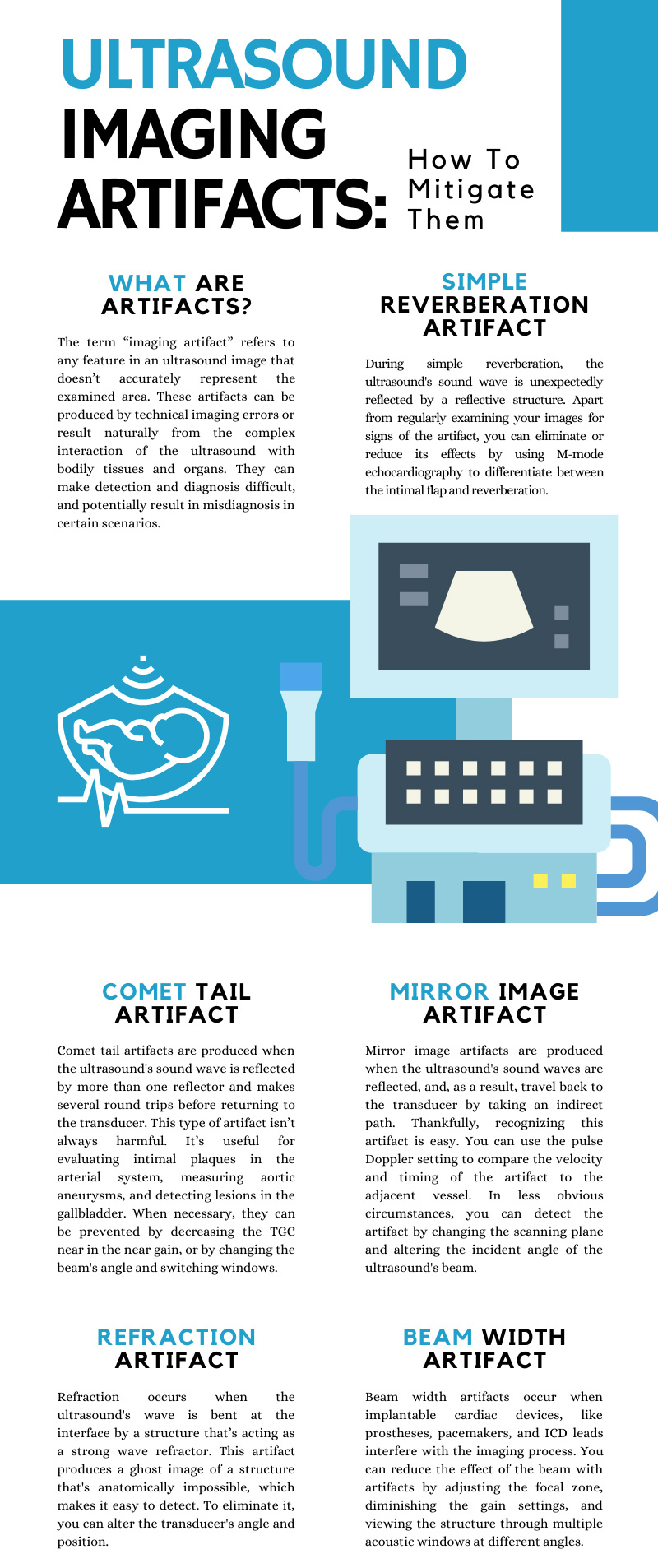
Ultrasound images can be difficult to read due to imaging artifacts. They can lead to a misdiagnosis and the improper treatment of certain conditions or diseases. Understanding how to prevent these artifacts is critical to receiving accurate, unaltered images and providing patients with the appropriate level of care. This guide to ultrasound imaging artifacts can help you reduce or eliminate unwanted artifacts.
What are Artifacts?
The term "imaging artifact" refers to any feature in an ultrasound image that does not accurately represent the examined area. These artifacts can be produced by technical imaging errors or occur naturally from the complex interaction of the ultrasound with bodily tissues and organs. They can make detection and diagnosis difficult, potentially leading to a misdiagnosis that results in patients receiving inaccurate treatment, including unhelpful or harmful medication and unnecessary surgical procedures. To prevent the negative consequences of imaging artifacts on the diagnostic process, it's critical to mitigate their likelihood of appearing and distorting an image. There are multiple types of artifacts, ranging from common, easy-to-find alterations to rarer distortions that can integrate seamlessly into an image.
The Different Types of Artifacts and How to Mitigate Them
Different artifacts present themselves in unique ways. They can be caused by problems in the ultrasound's operation, unwanted interaction with bodily tissues and organs, interference from pacemakers, prosthetics, and other metal devices, and more. Here are some common imaging artifacts and how to mitigate their effects.
Simple Reverberation Artifact
During simple reverberation, the ultrasound's sound wave is unexpectedly reflected by a reflective structure. It makes multiple round trips before returning to the transducer. Apart from regularly examining your images for signs of the artifact, you can eliminate or reduce its effects by using M-mode echocardiography to differentiate between the intimal flap and reverberation. You can also rely on different types of imaging technology to act as a complementary measure to the ultrasound or, alternatively, as the initial procedure. To decrease the likelihood of a misdiagnosis, you can use a multiplane TEE to analyze the image from multiple angles and windows.
Comet Tail Artifact
Comet tail artifacts are produced when the ultrasound's sound wave is reflected by more than one reflector and makes several round trips before returning to the transducer. This type of artifact isn't always harmful. It is useful for evaluating intimal plaques in the arterial system, measuring aortic aneurysms, and detecting lesions in the gallbladder. When necessary, they can be prevented by decreasing the TGC in the near gain, or by changing the beam's angle and switching windows.
Mirror Image Artifact
Mirror image artifacts are produced when the ultrasound's sound waves are reflected and, as a result, travel back to the transducer by taking an indirect path. Thankfully, recognizing this artifact is easy. You can use the pulse Doppler setting to compare the velocity and timing of the artifact to the adjacent vessel. In less obvious circumstances, you can detect the artifact by changing the scanning plane and altering the incident angle of the ultrasound's beam.
Acoustic Shadowing and Enhancement Artifact
Acoustic shadowing occurs when calcium-containing structures, prosthetic valves, or silicone implants interfere with the imaging process. To prevent the underestimation or displacement of regurgitant jets, you can increase the aliasing velocity to examine them. In addition, you can use a deep transgastric long-axis view to achieve improved imaging of the LVOT and aortic valve, and reposition the short-axis view to examine any aortic leaflets. As a supplementary procedure, cardiac CT and intra-cardiac echocardiography will work.
Acoustic enhancement is commonly seen when while evaluating organs that are filled with fluid, like the gallbladder and urinary bladder. Mitigating it is simple—you can alter the angle of the beam to eliminate traces of the artifact if it’s interfering with the diagnostic process.
Refraction Artifact
Refraction occurs when the ultrasound's wave is bent at the interface by a structure that's acting as a strong wave refractor. This causes the speed of the wave to alter depending on the material of the structure it was refracted from. This artifact produces a ghost image of a structure that is anatomically impossible, which makes it easy to detect. To eliminate it, you can alter the transducer's angle and position.
Beam Width Artifact
Beam width artifacts occur when implantable cardiac devices, like prostheses, pacemakers, and ICD leads, interfere with the imaging process. You can reduce the effect of beam width artifacts by adjusting the focal zone, diminishing the gain settings, and viewing the structure through multiple acoustic windows at different angles. Since artifacts aren't reproducible in more than one or two planes, you can get rid of the artifact by making use of several techniques.
Side-Lobe Artifact
Side-lobe artifacts occur when smaller lobes of energy encounter a strong reflector, which creates echoes that appear to originate from the ultrasound's central beam. This type of artifact is commonly found in fundamental imaging. As an alternative to fundamental imaging, you can use harmonic imaging. The nonlinear relationship between the harmonic generation and the original ultrasound wave can improve visualization and decrease the amount of side-lobe clutter.
Aliasing Artifact
Aliasing artifacts occur when the structure being examined exceeds the maximum velocity set by the Nyquist limit. You can reduce the effects of the artifact by decreasing the transducer's frequency, decreasing depth, and choosing an anatomic structure with a velocity below the Nyquist limit. This is accomplished by using a low-frequency probe and examining the structure from a window that's located close to the probe. When you're examining structures that exceed the Nyquist limit, you can avoid disruption from the artifact by using a continuous-wave Doppler. Like the comet tail artifact, aliasing artifacts are useful for diagnosing certain diseases and conditions. You can use them to assess the severity of valvular regurgitation, among other things.
Now that you understand what ultrasound imaging artifacts are and how to mitigate them, you'll be well-equipped to prevent any future misdiagnosis.