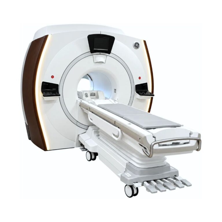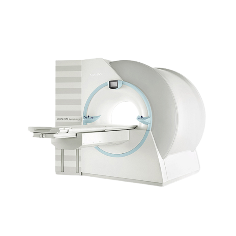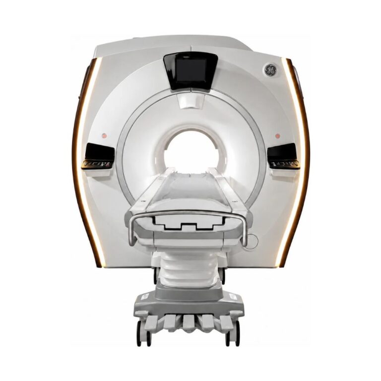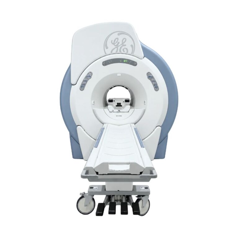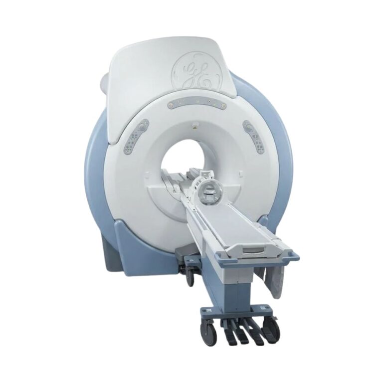
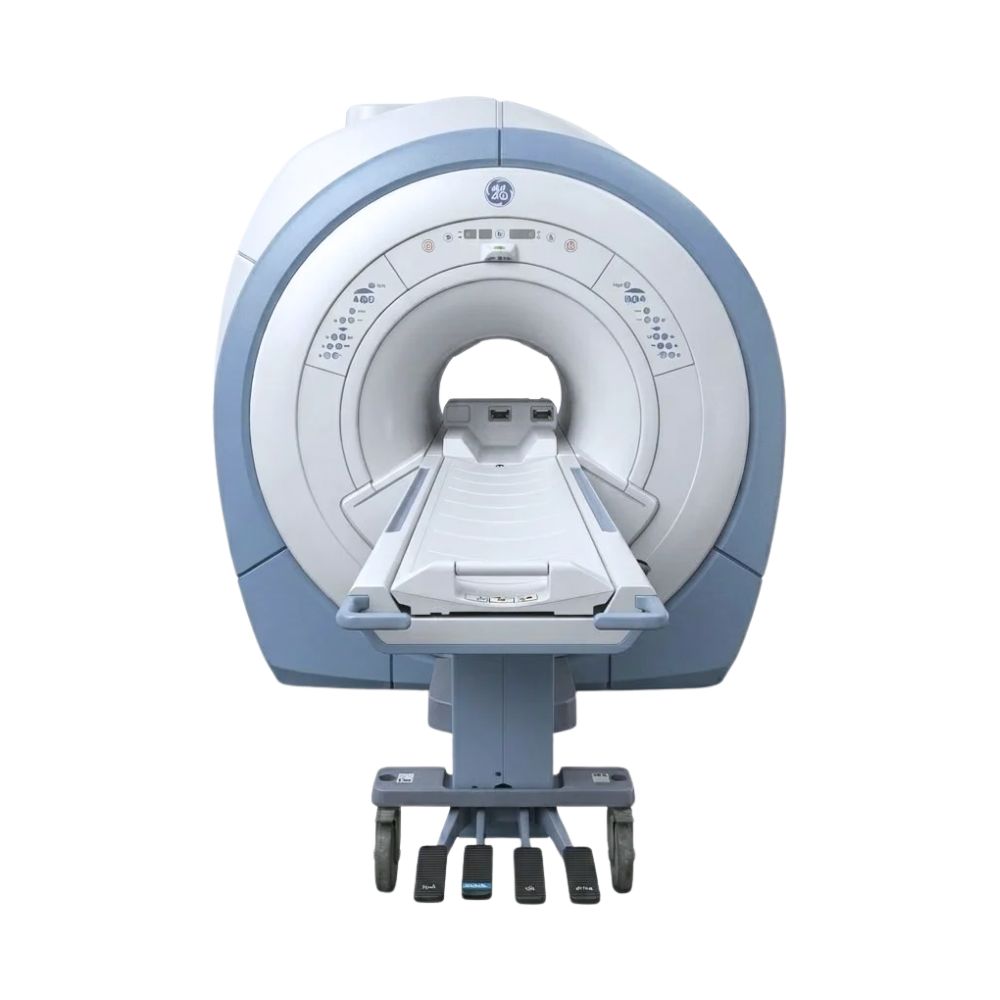
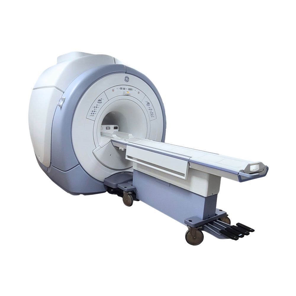
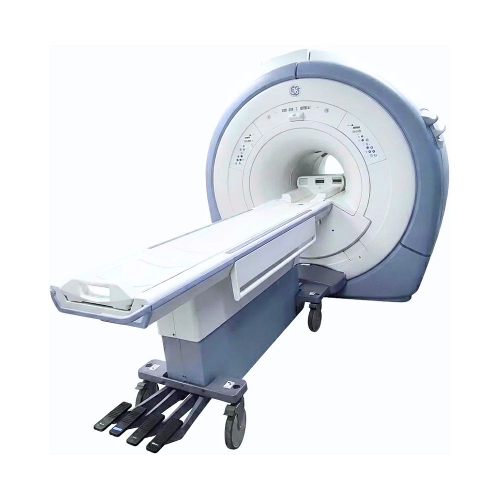
GE Signa HDxt 1.5T MRI
- Category:
- MRI Systems
- Brand:
- GE
The GE SIGNA HDxt 1.5T MRI is a 60 cm bore scanner well suited for hospitals, imaging centers, and specialty practices. Its high-fidelity gradients, multichannel receiving coils, and advanced imaging options support detailed neurological, musculoskeletal, and body examinations with efficient clinical workflows.
SIGNA HDxt MRI Highlights & Benefits
The GE SIGNA HDxt combines established 1.5T imaging performance with high-definition reconstruction, adaptable clinical applications, and efficient patient preparation. Its configurable coil technology and advanced imaging software support comprehensive diagnostic MRI across multiple specialties.
Key Features & Benefits
-
- High-Definition 1.5T Imaging: Supports detailed brain, spine, musculoskeletal, abdominal, and vascular examinations with a 48 cm imaging field of view.
- High-Fidelity Gradients: Provide precise magnetic-field control for detailed anatomical imaging and advanced techniques, including diffusion MRI.
- Multichannel Imaging Technology: Available 8-, 16-, and 32-channel configurations support compatible high-density receiving coils for detailed image acquisition.
- Advanced Imaging Applications: Compatible Cube, IDEAL, and PROPELLER technologies support three-dimensional imaging, consistent fat suppression, and reduced motion artifacts.
- 60 cm Bore & Patient Comfort: In-bore lighting, ventilation, and compatible detachable tables support patient positioning and preparation.
- Efficient Clinical Workflows: High-definition reconstruction, automated scan preparation, and configurable examination protocols help streamline daily operation.
SIGNA HDxt High-Definition Imaging Performance
The SIGNA HDxt combines a superconducting 1.5T magnet, high-fidelity gradients, and multichannel signal acquisition to produce detailed diagnostic images. These technologies support routine MRI examinations and specialized imaging techniques requiring precise anatomical coverage.
1.5T Magnet for Whole-Body MRI
The HDxt’s actively shielded superconducting magnet produces a stable 1.5 Tesla magnetic field for whole-body diagnostic imaging.
Its maximum 48 cm (18.9 in) field of view supports broad anatomical coverage, including the brain, spine, abdomen, pelvis, and extremities.
The magnet’s field uniformity helps maintain consistent tissue contrast and supports imaging techniques requiring reliable fat suppression.
This makes the HDxt well suited for comprehensive 1.5T MRI services, from routine examinations to more demanding specialty studies.
High-Fidelity Gradient Performance
The HDxt’s high-fidelity gradients control the magnetic fields that determine where MRI signals originate within the body. Their speed and precision support detailed anatomical imaging and advanced acquisition techniques.
The original HDxt EchoSpeed gradient configuration delivers:
-
- 33 mT/m Gradient Amplitude: Supports precise spatial imaging and detailed visualization of smaller anatomical structures.
- 120 T/m/s Slew Rate: Enables rapid magnetic-field changes during compatible scan sequences.
- Actively Shielded Gradient Design: Reduces unwanted magnetic-field effects that can interfere with image quality.
These capabilities support diffusion-weighted imaging, detailed musculoskeletal examinations, and other techniques requiring precise signal acquisition.
Installed gradient specifications may differ on subsequently modified systems.
Multichannel Imaging Architecture
The HDxt supports 8-, 16-, and 32-channel receiving configurations.
Receiving coils collect signals from the patient’s anatomy. High-density coils use multiple elements positioned close to the imaging area to capture detailed anatomical information.
Compatible parallel imaging technologies, including ASSET and ARC, use information from multiple coil elements to reconstruct images from fewer acquired measurements. This can shorten selected MRI examinations while maintaining useful anatomical detail.
The HDxt’s XVRE (Volume Reconstruction Engine) processes the acquired information into diagnostic images, including large three-dimensional datasets.
Together, these technologies support detailed imaging, efficient reconstruction, and flexible examination protocols.
SIGNA HDxt Neurological Imaging
The GE SIGNA HDxt supports advanced imaging technologies that help radiologists visualize anatomy, distinguish different tissues, and manage patient motion.
These applications expand the scanner’s capabilities across neurological, musculoskeletal, abdominal, vascular, and specialty examinations. Available technologies depend on the installed software and system configuration.
The HDxt supports detailed neurological examinations through imaging techniques that provide different types of information about brain anatomy, tissue characteristics, and function.
Compatible technologies include:
- PROPELLER Motion Correction: Acquires MRI information in overlapping, rotating sections rather than following a conventional line-by-line pattern. This makes the images less sensitive to certain types of patient movement, helping produce more consistent brain images and potentially reducing repeat acquisitions when motion affects image quality.
- Diffusion-Weighted Imaging (DWI): Measures how water molecules move within tissue. Changes in that movement help radiologists identify abnormalities that may be difficult to distinguish using conventional MRI, particularly during neurological examinations.
- SWAN: Combines information from multiple MRI echoes to emphasize differences in magnetic susceptibility between tissues. This improves visualization of small veins, microbleeds, and certain blood-product or mineral-related changes within the brain.
- BRAVO: Acquires a high-resolution three-dimensional set of T1-weighted brain images with strong differentiation between gray and white matter. The resulting images can be reviewed in multiple planes for detailed anatomical assessment.
- BrainWave: Supports functional MRI by identifying changes in blood oxygenation associated with brain activity during specific tasks. Compatible mapping tools help clinicians and researchers examine functional brain regions and their relationship to surrounding anatomy.
- 3D Arterial Spin Labeling (ASL): Available on compatible configurations, this technique magnetically labels blood flowing into the brain and measures its movement through tissue. It produces maps of cerebral blood flow without requiring injected contrast material.
Compatible diffusion tensor imaging can also evaluate the directional movement of water along white-matter pathways, supporting specialized neurological assessment and brain-mapping applications.
These technologies make the HDxt a versatile option for facilities performing neurological and spine MRI, with specialized applications selected according to their clinical requirements.
SIGNA HDxt Image Reconstruction Technology
Cube 3D Imaging
Cube is GE’s three-dimensional MRI acquisition technology, designed to provide detailed anatomical coverage from a single scan.
Traditional two-dimensional MRI acquires individual image slices in prescribed directions. Cube instead collects a three-dimensional volume containing information throughout the selected anatomy.
Radiologists can reconstruct that volume into different viewing planes without repeating the original acquisition. Compatible Cube techniques also support thin sections and high-resolution image review, helping visualize structures that extend across multiple conventional slices.
Cube is particularly useful for brain, spine, and joint examinations where detailed anatomy and flexible viewing angles are important.
Compatible ARC acceleration can reduce the data required for the acquisition, helping balance image detail with examination time.
IDEAL Fat Suppression
IDEAL is GE’s water-fat separation technology, designed to produce more consistent images when fat signals interfere with anatomical detail.
Fat and water generate MRI signals with slightly different frequencies. IDEAL acquires information at multiple echo times and uses those differences to separate the signals during image reconstruction.
A compatible acquisition can produce water-only, fat-only, and combined tissue images, giving radiologists different ways to evaluate the same anatomy.
This supports clearer visualization of soft tissue, joints, and abdominal structures, particularly where conventional fat suppression can be challenging.
By reducing uneven fat suppression, IDEAL can also help minimize image artifacts and the need to repeat affected sequences.
SIGNA HDxt Specialized Body Imaging
The HDxt also supports clinical applications designed for anatomical regions where breathing, blood flow, tissue contrast, or examination time create additional imaging challenges.
Compatible technologies include:
-
- LAVA: Acquires detailed three-dimensional abdominal images during short breath-holds. Rapid acquisition helps limit breathing-related motion while supporting liver and other abdominal examinations.
- LAVA Flex: Extends abdominal imaging by separating fat and water information from a single acquisition. It can generate water-only, fat-only, in-phase, and out-of-phase images, providing additional tissue information without requiring separate scans for every contrast.
- FIESTA: Uses a rapid MRI acquisition technique that produces strong signals from fluid-containing structures. This supports detailed visualization of selected anatomy, including compatible cardiac, vascular, and other fluid-sensitive examinations.
- Magnetic Resonance Angiography (MRA): Uses MRI signal differences to visualize blood vessels. Compatible techniques can provide vascular imaging with or without injected contrast material, depending on the examination.
- VIBRANT: Supports dedicated breast MRI using rapid three-dimensional imaging and techniques designed for consistent fat suppression. With compatible coils and software, it helps visualize breast tissue during contrast-enhanced examinations.
- Cardiac MRI: Uses specialized acquisitions synchronized with the cardiac cycle to capture the heart during motion, supporting assessment of cardiac anatomy and function on appropriately equipped systems.
These applications allow facilities to configure the HDxt for different specialties while balancing image quality, examination time, and patient requirements.
SIGNA HDxt Patient Access
The HDxt features a conventional 60 cm bore with patient-comfort and positioning options designed to support efficient diagnostic examinations.
60 cm Bore & Patient Table Options
The 60 cm (23.6 in) bore includes lighting and ventilation to improve the scanning environment.
Compatible detachable patient tables allow technologists to position patients outside the scanner and transfer them into place for imaging.
This can simplify patient preparation, assist with patient transfers, and support efficient room utilization between examinations.
The original HDxt patient table supports a maximum scanning weight of 350 lb (159 kg). Other documented HDxt configurations support up to 500 lb (227 kg), depending on the installed table.
Available table designs, movement capabilities, and positioning accessories vary by equipment configuration.
SIGNA HDxt Exam Workflow
The HDxt includes exam-planning and reconstruction technologies designed to simplify daily scanning, improve protocol consistency, and support efficient image management.
Automated Exam Preparation
The GE SIGNA HDxt uses automated calibration, patient communication, and image-processing technologies to reduce repetitive setup tasks and maintain consistent examination workflows.
-
- SmartPrescan: Automatically adjusts key scanner settings before image acquisition to optimize the system for the examination. By performing these calibration steps consistently, SmartPrescan reduces manual adjustments and helps technologists maintain reliable image quality across different patients and scanning protocols.
- AutoVoice: Delivers prerecorded breathing and breath-hold instructions at designated points during an examination. These instructions are coordinated with the scan sequence, helping patients follow consistent breathing patterns while reducing motion-related artifacts during abdominal and other breath-hold examinations.
- Integrated Image Reconstruction: The HDxt’s parallel processing architecture allows image reconstruction to take place while compatible scanning and workflow activities continue. Its XVRE reconstruction engine converts acquired MRI data into diagnostic images, helping technologists review results without waiting for every processing task to finish.
Together, these technologies simplify routine scanner operation, reduce interruptions between imaging sequences, and help facilities maintain consistent workflows throughout the day.
Protocol Management and Image Transfer
The HDxt’s operator workstation provides a centralized interface for examination setup, image acquisition, and review.
ProtoCopy allows compatible examination protocols to be transferred between systems, helping facilities maintain consistent scanning procedures.
DICOM connectivity supports image transfer to compatible picture archiving and communication systems (PACS) for storage, radiologist review, and clinical distribution.
Available workflow and networking capabilities depend on the installed workstation and software.
Clinical Environments & Applications
The SIGNA HDxt supports a broad range of diagnostic imaging services, from routine examinations to specialized neurological, musculoskeletal, and body imaging.
Clinical Environments
-
- Hospitals and diagnostic imaging departments
- Outpatient imaging centers
- Multispecialty medical practices
- Neurology and spine imaging centers
- Orthopedic and sports medicine facilities
- Oncology and cancer care centers
- Cardiac and vascular imaging programs
- Academic and teaching hospitals
Clinical Specialties
-
- Neurology & Neurosurgery: Brain, spine, diffusion, and compatible advanced neurological imaging.
- Orthopedics & Sports Medicine: Joint, ligament, tendon, cartilage, and soft-tissue examinations.
- Oncology: Tumor imaging, follow-up examinations, and treatment monitoring.
- Gastroenterology & Hepatology: Abdominal, liver, and pancreatic imaging.
- Cardiology: Cardiac anatomy and function using compatible applications.
- Vascular Medicine: Magnetic resonance angiography and vascular assessment.
- Urology: Renal, pelvic, and prostate MRI.
- Women’s Health: Pelvic and breast examinations with appropriate equipment.
Common MRI Applications
-
- Brain and spine MRI
- Diffusion-weighted neurological imaging
- Joint and extremity examinations
- Three-dimensional anatomical imaging
- Abdominal and pelvic MRI
- Liver and pancreatic imaging
- Magnetic resonance angiography
- Motion-corrected examinations
- Cardiac and breast MRI with compatible applications
SIGNA HDxt Equipment Configurations
The HDxt supports multiple hardware generations, receiving-channel configurations, and clinical software options.
These variations allow facilities to select an MRI scanner suited to their planned examinations, imaging volume, and operating requirements.
System Configurations
-
- GE SIGNA HDxt systems may include:
- 8-, 16-, or 32-channel receiving architectures
- Different gradient and reconstruction hardware configurations
- Compatible high-density coil packages
- Multiple software generations and clinical applications
- Different patient-table arrangements
- Operator workstation and accessory variations
The HDxt Optima Edition introduced additional imaging and workflow capabilities for compatible systems.
The later SIGNA HDxt SIGNA Works Edition represents a separate modernization pathway for eligible equipment. These upgraded capabilities should not be assumed to be included with every original HDxt scanner.
RF Coils
Compatible radiofrequency (RF) receiving coils collect MRI signals from different anatomical regions.
Available coil types include:
-
- Head & Head-Neck-Spine Coils: Neurological and cervical examinations.
- Spine Arrays: Thoracic, lumbar, and broader spinal imaging.
- Body & Torso Coils: Abdominal, pelvic, and vascular applications.
- Knee & Shoulder Coils: Detailed joint and musculoskeletal imaging.
- Wrist, Foot & Ankle Coils: Small-joint and extremity examinations.
- Breast & Cardiovascular Coils: Specialized imaging with compatible software.
High-density arrays support detailed signal acquisition and compatible accelerated imaging techniques.
MXR Imaging also provides MRI coil repair services for supported equipment.
Software & Clinical Options
Available software packages can support:
-
- Advanced neurological and diffusion imaging
- Cube three-dimensional acquisitions
- IDEAL fat-water separation
- PROPELLER motion reduction
- SWAN and BRAVO brain imaging
- Cardiac and vascular MRI
- Abdominal and breast imaging
- Specialized reconstruction and post-processing
Clinical application availability depends on the installed software licenses, system generation, and hardware compatibility.
SIGNA HDxt Magnet Cooling
The HDxt uses a superconducting magnet maintained at cryogenic temperatures with liquid helium and mechanical refrigeration.
The refrigeration system helps maintain the conditions required for stable 1.5T imaging while reducing helium loss during normal operation.
Reliable long-term performance depends on helium management, refrigeration condition, preventive maintenance, and monitoring of the magnet’s supporting equipment.
MXR Imaging provides MRI refrigeration and helium services for compatible scanners, including helium fills, refrigeration maintenance, and related service support.
MRI Installation Requirements
Installing a GE SIGNA HDxt requires planning for the magnet, patient table, operator area, equipment cabinets, and supporting infrastructure.
Important site considerations include:
- Electrical Service: Dedicated power for the MRI scanner and supporting equipment.
- Equipment Cooling: Cooling infrastructure for the gradient system, electronics, and refrigeration components.
- RF Shielding: Protection against external radiofrequency interference that can affect image quality.
- Cryogenic Safety: Appropriate venting and emergency provisions for the superconducting magnet.
- Structural Requirements: Suitable floor support, delivery access, and equipment clearances.
- Magnetic Safety: Controlled access and protection around the magnet’s fringe field.
Room dimensions and utility requirements depend on the installed equipment configuration and facility layout.
MXR Imaging supports MRI projects with custom MRI shielding solutions and turnkey imaging project management, helping coordinate equipment selection, site preparation, and installation.
GE SIGNA HDxt vs. Other SIGNA MRI Models
GE’s SIGNA MRI family includes multiple generations of 1.5T and 3T scanners with different imaging capabilities, patient-access designs, and operating requirements.
Differences Between the HD, HDx, & HDxt
-
- GE SIGNA HD 1.5T: An earlier high-definition MRI platform offering whole-body diagnostic imaging, multichannel coil technology, and established clinical applications.
- GE SIGNA HDx 1.5T: Builds on the HD platform with updated signal acquisition, reconstruction technology, and compatible multichannel coil connections.
- GE SIGNA HDxt 1.5T: Extends the HD-generation platform with advanced reconstruction, high-density receiving-coil options, and compatible clinical technologies such as Cube, IDEAL, and PROPELLER.
All three support 1.5T imaging, but their clinical capabilities depend on the individual system’s hardware, software generation, and included accessories.
The HDxt is particularly well suited for facilities seeking an established 1.5T scanner with flexible specialty imaging capabilities.
Comparing HDxt with Voyager, Pioneer, & Architect
Newer SIGNA platforms provide alternatives with different magnetic-field strengths, patient-access dimensions, and imaging technologies.
-
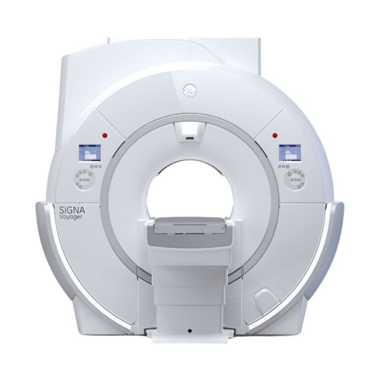
- GE SIGNA Voyager 1.5T: Offers a newer 1.5T platform with a spacious 70 cm wide bore, updated imaging technology, and efficient operating characteristics.
- GE SIGNA Pioneer 3T: Combines 3T imaging performance with energy-efficient gradient technology and wide-bore patient access.
- GE SIGNA Architect 3T: Supports advanced 3T imaging with a 70 cm bore, flexible coil technology, and comprehensive specialty applications.
- GE SIGNA Premier 3T: Emphasizes high-performance gradients and advanced imaging capabilities for demanding clinical and neuroscience applications.
The HDxt remains an established 1.5T alternative for facilities prioritizing versatile diagnostic imaging and configurable pre-owned equipment.
MXR Imaging can help compare these systems according to clinical requirements, patient volume, facility infrastructure, and budget.
Available SIGNA HDxt Equipment Packages
Pre-owned GE SIGNA HDxt systems can differ in hardware generation, receiving-channel architecture, software, patient tables, and accessories.
Available equipment packages may vary in:
- Imaging Hardware: Magnet configuration, gradients, receiving channels, and reconstruction equipment.
- Clinical Software: Installed applications, licenses, and compatible upgrades.
- RF Coils: Head, spine, body, extremity, and specialty coil packages.
- Patient Equipment: Table type, positioning accessories, and weight capacity.
- System History: Model year, maintenance records, magnet condition, and refrigeration performance.
MXR Imaging works with healthcare facilities to identify equipment configurations aligned with their examination requirements and installation needs.
SIGNA HDxt MRI Price & Availability
MXR Imaging offers pre-owned GE SIGNA HDxt 1.5T MRI scanners for healthcare facilities seeking high-definition diagnostic imaging with flexible equipment configurations.
Pricing depends on the selected system’s age, condition, gradient and RF hardware, installed software, receiving coils, patient table, and service history.
Eligible scanners may be available through MXR Imaging’s Platinum Certified program, which includes a 100-point reconditioning process, installation support, clinical applications training, and a full one-year warranty for qualifying equipment.
MXR Imaging maintains an ISO 13485:2016-certified quality management system and provides nationwide equipment, parts, installation, and service support to help healthcare facilities manage their imaging investment.
The total project investment may include transportation, installation, shielding, room preparation, and ongoing service.
MXR Imaging also provides MRI maintenance and repair services to support long-term equipment performance.
Contact MXR Imaging to discuss available GE SIGNA HDxt configurations, pricing, and installation requirements.
GE SIGNA HDxt Specifications
The specifications below describe the HDxt 1.5T platform and representative equipment configurations. Hardware generations, patient tables, software, and accessories can vary between systems.
Magnet System
-
- Field Strength: 1.5 Tesla
- Magnet Type: Superconducting
- Magnet Design: Actively shielded
- Bore Diameter: 60 cm (23.6 in)
- Maximum Imaging Field of View: 48 × 48 × 48 cm (18.9 × 18.9 × 18.9 in)
- Magnet Cooling: Liquid helium and mechanical refrigeration
- Imaging Coverage: Whole-body diagnostic MRI
Gradient System
-
- Gradient Technology: GE high-fidelity EchoSpeed gradients
- Maximum Gradient Amplitude: 33 mT/m for the original HDxt gradient configuration
- Maximum Slew Rate: 120 T/m/s for the original HDxt gradient configuration
- Gradient Shielding: Actively shielded
- Installed Gradient Package: Configuration-dependent
RF System & Reconstruction
-
- Receiving Architecture: 8-, 16-, or 32-channel configurations
- RF Coil Technology: Compatible high-density and phased-array coils
- Image Reconstruction: XVRE volume reconstruction
- Parallel Imaging: Supported with compatible ASSET and ARC applications
- Included Receiving Coils: Equipment-package dependent
Bore & Patient Table
-
- Bore Diameter: 60 cm (23.6 in)
- Patient Weight Capacity: 350 lb (159 kg) on the original documented table; up to 500 lb (227 kg) on applicable configurations
- Patient Table: Detachable, powered table in the original configuration; installed arrangements may vary
- Patient Entry: Head-first or feet-first for appropriate examinations
- Comfort Features: In-bore lighting and ventilation
- Positioning Accessories: Equipment-package dependent
Imaging Software
-
- Three-Dimensional Imaging: Compatible Cube applications
- Fat-Water Separation: IDEAL
- Motion Management: PROPELLER
- Neurological Imaging: Compatible diffusion, SWAN, BRAVO, BrainWave, and 3D ASL applications
- Body Imaging: LAVA, LAVA Flex, and additional supported techniques
- Vascular Imaging: Magnetic resonance angiography
- Cardiac & Breast Applications: Available with compatible software and coils
- Image Connectivity: DICOM support
Magnet Dimensions & Weight
Representative original HDxt magnet specifications:
-
- Magnet Width: Approximately 207 cm (81.5 in)
- Magnet Length: Approximately 173 cm (68.1 in)
- Magnet Height: Approximately 236 cm (92.9 in)
- Magnet Assembly Weight: Approximately 5,532 kg (12,198 lb), including cryogens and gradient coil
These dimensions describe the magnet assembly rather than the complete enclosed scanner footprint. Enclosures, patient-table equipment, and installation clearances increase the required space.
Installation & Utilities
-
- RF Shielding: Required
- Electrical Service: Dedicated supply appropriate to the equipment configuration
- Cooling Infrastructure: Required
- Cryogenic Refrigeration: Required
- Cryogenic Venting: Required
- Magnetic Safety Provisions: Required
- Installation Clearances: Determined by equipment configuration and room layout
Final equipment and installation specifications are matched to the selected system.