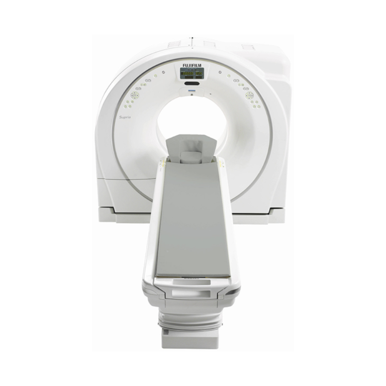
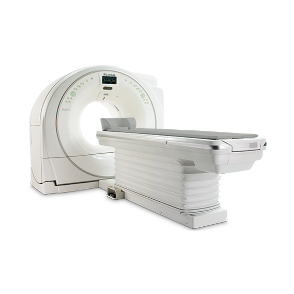
Fujifilm Supria Plus 32-Slice CT
- Category:
- CT Scanners
- Brand:
- Fujifilm
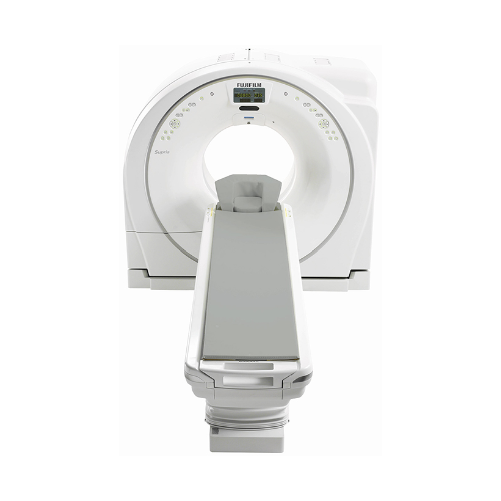
The Fujifilm Supria Plus is a compact 16-slice CT scanner with optional 32-slice reconstruction, well suited to hospitals, outpatient imaging centers, and specialty practices. Its 75 cm bore, 500 lb patient table, 180 cm scan range, and advanced dose management support versatile diagnostic imaging and patient access.
Supria Plus CT Highlights & Benefits
The Fujifilm Supria Plus CT combines routine diagnostic imaging, patient-focused design, and advanced dose management in a compact system. Its 16-channel acquisition platform supports optional 32-slice reconstruction, giving facilities flexibility for thin-section imaging without the installation requirements of larger CT platforms.
Key Supria Plus CT Capabilities
-
- 16-slice acquisition with optional 32-slice reconstruction: Supports detailed anatomical imaging and multiplanar views.
- 75 cm (29.5 in) gantry aperture: Provides additional room for patient positioning and accessibility.
- 500 lb (227 kg) patient table capacity: Accommodates a broad range of adult and pediatric patients.
- 180 cm (70.9 in) scannable range: Supports extended anatomical coverage and imaging of taller patients.
- Intelli IP and Intelli EC technology: Helps manage image noise and radiation exposure.
- Compact installation footprint: Supports minimum room layouts as small as 265 sq ft (24.6 m²), subject to site requirements.
- Eco Mode: Reduces idle power consumption by up to 35% compared with operation without Eco Mode.
Fujifilm Supria CT Video Preview
See the Supria family product tour for a closer look at Fujifilm’s CT design, patient access, and imaging workflow.
CT Imaging Detail & Scan Efficiency
The Fujifilm Supria Plus uses a 16-channel detector acquisition system with 20 mm of anatomical coverage per rotation. Its combination of thin-section imaging, selectable rotation speeds, and optional 32-slice reconstruction supports routine diagnostic examinations and detailed anatomical review.
16-Channel Detector & Thin-Slice Imaging
The solid-state ceramic detector uses 32 physical detector rows with 16-channel data output.
With a minimum slice thickness of 0.625 mm, the scanner captures thin-section data that can be reconstructed into detailed views of bones, joints, soft tissues, and other anatomical structures.
Rotation Speed & Extended Scan Coverage
The Supria Plus offers selectable rotation times of 0.75, 1.0, 1.5, and 2.0 seconds.
Its helical scanning mode supports continuous acquisition through the selected anatomical region, while the maximum 180 cm (70.9 in) scannable range accommodates extended-coverage examinations.
This combination is well suited to routine head-to-body CT and examinations requiring imaging across larger anatomical regions.
Optional 32-Slice Fine Recon Technology
The Supria Plus supports 16-slice image reconstruction, with optional Fine Recon technology enabling up to 32 reconstructed slices.
Fine Recon generates additional image sections from the acquired CT data, supporting smoother multiplanar views and improved anatomical visualization in examinations of the spine, joints, and other structures.
The scanner retains its underlying 16-channel acquisition hardware. The 32-slice designation refers to its optional reconstruction capability rather than 32-channel acquisition.
Supria Plus Image Quality & Radiation Dose Management
The Supria Plus combines image reconstruction, automatic exposure adjustment, and dose monitoring technologies to support consistent diagnostic image quality.
These capabilities help technologists tailor imaging protocols to patient size, anatomy, and examination requirements while managing radiation exposure.
Intelli IP Image Reconstruction
Intelli IP uses iterative reconstruction, a processing technique that reduces image noise by refining CT data during image generation.
Compared with conventional image reconstruction, Intelli IP can improve image clarity at a given exposure level. This supports lower-dose protocols when appropriate for the clinical examination.
Intelli EC Automatic Exposure Control
Intelli EC uses three-dimensional automatic exposure control to adjust X-ray tube current according to patient anatomy and size.
Instead of maintaining uniform exposure throughout an examination, the system adjusts output to help achieve the selected image quality while reducing unnecessary radiation exposure.
Dose Monitoring & Patient-Specific Protocols
The Supria Plus incorporates dose management capabilities aligned with the XR-29 Smart Dose Standard.
Key features include:
-
- Adult and pediatric protocols: Provide age-appropriate starting points for exposure settings.
- CT Dose Check: Alerts technologists when planned exposure exceeds established reference levels.
- DICOM dose reporting: Documents examination dose information for review and monitoring.
- Reduced-kV imaging: Supports selectable 80, 100, 120, and 140 kV settings.
- Locked protocols: Helps prevent unintended changes to approved examination settings.
Actual radiation exposure depends on the examination, patient characteristics, and selected imaging protocol.
Patient Accommodation & Exam Setup
The Fujifilm Supria Plus combines a spacious gantry opening, high patient weight capacity, and accessible table positioning in a compact design.
These features help facilities accommodate different patient sizes and mobility needs while supporting efficient examination setup.
75 cm Bore & Gantry Positioning
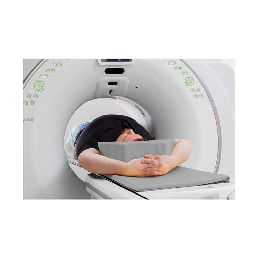
The 75 cm (29.5 in) gantry aperture provides additional positioning room compared with systems using 70 cm openings.
The gantry supports ±30° tilt, allowing adjustments for selected examinations, including head imaging.
A front-mounted digital display shows gantry tilt and table position, while integrated controls allow technologists to make positioning adjustments directly at the patient’s side.
500 lb Capacity Patient Table & Low Entry Height
The Supria Plus patient table supports up to 500 lb (227 kg) and offers an adjustable height range of approximately 17.7–39.4 in (450–1,000 mm).
The low minimum table height facilitates transfers for older adults, patients with limited mobility, and individuals requiring additional positioning assistance.
A 19 in (475 mm) tabletop and 75.2 in (1,910 mm) horizontal travel range support a broad range of examinations.
Patient Positioning & Communication
The system incorporates laser alignment, gantry-side controls, foot pedals, and automated patient instructions to simplify examination setup.
Integrated breathing lights and an intercom support communication throughout scanning.
Clinical Environments & Applications
The Supria Plus is best suited for facilities needing general diagnostic CT, emergency imaging, and selected advanced noncardiac examinations. Its combination of compact installation requirements, patient accessibility, and 16/32-slice imaging capabilities supports a broad range of clinical workflows.
Clinical Environments
-
- Hospitals and diagnostic imaging departments
- Outpatient imaging centers
- Emergency departments
- Community and regional healthcare facilities
- Multispecialty and specialty imaging practices
- Veterinary hospitals and specialty referral centers
Clinical Specialties
-
- Radiology: General diagnostic CT examinations, including head, neck, chest, abdominal, and pelvic imaging.
- Neurology and Neurosurgery: Brain imaging, head CT, and spine assessment.
- Orthopedics: Bone, joint, fracture, and implant evaluation.
- Oncology: Tumor assessment, staging, treatment planning support, and follow-up imaging.
- Pulmonology: Chest CT, lung evaluation, and low-dose chest examinations.
- Gastroenterology: Abdominal imaging and CT colonography.
- Urology: Renal, urinary tract, and pelvic imaging.
- Emergency Medicine: Evaluation of acute injuries and other urgent diagnostic conditions.
- Vascular Medicine: Selected contrast-enhanced vascular examinations.
- Veterinary Medicine: Diagnostic CT for animal patients, including soft-tissue, orthopedic, neurologic, and selected vascular examinations using appropriate veterinary positioning and protocols.
Common CT Applications
-
- Routine noncontrast and contrast-enhanced CT
- Head and brain examinations
- Chest and lung imaging
- Abdominal and pelvic imaging
- Spine and musculoskeletal CT
- Fracture and complex bone assessment
- Kidney stone and urinary tract evaluation
- Oncology staging and surveillance
- CT colonography
- Low-dose chest CT
- Selected CT angiography protocols
- Extended anatomical coverage examinations
- Veterinary head, spine, musculoskeletal, thoracic, abdominal, and selected vascular CT examinations
The Supria Plus also offers optional prospective ECG gating for coronary calcium scoring. However, it is primarily a general diagnostic CT platform rather than a dedicated cardiac CT angiography system.
Advanced Clinical & Software Capabilities
The Supria Plus supports additional clinical functionality through image-processing tools and optional software packages. These technologies help facilities expand anatomical review capabilities beyond standard axial imaging.
Multiplanar & 3D Image Reconstruction
Multiplanar reconstruction (MPR) allows CT data to be viewed in axial, coronal, sagittal, and other planes without repeating the examination.
The platform supports maximum and minimum intensity projection, surface rendering, and three-dimensional volume rendering.
MPR Spine Mode automatically selects reconstruction angles for spine examinations, reducing manual image setup.
Contrast Imaging & Metal Artifact Reduction
Predict Scan monitors contrast enhancement and can automatically initiate scanning when a selected threshold is reached, supporting consistent timing for contrast-enhanced examinations.
HiMAR metal artifact reduction processing helps minimize streaks and image distortion caused by metallic implants and other high-density objects.
These capabilities support more efficient review of vascular structures and anatomy near orthopedic hardware.
Optional Cardiac & Advanced Visualization Packages
Optional prospective ECG gating supports coronary calcium scoring examinations.
Compatible configurations may also incorporate TeraRecon advanced visualization software for expanded 3D image review and analysis.
Installed capabilities depend on the individual system’s software, licensing, and processing hardware.
Supria Plus CT Workflow & Connectivity
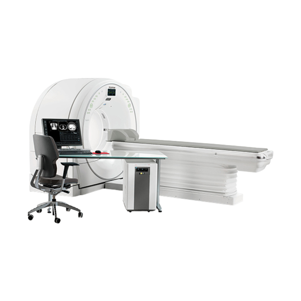
The Supria Plus combines patient registration, examination setup, image acquisition, and post-processing into a single operator workstation.
Its protocol-driven interface helps technologists reduce repetitive setup steps while maintaining consistent examination procedures.
Automated Protocol Selection & Image Review
The operator console uses a 24-inch widescreen display with 1920 × 1200 resolution.
Quick-entry controls, patient preregistration, and predefined examination protocols simplify workflow.
Immediate preview reconstruction allows images to be reviewed shortly after acquisition, with reconstruction performance up to 15 images per second depending on selected settings.
DICOM & Network Integration
DICOM connectivity supports integration with Picture Archiving and Communication Systems (PACS) and compatible imaging information systems.
Capabilities include DICOM Modality Worklist, Modality Performed Procedure Step (MPPS), and dose reporting. DICOM Query/Retrieve is available as an option.
Compatible configurations may also include a direct contrast injector interface and Fujifilm Sentinel remote diagnostic capability.
Available Supria Plus Configurations & Installed Options
The Fujifilm Supria Plus is available new through MXR, with configuration options to support different clinical imaging needs. Pre-owned systems may also be available, offering additional purchasing flexibility.
MXR works with customers to identify the appropriate reconstruction capabilities, software, accessories, and equipment configuration for their facility. Available features depend on the selected system, with pre-owned units also varying by equipment history and installed options.
16/32-Slice Configuration Differences
The standard Supria Plus platform provides 16-channel acquisition and supports 16-slice reconstruction.
Systems equipped with the Fine Recon option can generate up to 32 reconstructed slices, making the 16/32-slice designation relevant when evaluating available configurations.
Other configuration considerations include:
-
- Optional ECG-gated calcium scoring
- TeraRecon advanced visualization software
- Contrast injector integration
- DICOM networking and installed software licenses
- Operator workstation and processing hardware
- X-ray tube history, service condition, and included accessories
Supria Plus vs. Supria True64
Facilities requiring higher acquisition performance may also consider the Fujifilm Supria True64.
The Supria Plus uses 16-channel acquisition with optional 32-slice reconstruction, while the Supria True64 offers 64-slice acquisition with up to 128-slice reconstruction.
Both platforms emphasize patient accessibility and compact design, but their acquisition capabilities serve different performance requirements.
Compact Installation & Site Requirements
The Supria Plus was designed for space-efficient installation, making it a practical consideration for facilities replacing older CT equipment or introducing CT imaging within an existing building.
Minimum Room Size & Equipment Footprint
Fujifilm documents minimum room layouts of approximately 265 sq ft (24.6 m²), with typical layouts of approximately 342 sq ft (31.8 m²).
The system uses a compact three-module design consisting of the gantry, patient table, and image-processing cabinet.
Actual installation requirements depend on room dimensions, equipment clearances, shielding, patient access, and applicable regulations.
Electrical Requirements & Energy Efficiency
The documented electrical requirements include 208 VAC power, 200-amp service, and peak power capacity of 75 kVA.
The X-ray tube uses oil/air cooling without requiring a separate tube chiller.
Eco Mode can reduce power consumption by up to 35% during non-scanning periods compared with operation without Eco Mode.
MXR offers turnkey imaging project management to help coordinate CT equipment planning, installation, and facility requirements.
Supria Plus CT Price & Availability
MXR offers the Fujifilm Supria Plus as a new CT scanner, with pre-owned systems potentially available depending on inventory.
Pricing depends on the selected configuration, reconstruction capabilities, installed software, accessories, and installation requirements. For pre-owned equipment, system condition and X-ray tube history may also influence pricing.
MXR Imaging works with hospitals, imaging centers, and specialty practices to identify solutions aligned with their clinical needs, facility requirements, and budget.
We also provide CT maintenance and repair services to help support reliable equipment performance throughout the scanner’s operational life.
Contact MXR Imaging to discuss Fujifilm Supria Plus pricing, available configurations, and the best fit for your facility.
Fujifilm Supria Plus CT Specifications
The following specifications describe the manufacturer’s documented Supria Plus platform. Optional features and installed equipment configurations may vary.
CT Acquisition & Detector
-
- Acquisition channels: 16
- Maximum reconstruction: 32 slices with optional Fine Recon
- Detector type: Solid-state ceramic
- Detector arrangement: 32 rows × 880 elements
- Detector output: 16 rows × 880 elements
- Anatomical detector coverage: 20 mm (0.79 in) per rotation
- Minimum slice thickness: 0.625 mm
- Rotation times: 0.75, 1.0, 1.5, and 2.0 seconds
- Data collection: 1,200 views/second
- Volume scan pitch: 0.578–1.578
- Maximum scan range: 1,800 mm (70.9 in)
- Maximum scan rotations: 100
- Acquisition modes: Axial, helical, dynamic, and scanogram
- Scanogram directions: AP and lateral
Gantry & Patient Table
-
- Gantry aperture: 750 mm (29.5 in)
- Gantry tilt: ±30°
- Gantry dimensions (H × D × W): 1,843 × 920 × 1,990 mm (72.6 × 36.2 × 78.3 in)
- Gantry weight: Approximately 1,600 kg (3,527 lb)
- Table weight capacity: 227 kg (500 lb)
- Tabletop width: 475 mm (18.7 in)
- Table length: 2,688 mm (105.8 in)
- Table height range: 450–1,000 mm (17.7–39.4 in)
- Horizontal travel: 1,910 mm (75.2 in)
- Tabletop travel speed: 5–150 mm/second
- Horizontal positioning accuracy: ±0.25 mm
- Table weight: Approximately 423 kg (933 lb)
- Patient communication: Intercom and automated voice
- Positioning aids: Laser alignment and gantry-side controls
X-Ray Generator & Tube
-
- Generator type: High-frequency inverter
- Generator output: 48 kW
- Tube voltage: 80, 100, 120, and 140 kV
- Tube current: 10–400 mA in 5 mA steps
- Maximum tube current: 400 mA at 120 kV or below; 340 mA at 140 kV
- X-ray tube heat capacity: 5.0 MHU
- Maximum anode cooling rate: 748 kHU/min
- Tube cooling: Oil/air
- Focal spot sizes: 0.7 × 0.8 mm and 1.2 × 1.4 mm
Image Reconstruction & Performance
-
- Reconstructed slice thickness: 0.625–10 mm
- Reconstruction matrix: 512 × 512
- Display matrix: Up to 1024 × 1024
- Reconstruction speed: Up to 15 images/second, depending on settings
- Reconstruction field of view: 20–500 mm (0.8–19.7 in)
- Spatial resolution: 17.2 lp/cm at 0% MTF, 50 mm FOV
- Low-contrast resolution: 2.5 mm at 0.25% contrast, 160 mm FOV
- Iterative reconstruction: Intelli IP
- Automatic exposure control: Intelli EC
- Metal artifact reduction: HiMAR
- Image processing: MPR, MIP, MinIP, surface and 3D volume rendering
- Optional cardiac application: Prospectively ECG-gated calcium scoring
- Optional reconstruction: 32-slice Fine Recon
Operator Console & Connectivity
-
- Console display: 24-inch LCD widescreen
- Console resolution: 1920 × 1200
- Image storage: 110 GB, approximately 200,000 images
- Raw data storage: 200 GB, more than 6,000 scans
- DICOM connectivity: Modality Worklist, MPPS, Dose Structured Reporting
- DICOM Query/Retrieve: Optional
- Contrast injector interface: Optional
- Remote diagnostic capability: Fujifilm Sentinel
- Console and image processor: UPS-supported
System & Site Requirements
-
- Minimum documented room layout: 265 sq ft (24.6 m²)
- Typical documented room layout: 342 sq ft (31.8 m²)
- Electrical input: 208 VAC, ±10%
- Main breaker: 200 A
- Peak power capacity: 75 kVA
- Input frequency: 50/60 Hz
- Operating temperature: 20–28°C (68–82°F)
- Operating relative humidity: 35–80%
- Energy-saving capability: Eco Mode, up to 35% reduced idle power consumption
For additional information and specifications, access the PDFs provided below.