Towards the end of the 19th century, German physicist Wilhelm Rontgen studied the effects of passing an electrical current through gases at low pressure. What he ended up doing, on accident of course, was discovering X-rays. X-ray is energetic radiation capable of penetrating almost any solid object. This new discovery changed medicine almost overnight.
The first radiology department opened in a Glasgow hospital within a year. They were able to produce clear X-ray images of a kidney stone and a penny lodged in a child’s throat. Ever since, the medical industry has been using X-rays and medical imaging technology to explore the human body on the inside. The benefits of being able to see internal organs, tissues, bones, and systems is invaluable. Doctors are able to find broken bones and tumors, view brain activity, and perform a host of other applications thanks to the discovery.
Let’s take a deep dive and explore the world of medical imaging and the technology that makes it possible. Imaging technology explained: different types and their purpose.
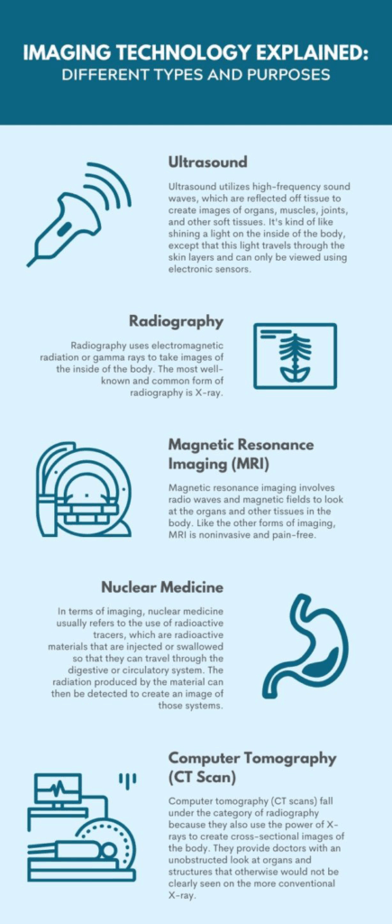
Ultrasound
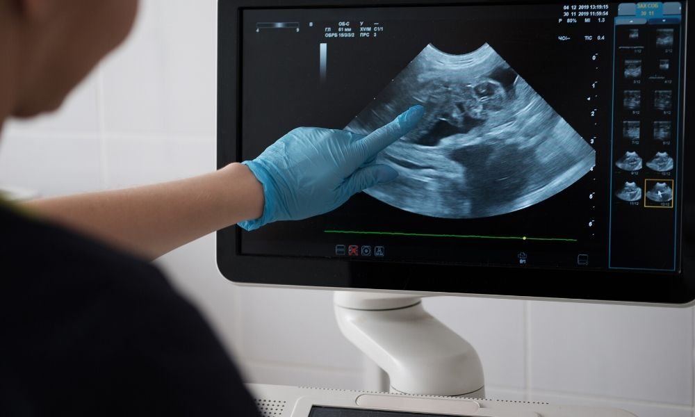
Ultrasound as a principal and concept predates X-rays. It was first documented in the late 18th century by someone studying echolocation in bats. That’s really the basis of ultrasound. Ultrasound utilizes high-frequency sound waves, which are reflected off tissue to create images of organs, muscles, joints, and other soft tissues. It’s kind of like shining a light on the inside of the body, except that this light travels through the skin layers and can only be viewed using electronic sensors.
It wasn’t until the 20th century that ultrasound as we know it came to be. Ultrasound was first used to detect tumors in the brain and then became the major diagnostic tool to monitor fetal growth in pregnant mothers. Ultrasound has become an invaluable diagnostic tool for doctors to examine patients and diagnose a variety of illnesses. They still use it to find tumors, but they also use it to promote healing and caring for strained muscles and chronic pain.
Today, there are dozens of ultrasound machines available, like the GE Logiq E9, and they continue to improve every year.
Radiography (X-Ray)
Radiography is the fancier, more technical-sounding name for X-rays. Radiography uses electromagnetic radiation or gamma rays to take images of the inside of the body. The most well-known and common form of radiography is X-ray. At one point or another, most of us have had an X-ray. It is a very versatile imaging technology that can capture an image of any part of the body.
For this procedure, an X-ray machine beams high-energy waves onto the body. The soft tissues, such as skin and organs, do not absorb the waves, whereas hard tissues, like bones, do. The machine transfers the results of the X-ray onto a film, showing the parts of the body that absorbed the waves in white and leaving the unabsorbed materials in black. Over the years the images have gotten better and clearer, making them more effective in discovering disease.
Magnetic Resonance Imaging (MRI)
Any sports fan is familiar with the MRI. Athletes get them daily, it seems, to find out what ligament they pulled and what muscle they tore during the game. Magnetic resonance imaging involves radio waves and magnetic fields to look at the organs and other tissues in the body. Like the other forms of imaging, MRI is noninvasive and pain-free.
The procedure requires an MRI scanner, which is a large tube that contains a massive circular magnet. The patient lies on the table and is fed into the futuristic-looking machine while the magnet circles around them, gathering images. This magnet creates a powerful magnetic field that aligns the protons of hydrogen atoms in the body. Those protons are then exposed to radio waves, causing the protons to rotate. When the radio waves are turned off, the protons relax and realign themselves, emitting radio waves in the recovery process that can be detected by the machine to create an image. The faster the protons realign, the clearer the image is.
Because MRI doesn’t use any kind of radiation to produce the images, it is the modality of choice when frequent imaging is needed, especially in the brain.
Nuclear Medicine
Nuclear medicine is a rather general term that involves any medical use of radioactive materials. But in terms of imaging, it usually refers to the use of radioactive tracers, which are radioactive materials that are injected or swallowed so that they can travel through the digestive or circulatory system. The radiation produced by the material can then be detected to create an image of those systems. As the tracer travels through the area of examination, it leaves an energy trail in the form of gamma rays.
A special camera detects the rays and sends the information to a computer, where an image is created. Nuclear imaging provides unique information that can’t be obtained with other imaging procedures. It has the ability to catch terminal diseases in their earliest stages. It is most often used when a doctor wants to see the structure and function of a bone, organ, tissue, or system within the body. The only pain involved is when the tracer is injected via needle. There are no known side effects to the use of the tracer.
Computer Tomography (CT Scan)
Last on our explanations of different types and purposes of imaging technology is computer tomography. Computer tomography (CT scans) fall under the category of radiography because they also use the power of X-rays to create cross-sectional images of the body. They provide doctors with an unobstructed look at organs and structures that otherwise would not be clearly seen on the more conventional X-ray. During a CT scan, the patient lies on a motorized table inside a large circular chamber that is similar to an MRI machine. The low-dose X-ray source and its detector or receiver rotate around the patient to create a series of 3D slides of the inside of the body.
Stacked together, these slices create a virtual, 3D representation of the affected area and can be viewed as a whole or by themselves. Once compiled together, the entire human body can be viewed from head to toe, and individual slides are accessible. Thousands of the slides are cataloged, and a doctor can view a specific one to make a diagnosis.
MXR Imaging is the largest independent distributor of imaging devices, providing imaging equipment, service, parts, and supplies for CT, MR, X-Ray, PET/CT, and Ultrasound.