In the medical field, there is a lot of information being gathered and shared. Doctors and nurses gather patient data every day so that they can find out what is ailing them. Diagnostic tools in hospitals and clinics all over the world allow medical professionals to gather that data, analyze it, decide, and share it with the patient. What kind of symptoms a patient describes dictates what diagnostic tool a doctor will use to find the cause of them. One of the most trusted and valuable diagnostic tools at their disposal is the ultrasound machine. These are used to examine the internal organs and structures of a person without having to make an incision. Most ultrasounds are completely noninvasive and causes no pain to the patient.
Ultrasound machines have been in clinical use since the 1950’s and continue to be the go-to device for diagnosing patients. There are different kinds of ultrasound examinations, like the echocardiogram. As the name implies, it uses sound waves from an ultrasound machine and the resulting echoes ping back and forth to create an image of the heart. Echocardiograms are used by cardiologists to gauge the existence and progression of heart disease in their patients.
Heart Disease Defined
Heart disease is an umbrella term used to describe many types of heart conditions. There are dozens of heart related conditions that could fall under this open definition. One of the most common forms is the narrowing of the arteries due to a build-up of cholesterol. The constant narrowing leads to full blockages and eventually a heart attack. It is among the most common type of heart disease in the world. Most people are unaware of the onset of heart disease until systems become life threatening.
According to the Centers for Disease Control, in the U.S.:
- Heart disease is the leading cause of death for men and women of all racial and ethnic groups.
- Every 37 seconds a person dies from cardiovascular disease
- One in every four deaths is due to heart disease
Suffice it to say that heart disease is a big problem in the U.S. and the modern world. The abundance of fast, easy, and high cholesterol food and a sedentary lifestyle contributes to the declining health of the world. Heart disease will likely rise until educational programs on healthy living and eating can be implemented across generations.
Causes of Heart Disease
Heart disease is the long term effect of a lifestyle choice that can be overcome. There are many physical warning signs that alert us all to the dangers. Things like obesity, diabetes, smoking, and excessive alcohol use all contribute to heart disease and increase the risk of heart attacks. Almost half, 47%, of Americans have one of the three risk factors; smoking, high cholesterol, and high blood pressure.
Bad habits like smoking and drinking can be overcome by stopping those behaviors. Obesity can be harder to defeat due to lifestyle choices and genetics. Some people are predisposed to being overweight due to their genetic makeup. It is harder for them to shed pounds and maintain a healthy lifestyle. The symptoms of heart disease can provide warning signs of an impending heart attack. Things like chest pain, upper back or neck pain, indigestion, heartburn, nausea or vomiting, fatigue, upper body discomfort, and shortness of breath are all signs that you have heart disease and should see a doctor.
What’s an Echocardiogram?
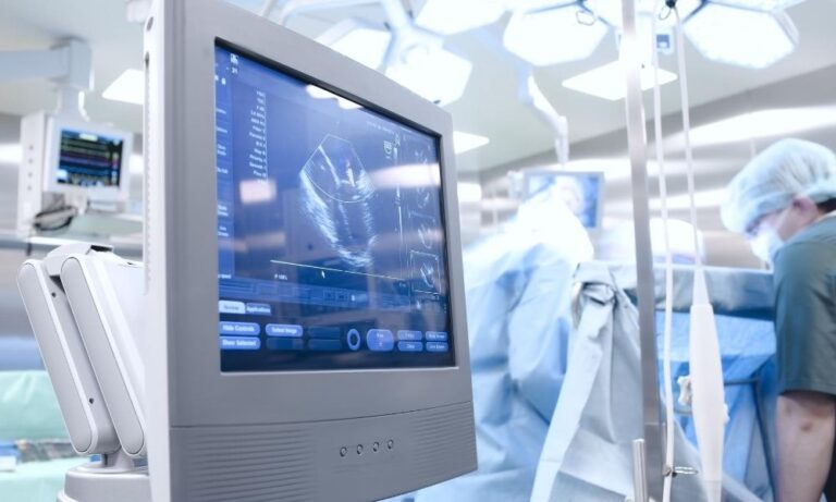
An echocardiogram is a diagnostic procedure that doctors use to examine the heart. An ultrasound machine will produce sound waves, transmit them through a probe into your body to create an image of your heart in real time. It can capture real time images, making it possible for you to view your beating heart. The test is so the doctor can view the blood flow in and out of your heart and look for blockages.
They are also able to look for problems within the heart chambers and valves and look for the problems that create shortness of breath. Doctors also utilize the echocardiogram to check for congenial heart defects in babies before birth, called a fetal echocardiogram. Depending on what kind of information a doctor needs about you, they can order different kind of echocardiograms.
Different Kinds of Echocardiograms
Transthoracic Echocardiogram
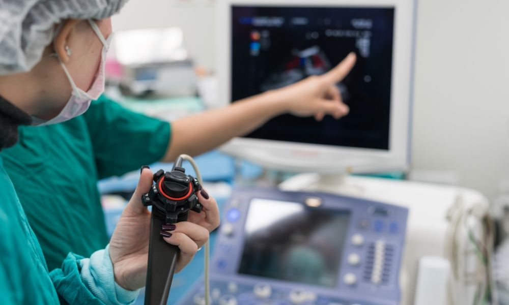
This is the most common type employed by doctors. It is the least invasive of the group and causes the least amount of discomfort for patients. A sonographer will rub a gel on the chest of the patient to ease movement of the probe and help the sound waves. They will press the transducer probe against the skin and slowly move it around.
The waves pulse back and forth within the body and the ultrasound machine translates the information into an image. The image of the heart will appear on the screen for observation. If there is difficulty viewing the heart and blood vessels due to the rib cage, the patient can be given an enhancing agent through an IV line. The agent will make the heart and blood vessels easier to see on the screen.
Transesophageal Echocardiogram
If the doctor wants more detailed images of the heart, they will order a transesophageal echocardiogram. This involves a transducer being snaked down the patient’s throat to get a better image. The patient’s throat is numbed, and they are given medication to help them relax. A flexible tube containing a transducer probe is guided down the throat and into the esophagus connecting the mouth to the stomach.
The probe sends the sound waves towards the heart and records the information and creates the image. Sending the probe into the stomach prevents the ribs from blocking the sound waves and can make a clearer picture for the doctor so view.
Stress Echocardiogram
Some heart problems only manifest during physical activity. Symptoms that involve the arteries that supply blood to the coronary arteries. The doctor would order this kind of test to check for coronary artery problems.
This kind of test is limited though because it can’t show any blockages in the heart. Ultrasound images of the heart are taken before and immediately after a patient walks on a treadmill or rides a stationary bike for a period. The doctor will compare the sets of information and look for abnormalities and differences between the two.
Contact MXR Imaging today and find out what kind is right for your practice.