Since the invention of the ultrasound, or sonogram, in the middle of the twentieth century, it has been closely associated with pregnancy. The first thing that comes to mind when you think of an ultrasound is probably fetal health and monitoring. Even though they’re often associated with pregnancy and prenatal care, ultrasounds can do much more. In fact, the first medical application of the ultrasound was to detect brain tumors—their use has expanded from there.
Sonograms are invaluable tools in preventative screenings. Medical device companies have spent many years and millions of dollars developing these machines. Their ability to detect diseases before they become fatal has saved countless lives. Ultrasound screenings help doctors find diseases before any symptoms are present. If caught early enough, these diseases can be treated before they become life-threatening.
Many different types of ultrasound machines can detect a variety of conditions before they become more serious. Most ultrasounds are noninvasive and pain-free, and they’re used in doctors’ offices by trained professionals so that you get the best possible care.
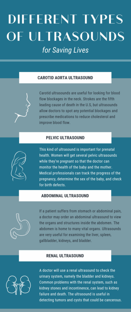
Different Types Of Ultrasounds
Carotid Aorta Ultrasound
Simply stated, this is an ultrasound of the neck, but with a specific target. The human brain gets blood from the main artery in the body, the aorta. Stemming from the heart, the aorta branches off into four smaller arteries. The two smaller arteries travel deep to the back of the neck, where they cannot be touched. They supply blood to the rear of the brain. The other two arteries, known as the carotid arteries, are the two main blood vessels in the neck. When you feel your pulse in your neck, you’re feeling those carotid arteries. They’re responsible for transporting oxygenated blood to the front part of the brain. This is the part of the brain that controls speech, thinking, personality, and sensory and motor functions.
The more clinical name for carotid artery disease is carotid artery stenosis. Stenosis refers to the narrowing and hardening of the arteries from the inside. Cholesterol and fatty deposits build up in the arteries and begin to restrict the flow of blood. As the plaque builds up, it can lead to an occlusion, or the total blockage of the artery. A stroke will occur if the flow of blood to the brain experiences any interruptions. Carotid ultrasounds are useful for looking for these blood flow blockages in the neck. Strokes are the fifth leading cause of death in the U.S, but ultrasounds allow doctors to spot any potential blockages and prescribe medications to reduce cholesterol and improve blood flow.
Pelvic Ultrasound
Most people are familiar with this kind of ultrasound because it’s important for prenatal health. Women will get several pelvic ultrasounds while they’re pregnant so that the doctor can monitor the health of the baby and the mother. With this tool, medical professionals can track the progress of the pregnancy, determine the sex of the baby, and check for birth defects. These ultrasounds are good for more than just examining the progress of a fetus: they also can help the doctor examine the ovaries, uterus, and bladder to spot any cancer or developing diseases. The images are useful in detecting ovarian cancer, internal bleeding, and occult hernias in women.
Pelvic ultrasounds aren’t only for pregnant women: men can also receive pelvic ultrasounds as a preventative measure against disease. The prostate is in the pelvic region, and it’s a source of trouble for a lot of men. Many men over the age of 50 suffer from an enlarged prostate and the associated problems—mostly trouble with urination. Prostate cancer is the most common type of cancer in men after skin cancer, but the mortality rate for prostate cancer is low. A pelvic ultrasound will help the doctor spot any irregularities early on and treat any diseases.
Abdominal Ultrasound
If a patient suffers from stomach or abdominal pain, a doctor may order an abdominal ultrasound to view the organs and structures inside the abdomen. The abdomen is home to many vital organs—as well as many potential problems. Ultrasounds are very useful for examining the liver, spleen, gallbladder, kidneys, and bladder. An ultrasound can spot kidney stones, gall stones, cancerous tumors, and cysts in any organ.
Sonograms are also the preferred screening method for an abdominal aortic aneurysm. That’s a bulging or weakened spot in the abdominal aorta, the major blood vessel that transports blood throughout the body. If you are or have ever been a smoker, or if you’re a man between 65 and 75 years old, doctors will recommend this kind of ultrasound to check for an aneurysm. Because the abdominal aorta is the largest blood vessel in the body, a rupture in it could be life-threatening. The survival rate for an abdominal aortic aneurysm is quite low. 50 percent of people with an aneurysm won’t make it to the hospital. If they do make it to the emergency room, the survival rate drops one percent every minute. With such a high mortality rate, regular screening for this condition is important.
Renal Ultrasound
The renal system is also known as the urinary system. It’s responsible for the creation, storage, and elimination of urine within the body. It includes the kidneys, the ureters, the bladder, and the urethra. The kidneys filter waste within the body to create urine, and the bladder stores it until the urethra excretes it. Kidney disease increases the risk of other life-threatening problems such as stroke or heart attacks. Major risk factors for kidney disease include diabetes, high blood pressure, and a family history of kidney problems.
A doctor will use a renal ultrasound to check the urinary system, namely the bladder and kidneys. Common problems with the renal system, such as kidney stones and incontinence, can lead to kidney failure and death. The ultrasound is useful in detecting tumors and cysts that could be cancerous. Abscesses, infections, and obstructions within the renal system are also visible with ultrasound. Ultrasound images are useful as a roadmap of sorts so that doctors can remove liquid from the kidneys and obtain samples. Kidney stones will show up on ultrasound images as well.