The invention of ultrasound transformed the way we evaluate, diagnose, and treat certain medical conditions. It allowed for a more in-depth examination of lesions, masses, tissues, and organs, such as the heart. Today, ultrasounds are widely used as a medical imaging technique and a useful diagnostic tool.
There are several common medical procedures that use ultrasound — here are just a few.
What is an Ultrasound?
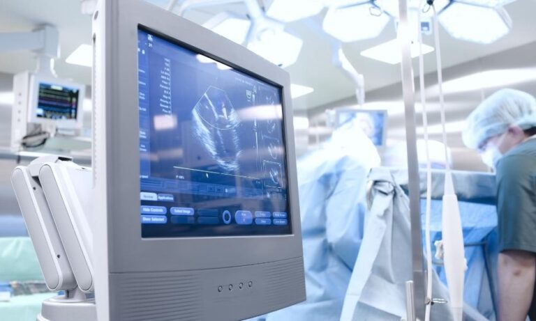
An ultrasound uses high-frequency sound waves to visualize muscles, tendons, and internal organs and detect abnormalities in their structure or operation. It can detect certain diseases, conditions, injuries, and cancerous lumps, making it a valuable tool for diagnosis. Ultrasounds are quick, safe, and, in most cases, non-invasive.
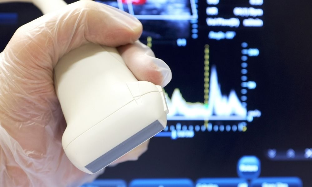
During the procedure, a transducer probe is outfitted with a thin layer of gel and placed on or within the body. Waves are transmitted through the gel and reflect back to the transducer. As these reflected waves hit the transducer, they generate electrical signals that transfer to the scanner. The scanner uses these signals to generate a two-dimensional image of tissues and organs.
What is Ultrasound Used For?
Ultrasounds are a common diagnostic tool, used for evaluating, diagnosing, and treating various medical conditions. They’re used in wide range of procedures, including the following.
Abdominal Ultrasound
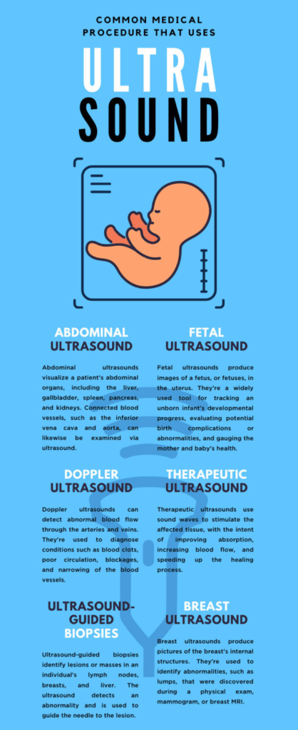
Abdominal ultrasounds visualize a patient’s abdominal organs, including the liver, gallbladder, spleen, pancreas, and kidneys. Connected blood vessels, such as the inferior vena cava and aorta, can likewise be examined via ultrasound. These ultrasounds can help professionals evaluate the cause of stomach pain or bloating and check for kidney stones, liver disease, and tumors.
These ultrasounds can help professionals evaluate the cause of stomach pain or bloating and check for kidney stones, liver disease, and tumors. For patients at risk of an abdominal aortic aneurysm, a one-time screening is recommended. Men between the ages of 65 to 75, especially those who are smokers, are the most susceptible to the condition.
Bone Sonometry
Bone sonometry, a procedure that relies on specialized ultrasound machines, is used to determine a bone’s level of fragility. It provides information about the bone’s strength, structure, and elasticity. The procedure detects conditions such as osteopenia and osteoporosis, in which the bone has few minerals, poor density, and an increased risk of fractures. Bone sonometry is commonly used to examine the heel, fingers, wrist, or tibia.
Breast Ultrasound
Breast ultrasounds produce pictures of the breast’s internal structures. They’re used to identify abnormalities, such as lumps, that were discovered during a physical exam, mammogram, or breast MRI. This procedure can determine the lump’s status as either non-cancerous or cancerous, fluid-filled, or cystic. Ultrasounds make it easier to detect lesions in pregnant women, women with dense breast tissue, or individuals who are unable to undergo an MRI.
Doppler Ultrasound
Doppler ultrasounds can detect abnormal blood flow through the arteries and veins. They’re used to diagnose conditions such as blood clots, poor circulation, blockages, and narrowing of the blood vessels. The detection and prevention of blocked and reduced blood flow are critical for preventing a stroke. Individuals who show signs of deep vein thrombosis, superficial thrombophlebitis, arteriosclerosis, thromboangiitis obliterans, or vascular tumors should consult their doctor. Their doctor may recommend an ultrasound to assess the issue.
Echocardiogram
Echocardiograms, performed with high-quality ultrasound equipment, monitor the movement of the heart’s valves and chambers. They’re used to assess the overall function of your heart and detect conditions such as valve disease, myocardial disease, pericardial disease, cardiac masses, and congenital heart disease. Echocardiograms can track the progress of a patient’s disease and gauge the effectiveness of their medical or surgical treatments over time. Physicians frequently pair them with color Doppler and Doppler ultrasounds, which allow for the evaluation of blood flow across the heart valves.
Fetal Ultrasound
Fetal ultrasounds produce images of a fetus, or fetuses, in the uterus. They’re a widely used tool for tracking an unborn infant’s developmental progress, evaluating potential birth complications or abnormalities, and gauging the mother and baby’s health. Depending on the pregnancy’s risk level, physicians may offer them during the first or second trimester. Third-trimester ultrasounds are rarer but do exist. Among other things, fetal ultrasounds can confirm a pregnancy, determine a baby’s gestational age, identify multiples, and, later, confirm the sex of the baby. Genetic conditions, such as Down syndrome, can be predicted with ultrasounds, as can heart defects.
There are two types of fetal ultrasound: transvaginal and transabdominal. The former is recommended during early pregnancy when the pea-sized fetus is difficult to detect through tissue and skin. The latter is intended for pregnancies that are further along.
Doppler Fetal Heart Rate Monitors
These monitors are small, handheld devices that can detect and monitor the fetal heartbeat. If there are concerns over a fetus’s health, a monitor will track the heartbeat and ensure everything’s functioning normally. An abnormal heart rate can signify a lack of oxygen, amongst other issues. Doppler fetal heart rate monitors are commonly used in the third trimester or during labor and birth.
Ultrasound-Guided Biopsies
Another common medical procedure that uses ultrasound is, as the name suggests, the ultrasound-guided biopsy. Ultrasound-guided biopsies identify lesions or masses in an individual’s lymph nodes, breasts, and liver. During the procedure, local anesthesia is given to numb the target area. The ultrasound detects an abnormality and is used to guide the needle to the lesion. A tissue sample is removed for testing. Specialists determine whether a mass is cancerous or benign, and treatment proceeds from there. If a patient reports unusual lumps or if a different test, such as a mammogram or MRI, unearths suspicious findings, an ultrasound-guided biopsy may be recommended.
Ophthalmic Ultrasound
Ophthalmic ultrasounds assess the pathology and structure of the eye. They’re useful for patients with complicated pathologies, such as dense cataracts or large corneal opacities, that prevents ophthalmoscopy. Ophthalmic ultrasounds can identify the cause of acute changes in vision and eye pain, as well as an elevation in intracranial pressure. Globe rupture, retrobulbar hematoma, lens detachment, and vitreous hemorrhage are some of the different conditions and traumas that ophthalmic ultrasounds can detect.
Therapeutic Ultrasound
Ultrasounds have been used to treat certain musculoskeletal conditions and injuries, such as muscle strains or runner’s knee. Therapeutic ultrasounds use sound waves to stimulate the affected tissue, with the intent of improving absorption, increasing blood flow, and speeding up the healing process.
The use of ultrasound for therapeutic purposes is highly debated, and until further research emerges, its efficacy is doubtable. If you’re looking for effective, high-quality ultrasound equipment and machines, browse through MXR Imaging’s impressive inventory for all the latest technology.
Your Partner for Quality Ultrasound Equipment and Service
Whether you’re in need of high-quality ultrasound equipment for sale, a specialized transducer probe, or professional ultrasound repair service, a reliable partner is essential. Our team is dedicated to helping you find the right ultrasound machines and providing expert ultrasound maintenance, including comprehensive ultrasound probe repair and access to a wide range of parts for sale, ensuring your practice runs smoothly and efficiently.