
An abnormal collection of fluid in the lungs is a sign of pleural effusion, which can complicate a patient's breathing and cause sharp, stabbing chest pains. To remove excess fluid in the lungs, a professional might recommend an ultrasound-guided thoracentesis. You might wonder, what is ultrasound-guided thoracentesis? This guide will outline what ultrasound-guided thoracentesis is, how it's performed, and some of the complications associated with the procedure.
What Is Thoracentesis?
Thoracentesis is used to determine the cause of fluid in the lungs or to remove large amounts of pleuritic fluid. It can be both diagnostic and therapeutic for the patient. Removing excess fluid can decrease or eliminate chest pain, coughs, shortness of breath, and other symptoms associated with pleural effusion. In many cases, thoracentesis is performed with ultrasound. This makes it easier for the radiologist to select the insertion site and monitor the patient's lungs during the procedure. Since ultrasound is in real-time, the physician can accurately guide the needle placement into the pleural effusion area.
What Is the Procedure Like?
To fully understand what ultrasound-guided thoracentesis is, you’ll need to learn how it’s performed. During the procedure, the patient is seated in an upright position and asked to lean forward. Although, depending on where the pleural area is located, the patient may lie flat on their back.
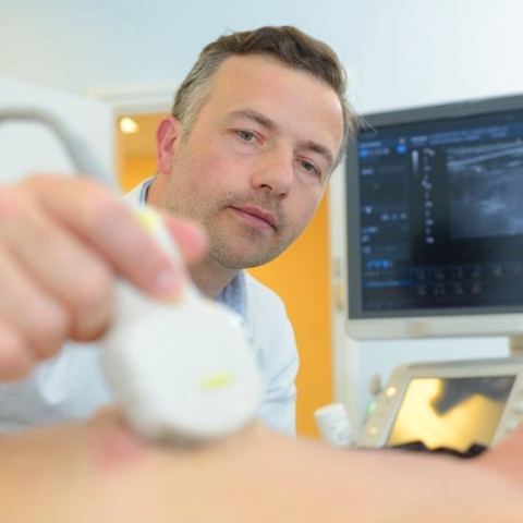
An ultrasound transducer is placed on their back in a sagittal or transverse position, which allows the ultrasound technician to visualize the lungs and other internal organs. Using ultrasound as a guide, the radiologist locates the best site to insert the needle. They clean the site with an antiseptic solution and administer a local anesthetic to the patient. The radiologist will then insert a needle through the patient's chest wall and guide it into the pleural effusion area (being careful not to puncture the lungs or other internal organs), where it will remove or collect fluid accumulation.
The collected fluid is sent to the lab for examination. Once the results are collected, they’re sent to a physician for analysis. The entire exam can last anywhere from 20 to 30 minutes.
Are There Risks Involved?
There’s a high complication rate associated with thoracentesis. Using ultrasound to guide the procedure decreases the risk of damage or unwanted side effects. Two major complications of ultrasound guided thoracentesis are pneumothorax and the puncturing of lung tissue, cystic masses, empyema, or mediastinal structures. A chest x-ray will follow the patient's procedure, and they'll be monitored for one to two hours following the exam.
Conquest Imaging is the best place to find efficient, high-quality ultrasound equipment and accessories. We offer some of the latest ultrasound machines, such as the Philips iE33™, along with transducer probes, machine parts, and repair services.
Authored by Jacqueline Guerra
Ultrasound Imaging Expert
Former industry expert for Conquest Imaging and MXR Imaging