Magnetic Resonance Imaging: a Brief MRI History
The invention of the MRI (magnetic resonance imaging) has greatly improved the ability to accurately diagnose a wide range of medical conditions. As a result, it's had an enormous impact on the healthcare industry as a whole. These days we are heavily dependent on MRIs to evaluate certain problems that patients are having, yet they haven't been around for that long.
The following explores what an MRI system does, the type of technology it uses, how it was invented, and what kind of changes have been made to improve the MRI system's capabilities over the years.
What is an MRI?
An MRI is a diagnostic imaging procedure that makes use of magnets, radio waves, and a computer to record high-resolution images of the inside of your body. It's different from other similar technology, such as CT scans and X-rays in that it doesn't use radiation. Doctors use MRIs to accurately diagnose diseases or injuries as well as to monitor treatment for specific diseases or injuries. MRIs can test every part of your body.
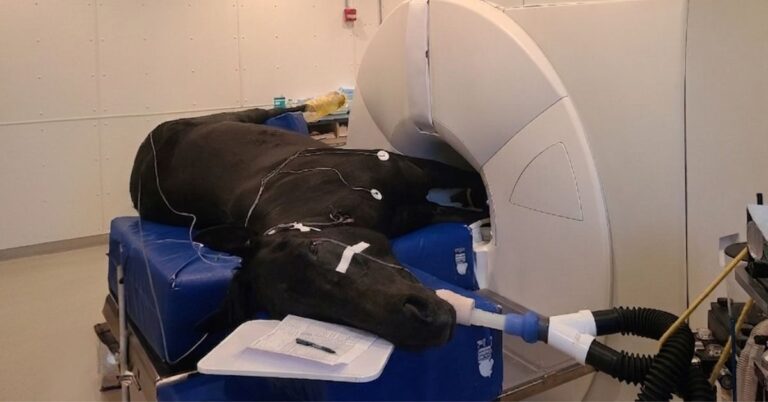
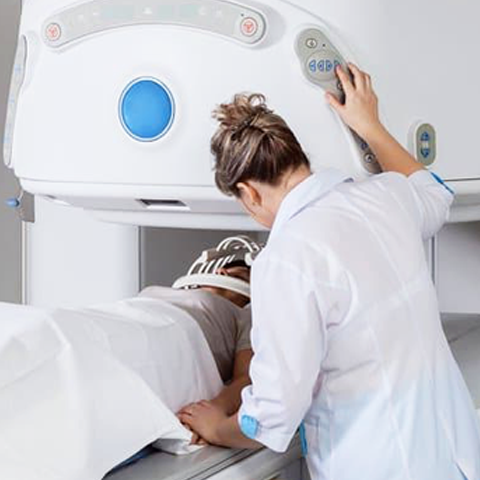
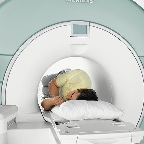
MRI scans are performed by first having the patient lie on the MRI table, and then moving the table through the MRI scanner. The traditional MRI system consists of a table that slides into a large tube. The patient lies in the mostly enclosed tube for anywhere from 15 to 90 minutes depending on the type of procedure is being performed and what part(s) of their body are being scanned. It’s important for the patient to hold still during the procedure to ensure the images turn out clear. If they move, it could blur the images, requiring the procedure to be repeated.
The way the MRI system works is by using magnets that produce a strong magnetic field. This causes the protons within the water molecules of the body to become aligned. When the magnetic field is turned off, the protons will slowly return back to their normal state. As this occurs, signals are picked up by the magnets RF coils, which are measured by the MRI scanner and produced into an image.
Technology That Led the Way to Modern MRI Being Possible
Although many scientists were working with magnetic fields and nuclear magnetic resonance (NMR) for many decades prior to the invention of the scanning device, the MRI system as we know it today would not be possible without the superconducting magnet, which was developed by Oxford Instruments.
Oxford Instruments Developed Superconducting Magnet
The MRI system's bore is inside of the magnet itself, which takes the shape of a large cylinder. The magnet is a superconducting magnet, which is an electromagnet that consists of coils made from superconducting wire which has no electrical resistance when cooled to cryogenic temperatures. Because the wire lacks electrical resistance, it's able to conduct greater electric currents than ordinary wire, thereby allowing it to produce intense magnetic fields. To operate safely, the magnet must be cooled to below their critical temperature using either liquid helium or a two-stage mechanical refrigeration.
In 1959, Oxford Instruments, the first commercial spin-off company from the University of Oxford, was founded to manufacture superconducting magnet for scientific research. Oxford Instruments had a pioneering role in the development of magnetic resonance imaging by providing the first superconducting magnets for this application.
Who Invented The MRI System
No single person can be credited for the invention of the MRI system. Instead, multiple scientists made discoveries over a period of time that all contributed to the development of the MRI system.
For example, the chemist Paul C. Lauterbur won the Nobel Prize in Physiology or Medicine in 2003 for his contribution to the use of MRI in medical research and diagnostics. It was his use of nuclear magnetic resonance in the study of molecules, solutions, and solids that led to the application of NMR technology to medicine, which eventually led to the development of the MRI system.
However, Raymond Damadian is often credited as one of the main contributors to the invention of the MRI system as well. Damadian discovered that the use of NMR could be used to distinguish tumors from normal tissue. In 1977, he became the first to perform a full body scan to diagnose cancer. Both he and Lauterbur played an important part in the development of the MRI, but there has been a lot of controversy over who played the more important part - with Damadian believing he was snubbed for the Nobel Prize when it was awarded to Lauterbur.
Several other individuals played an important part in discovering new technology that would eventually make the MRI system possible, including Nikola Tesla. The idea of applying a rotating magnetic field in an AC motor was attributed to him as well as Galileo Ferraris. Tesla claimed he came up with the idea in 1882 during a stroll in the park, while Ferraris claimed he wrote about the concept and built a working model in 1885. However, there is no independent verification for these claims. Tesla did receive a patent for his design in 1888 - and to this day, the strength of the magnetic field produced by MRI scanners is measured in units called Tesla.
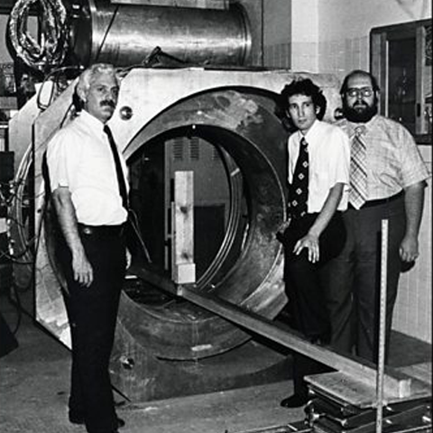
First Commercial MRI Whole Body Scanner Manufactured by Oxford in 1980
Building superconducting magnets isn't the only thing Oxford Instruments is known for. They were also the first company to build the first commercial whole-body MRI scanner back in 1980. It was built at their Osney Mead factory in Oxford and then installed at the Hammersmith Hospital in London. This eventually led to Oxford Instruments manufacturing superconducting magnets for MRI system manufactures such as General Electric, Siemens, and Philips.
How MRI Technology Has Changed since 1977
MRI technology has continued to advance over the past few decades. Not only have they made advancements in greatly reducing the noise levels of the systems, but a variety of different types of MRI systems are in use now as well. In addition to the standard closed MRI systems, the following are some of the different types of MRI systems now available:
- Open MRI Systems - Open MRI systems are designed so that, in addition to the opening in which the patient is slid, the sides are open as well. This makes getting an MRI scan much less uncomfortable for patients who might be suffering from an anxiety disorder or who are claustrophobic.
- Upright MRI Systems - Upright MRI systems allow patients to sit down or stand up during the procedure. They eliminate the need to lie underneath the system, making it much easier for patients with claustrophobia or anxiety to use. They're also more convenient for patients who may be a bit overweight as they can accommodate individuals upwards of 500 pounds. Additionally, they make it easier to diagnose certain conditions, such as joint issues, since it allows the patient to be scanned in a weight-bearing position.
- Extremity MRI Systems - Extremity MRI systems completely eliminate any kind of discomfort a patient might have since they only have to insert one of their hands, arms, legs, or feet at a time. These systems are built to perform MRI scans specifically on these areas of the body, which means that they cannot be used to diagnose conditions in the head or torso.
More recently, MRI advancements have come in the form of software advancements. New types of MRI software have allowed for faster scan times, simplified cardiac imaging workflows, lung scans, shortened prostate exams, and more.
How Magnetic Resonance Imaging Has Changed Healthcare
The use of MRIs has drastically changed healthcare for the better. MRI scans have saved countless lives by significantly reducing the risk of misdiagnosis. They allow doctors to see exactly what the problem is so that they can begin treating it right away -- and to see whether treatment is working so that they can make adjustments if necessary. For instance, MRI scans can help to detect cancer in the early stages when it is optimally treatable. Additionally, it's a non-invasive test that does not involve radiation, which means there are no potential side effects.
MRI scans can be used to diagnose issues in every part of the body. For example, MRI scans can be taken of the head and spinal cord in order to identify blood vessel damage, brain injuries, spinal cord injuries, multiple sclerosis, stroke occurrences, and cancer. MRIs can also be used to identify blocked blood vessels, heart disease, heart attack damage, and structural problems in the heart. These are just a few examples of some of the conditions that can be diagnosed.
What is the future of MRIs and medical physics
MRI systems are continuing to advance, especially in the strength of their magnets. Newer MRI systems have been recorded producing magnetic fields as high as 11.7 Tesla. This kind of magnetic strength could potentially reveal details about the body that were impossible to capture until now. Additionally, advancements in artificial intelligence (AI) have led to the development of image-guided, semi-autonomous robots that can guide a procedure using real-time MRI scanning. Although scientists still have to discover how to build robots that can fit within an MRI system to perform a procedure, the potential is there. The creation of MRI systems has led to significant medical breakthroughs and the continued advancement of MRI technology can be expected to continue impacting healthcare significantly in the future.
| Authored by Rex Lindsey Specialist – CT, MRI, and PET/CT MXR Imaging |